Download presentation
Presentation is loading. Please wait.

1
Peptic ulcer Presented by د. قصي العبيدي بورد ( دكتوراه ) جراحه عامه جامعة الكوفة - كلية طب
 جراحه عامه جامعة الكوفة - كلية طب")
3
Axises of lectures What is peptic ulcer? What are the causes? Which group of people more likely affected? How patient present? How we can reach to diagnosis? What are the lines of treatment? What are the complication?

4
Aims of lectures: 1.identification what is peptic ulcer? 2.understanding underlying causes for this disease. 3.identify which group of people more susceptible to this disease. 4.understanding clinical presentation. 5. explanation methods of diagnosis & treatment.

5
Definition: Ulceration of areas of the GI tract caused by exposure to gastric acid and pepsin. Most commonly gastric and duodenal (can also occur in oesophagus and Meckel’s diverticulum).
..")
7
Aetiology: Cause is an imbalance between damaging action of acid and pepsin and mucosal protective mechanisms. Common: Very strong association with Helicobacter pylori (present in 95% of duodenal and 70–80% of gastric ulcers), NSAID use. Rare: Zollinger–Ellison syndrome.
, NSAID use. Rare: Zollinger–Ellison syndrome..")
8
Associations/Risk factors: Weak association with smoking, alcohol, genetic susceptibility, blood group O.

9
Epidemiology: Common. Annual incidence is about 1–4/1000. More common in males. Duodenal ulcers have a mean age in the thirties, while gastric ulcers have a mean age in the fifties. H. pylori is usually acquired in childhood and the prevalence is roughly equivalent to age in years.

10
Signs & symptoms: History: Epigastric abdominal pain: Relieved by antacids. Symptoms have a variable relationship to food (e.g. if worse soon after eating, more likely to be gastric ulcers; if worse several hours later, more likely to be duodenal). May present with complications (e.g. haematemesis, melaena).
. May present with complications (e.g. haematemesis, melaena)..")
11
Examination: May be no physical findings. Epigastric tenderness. Signs of complications (e.g. anaemia, succession splash in pyloric stenosis).
..")
13
Investigation:

14
Bloods: FBC (for anaemia), amylase (to exclude pancreatitis), U&Es, clotting screen (if GI bleeding), LFT, cross match if actively bleeding. Endoscopy: Four quadrant gastric ulcer biopsies to rule out malignancy; duodenal ulcers need not be biopsied. Testing for H. pylori:

16
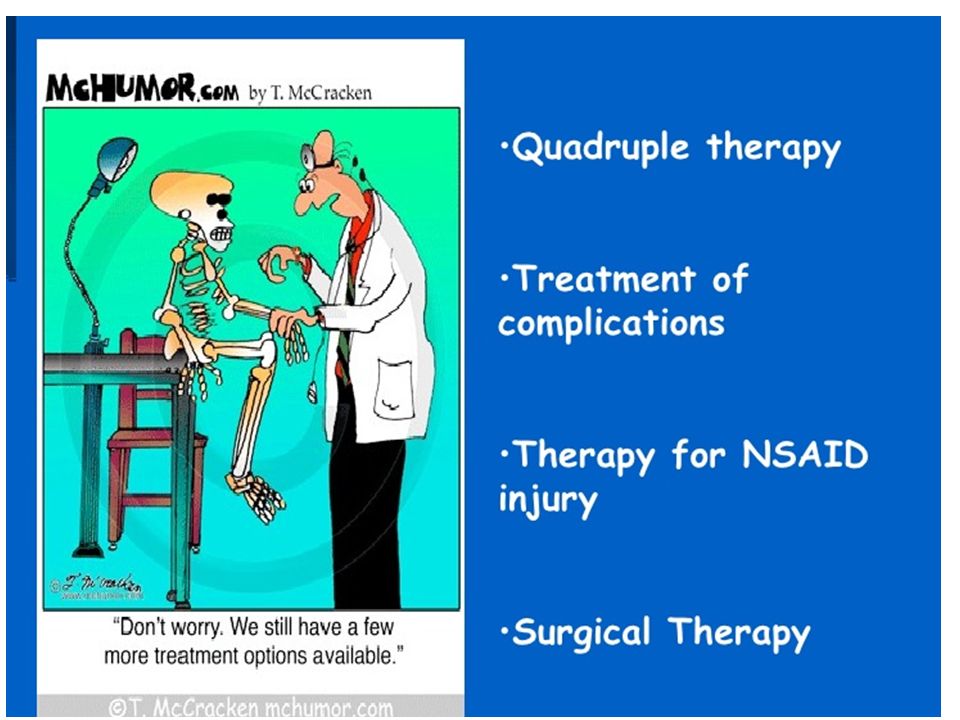
Management:

17
Acute: Resuscitation if perforated or bleeding, and proceeding endoscopic or surgical treatment. Endoscopy: Haemostasis by injection sclerotherapy, laser or electrocoagulation. Surgical: If perforated, ulcer can be over sewn or an omental patch can be placed over it. Haemorrhage is controlled by suturing the affected vessels (usually gastroduodenal artery).
..")
18
Medical: H. pylori eradication with ‘triple therapy’ for 1–2 weeks: Various combinations are recommended made up of one PPI/ranitidine bismuth sulphate and two antibiotics (e.g. clarithromycin amoxicillin, metronidazole ,tetracycline). If not associated with H. pylori: Treat with PPIs or H2-antagonists. Stop NSAID use (especially diclofenac), use misoprostol (prostaglandin E1 analogue), if NSAID use is necessary.
. If not associated with H. pylori: Treat with PPIs or H2-antagonists. Stop NSAID use (especially diclofenac), use misoprostol (prostaglandin E1 analogue), if NSAID use is necessary..")
20
Complication: Major complication rate: 1% per year including haemorrhage (haematemesis, melaena, iron- deficiency anaemia), perforation, obstruction/pyloric stenosis (due to scarring, penetration, pancreatitis). Prognosis: Overall lifetime risk _10%. Generally good as peptic ulcers associated with H.pylori can be cured by eradication.

21
thank you

Similar presentations


>")
![Peptic Ulcer Disease Dr Maha Arafah. Objectives Upon completion of this lecture the students will : A] Understand the Pathophysiology of acute and chronic.](/13/3809458/big_thumb.jpg "Peptic Ulcer Disease Dr Maha Arafah. Objectives Upon completion of this lecture the students will : A] Understand the Pathophysiology of acute and chronic.>")


>")
 Dr. Gehan Mohamed Dr. Abdelaty Shawky.>")


 2004 Elsevier Inc. All rights reserved. Drugs for Peptic Ulcer Disease Chapter 73.>")
