Download presentation
Presentation is loading. Please wait.

1
Keith Clements Introduction to Neuroscience
4/27/2017 Visual Perception Keith Clements Introduction to Neuroscience

2
Aims You should be able to
Describe the key features of the eye and the visual pathways. Describe how information about brightness and colour is processed from the retina to the cortex.

3
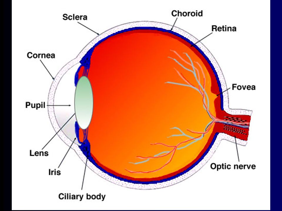
How the eye works Light reflects off objects. Reflected light passes through pupil & lens and is focussed onto retina. Ciliary muscles alter the shape of the lens to focus the image on the retina. Receptors in the retina, known as rods & cones, convert light into neural signals. Cones provide visual acuity. Small numbers of cones are connected to each ganglion cell, which give rise to the optic nerve. Rods provide night vision, they work better in dim light and information from large numbers of rods is pooled. Daytime vision is best at the fovea, where cones are most common.

6
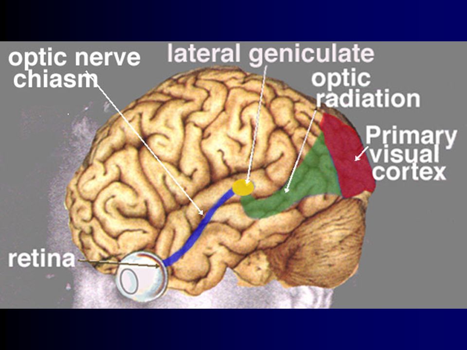
Visual pathways Visual information travels from retinal ganglion cells to brain via the Lateral Geniculate Nucleus of the Thalamus. The visual cortex is arranged topographically, but with 25% of area devoted to fovea. Cells are arranged in columns six deep.

8
Binocular vision Visual information from each eye crosses over at the optic chiasm. Each hemisphere sees one half of the visual field.

9
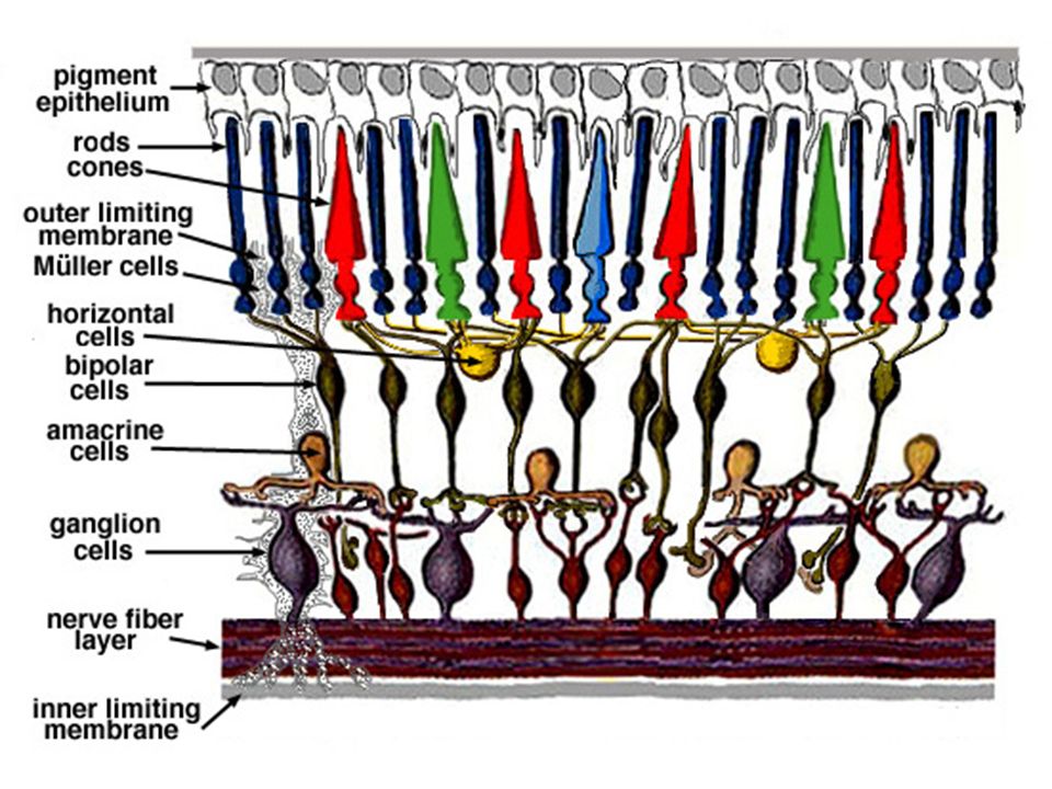
Visual processing in the retina
On-centre cell On response when light falls on centre. Off response with light in periphery Receptive field of ganglion cells (in retina) selectively respond to different patterns of light. Off-centre cell Off response when light falls on centre. On response with light in periphery
 selectively respond to different patterns of light. Off-centre cell. Off response when light falls on centre. On response with light in periphery.")
11
So What? Receptive fields enhance contrast
Contribute to brightness constancy

12
Visual processing beyond the retina
Receptive cells in LGN and visual cortex (Hubel & Weisel, 1958, 1963). LGN cells had same concentric fields as retinal cells. Cells in visual cortex had “oblong structure” and were sensitive to elongated areas of light. Cells were selectively responsive to lines of particular orientation (called these simple cells).
. LGN cells had same concentric fields as retinal cells. Cells in visual cortex had oblong structure and were sensitive to elongated areas of light. Cells were selectively responsive to lines of particular orientation (called these simple cells).")
13
Receptive fields in the cortex
In deeper levels of cortex Hubel and Weisel found complex and hypercomplex cells. Complex cells respond when line is anywhere in oblong so long as it has a particular orientation. Complex cells more sensitive to movement than simple cells. Hypercomplex cells respond to line of particular orientation and length. Some appear to be sensitive to angles (2 lines connecting).
.")
14
Theories of Colour Vision
Cones sensitive to light corresponding to blue, green and red. Trichromatic theory: 3 colours mix to produce other colours. Opponent process theory: receptors work in opposition, with red green and yellow-blue channels.

15
4/27/2017 Evidence Three types of cone exist in retina that are differentially sensitive to blue, green, red. After-image effects are consistent with opponent process. Both theories are correct - trichomatic account explains creation of yellow pathway for blue - yellow opponent process. Some Ganglion cells have colour-sensitive concentric receptive fields red-green & blue-yellow Mention that LGN cells have similar receptive fields

16
Reading Read chapter 2 in Wickens Most images from

Similar presentations


 2013.6.30.>")


