Download presentation
Presentation is loading. Please wait.

1
Chapter 4: The Visual Cortex and Beyond

2
Overview of Questions How can brain damage affect a person’s perception? Are there separate brain areas that determine our perception of different qualities?

3
Figure 4.1 (a) Side view of the visual system, showing the three major sites: the eye, the lateral geniculate nucleus, and the visual cortex. (b) Visual system showing how some of the nerve fibers from the retina cross over to the opposite side of the brain at the optic chiasm.
 Visual system showing how some of the nerve fibers from the retina cross over to the opposite side of the brain at the optic chiasm..")
4
Pathway from Retina to Cortex
Signals from the retina travel through the optic nerve to the Lateral geniculate nucleus (LGN) Primary visual receiving area in the occipital lobe (the striate cortex) And then through two pathways to the temporal lobe and the parietal lobe The instructor should also note the location of the superior colliculus which controls eye movements and receives about 10% of the fibers from the optic nerve.
 Primary visual receiving area in the occipital lobe (the striate cortex) And then through two pathways to the temporal lobe and the parietal lobe. The instructor should also note the location of the superior colliculus which controls eye movements and receives about 10% of the fibers from the optic nerve.")
5
Visual Areas

6
Areas V1 – V5 KW 8-17

7
Visual Cortex

8
How do we study visual cortex?
Hubel and Wiesel Single cell recording in visual cortex. Implant one cell in visual cortex. Shine light on retina. See is we can get that cell to respond. What does the single cell like to “see”.

9
Figure 2.17 Recording electrical signals from a fiber in the optic nerve of an anesthetized cat. Each point on the screen corresponds to a point on the cat’s retina.

10
Receptive fields are determined by monitoring single cell responses.
Area of receptors that affects firing rate of a given neuron in the circuit Receptive fields are determined by monitoring single cell responses. Research example for vision Stimulus is presented to retina and response of cell is measured by an electrode. Important to emphasize that the receptive field is on the retina. Students tend to forget this as you work your way through the explanation of more specifically tuned neurons further into the system.

11
Neural Activity KW 8-25

12
Overlap in Receptive Fields
KW 8-27

13
The Map on the Striate Cortex
Cortex shows retinotopic map. Electrodes that recorded activation from a cat’s visual cortex show: Receptive fields on the retina that overlap also overlap in the cortex. It might be helpful here to explain how the Hubel and Wiesel experiment works. Stimuli are shown on specific points on the retina to find the stimulus and the receptive field that stimulates a neuron in the cortex that is being monitored by an electrode. This is also a good place to discuss ethics in the use of the animals for research, if the instructor desires to do so.

14
Neurons in Striate Cortex
Simple cortical cells Side-by-side receptive fields Respond to spots of light Respond best to bar of light oriented along the length of the receptive field Orientation tuning curves Shows response of simple cortical cell for orientations of stimuli

15
Figure 4.6 (a) The receptive field of a simple cortical cell.
(b) This cell responds best to a vertical bar of light that covers the excitatory area of the receptive field. The response decreases as the bar is tilted so that it also covers the inhibitory area.
 This cell responds best to a vertical bar of light that covers the excitatory area of the receptive field. The response decreases as the bar is tilted so that it also covers the inhibitory area.")
16
Orientation tuning curve of a simple cortical cell for a neuron that responds best to a vertical bar (orientation = 0). (From Hubel & Wiesel, 1959.)
.")
17
Neurons in Striate Cortex - continued
Complex cells Like simple cells Respond to bars of light of a particular orientation Unlike simple cells Respond to movement of bars of light in specific direction

18
Figure 4.8 (a) Response of a complex cell recorded from the visual cortex of a cat. The stimulus bar is moved back and forth across the receptive field. The cell fires best when the bar is positioned with a specific orientation and is moved in a specific direction

19
Response of an end-stopped cell recorded from the visual cortex of the cat. The stimulus is indicated by the light area on the left. This cell responds best to a medium-sized corner that is moving up (*).
..")
20
Neurons in Striate Cortex – Edge detector
End-stopped cells Respond to: Moving lines of specific length Moving corners or angles No response to: Stimuli that are too long

21
Feature Detectors Neurons that fire to specific features of a stimulus Pathway away from retina shows neurons that fire to more complex stimuli Cells that are feature detectors: Simple cortical cell Complex cortical cell End-stopped cortical cell

22
Table 4.1 Properties of cortical neurons

23
Vision Visualized With FMRI Fovea Periphery
Figure 4.17 (a) Red and blue areas show the extent of stimuli that were presented while a person was in an fMRI scanner. (b) Red and blue indicates areas of the brain activated by the stimulation in (a). (From Dougherty et al., 2003.)
 Red and blue areas show the extent of stimuli that were presented while a person was in an fMRI scanner. (b) Red and blue indicates areas of the brain activated by the stimulation in (a). (From Dougherty et al., 2003.)")
24
Brain Imaging Techniques - fMRI
Functional magnetic resonance imaging (fMRI) Hemoglobin carries oxygen and contains a ferrous molecule that is magnetic Brain activity takes up oxygen, which makes the hemoglobin more magnetic fMRI determines activity of areas of the brain by detecting changes in magnetic response of hemoglobin Subtraction technique is used like in PET
 Hemoglobin carries oxygen and contains a ferrous molecule that is magnetic. Brain activity takes up oxygen, which makes the hemoglobin more magnetic. fMRI determines activity of areas of the brain by detecting changes in magnetic response of hemoglobin. Subtraction technique is used like in PET.")
25
Figure 4.14 The magnification factor in the visual system: The small area of the fovea is represented by a large area on the visual cortex.

26
Maps and Columns in the Striate Cortex
Cortical magnification factor Fovea has more cortical space than expected Fovea accounts for .01% of retina Signals from fovea account for 8% to 10% of the visual cortex This provides extra processing for high-acuity tasks.

27
Figure 4.24 How a tree creates an image on the retina and a pattern of activation on the cortex.

28
Other Cortical Areas Vision begins to processed by V1-V5
Then goes to other lobes of the brain for further processing. What we have seen. Object identification. Where we have it. Locating object in world.

29
Figure 4.27 The monkey cortex, showing the what and the where pathways. The where pathway is also called the how pathway. (From Mishkin, Ungerleider, & Macko, 1983.)
.")
30
What and Where (How) Pathways
Where pathway may actually be “How” pathway Dorsal stream shows function for both location and for action. Evidence from neuropsychology Single dissociations: two functions involve different mechanisms Double dissociations: two functions involve different mechanisms and operate independently Note that neuropsychology involves the study of the behavioral effects of brain damage.

31
Table 4.2 A double dissociation

32
What and How Pathways - Further Evidence
Rod and frame illusion Observers perform two tasks: matching and grasping Matching task involves ventral (what) pathway Grasping task involves dorsal (how) pathway Results show that the frame orientation affects the matching task but not the grasping task.
 pathway. Grasping task involves dorsal (how) pathway. Results show that the frame orientation affects the matching task but not the grasping task.")
33
Figure 4. 30 (a) Rod and frame illusion
Figure 4.30 (a) Rod and frame illusion. Both small lines are oriented vertically. (b) Matching task and results. (c) Grasping task and results.
 Rod and frame illusion. Both small lines are oriented vertically. (b) Matching task and results. (c) Grasping task and results.")
34
Modularity: Structures for Faces, Places, and Bodies
Module - a brain structure that processes information about specific stimuli Inferotemporal (IT) cortex in monkeys Responds best to faces with little response to non-face stimuli Temporal lobe damage in humans results in prosopagnosia.
 cortex in monkeys. Responds best to faces with little response to non-face stimuli. Temporal lobe damage in humans results in prosopagnosia.")
35
Figure (a) Monkey brain showing location of the inferotemporal (IT) cortex. (b) Human brain showing location of the fusiform face area (FFA), which is located under the temporal lobe.
 Human brain showing location of the fusiform face area (FFA), which is located under the temporal lobe..")
36
Figure 4.33 Size of response of a neuron in the monkey’s IT cortex that responds to face stimuli but not to nonface stimuli. (Based on data from Rolls & Tovee, 1995.)
.")
37
Monkey Face Cells

38
Evolution and Plasticity: Neural Specialization
Evolution is partially responsible for shaping sensory responses: Newborn monkeys respond to direction of movement and depth of objects Babies prefer looking at pictures of assembled parts of faces Thus “hardwiring” of neurons plays a part in sensory systems

39
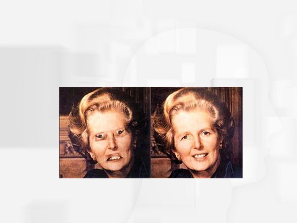
Margaret Thatcher Illusion

Similar presentations


 Lavanya Sharan January 24 th, 2011.>")


 Ch 4-6, 9.>")