Download presentation
Presentation is loading. Please wait.

1
The Respiratory System
Thomas Ackerman Juyoung Jang Liezel Riego

2
IB Syllabus Gas exchange:
6.4.1: Distinguish between ventilation, gas exchange, and cell respiration. 6.4.2: Explain the need for a ventilation system. 6.4.3: Describe the features of the alveoli that adapt them to gas exchange. 6.4.4: Draw and label a diagram of the ventilation system, including trachea, lungs, bronchi, bronchioles, and alveoli. 6.4.5: Explain the mechanism of ventilation of the lungs in terms of volume and pressure changes caused by the internal and external intercostal muscles, the diaphragm and abdominal muscles.

3
6.4.1: Distinguish between ventilation, gas exchange, and cell respiration.
Cell respiration (recall plant chapter) Occurs in the cytoplasm and the mitochondria. Releases energy in the form of ATP. Gas exchange Swapping of gases. Oxygen in, CO2 out. Occurs in alveoli in the lungs. Oxygen diffusion: from air in alveoli to blood in capillaries. CO2 diffusion: opposite direction of oxygen, capillaries to alveoli. Reason for diffusion: concentration gradients of oxygen and CO2 between air and blood are different. Ventilation Maintains the concentration gradient (6.4.2). Air in the alveoli must be refreshed frequently.
 Occurs in the cytoplasm and the mitochondria. Releases energy in the form of ATP. Gas exchange. Swapping of gases. Oxygen in, CO2 out. Occurs in alveoli in the lungs. Oxygen diffusion: from air in alveoli to blood in capillaries. CO2 diffusion: opposite direction of oxygen, capillaries to alveoli. Reason for diffusion: concentration gradients of oxygen and CO2 between air and blood are different. Ventilation. Maintains the concentration gradient (6.4.2). Air in the alveoli must be refreshed frequently.")
4
6.4.3: Describe the features of the alveoli that adapt them to gas exchange.
Gas exchange occurs in the alveoli. Surface area Alveolus is very small; millions of them in total. Results in large surface area. Alveolus wall Consists of a single layer of very thin cells. Capillary wall Also single layer of very thin cells. Gases only need to diffuse very short distance.

5
6.4.3: Alveolus (cont.) Covered by a dense network of blood capillaries with low oxygen and high CO2 concentrations. Due to that, oxygen diffuses into the blood, while CO2 diffuses out (concentration gradient). Secretion of fluid in alveolus wall Keeps inner surfaces of alveolus moist. Allows gases to dissolve. Fluid contains natural detergent, preventing the sides of the alveolus from sticking together.
. Secretion of fluid in alveolus wall. Keeps inner surfaces of alveolus moist. Allows gases to dissolve. Fluid contains natural detergent, preventing the sides of the alveolus from sticking together.")
6
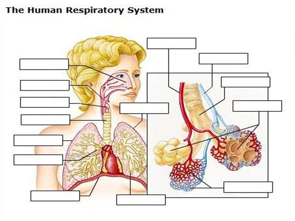
6.4.4: Diagram of Ventilation System
Nasal cavity – nose; takes in/releases air Pharynx – area in vertebrate throat where air and food passages cross Larynx – voicebox; containing the vocal cords Trachea – windpipe; airway extending from larynx to primary bronchi Lungs – invaginated respiratory surface that connects to atmosphere via narrow tubes

7
6.4.4: Diagram (cont.)* Bronchus – breathing tube branching from trachea into lungs Diaphragm – sheet of muscle forming the bottom wall of thoracic cavity in mammals; active in ventilating lungs Bronchiole – fine branch of bronchus transporting air to alveoli Alveoli – tiny air sacs at end of each bronchiole Capillary –microscopic blood vessel; allows exchange between blood and interstitial fluid.

9
Ventilation of lungs Air’s Journey Inhale – air enters lung
Travels to trachea Then to bronchi and bronchioles Exhale – same route Muscles are used to lower and raise pressure inside lungs Causes movement of air

10
Vocabulary Cell respiration Gas exchange Ventilation Oxygen diffusion
CO2 diffusion Alveolus/alveoli Exhalation/Expiration Inhalation/Inspiration Nasal cavity Pharynx Larynx Trachea Lungs Bronchus Diaphragm Bronchiole Alveoli Capillary

Similar presentations