Download presentation
Presentation is loading. Please wait.

1
Reproduction Lesson 5 Foetal development

2
1Fertilisation is when an egg cell joins with: Aanother egg cell. Ba sperm cell. Ca body cell. Droot hair cells. 2Before it is born, a calf is inside a cow’s: Astomach. Buterus. Coviduct. Dlarge intestine. 3One way that a sperm cell is adapted to its job is that: Ait dies quite quickly. Bit is hollow. Cit is surrounded by a liquid. Dit has a tail to allow it to swim.

3
4An egg cell is much larger than a sperm cell because: Ait needs to be able to hide from sperm cells. Bit needs to be big to allow it to have a streamlined shape. Cit contains a store of food that can be used if it is fertilised. Dthe nucleus in an egg cell is much larger than in a sperm cell. 5Fertilisation occurs in: Aan ovary. Ban oviduct. Cthe scrotum. Dthe penis. 6A fertilised egg cell develops into an embryo by: Acell division. Bpuberty. Covulation. Dfertilisation.

4
LO – To describe the conditions needed for fetal development. [Level 4] To describe how a baby develops in the uterus and is born. [Level 5]

5
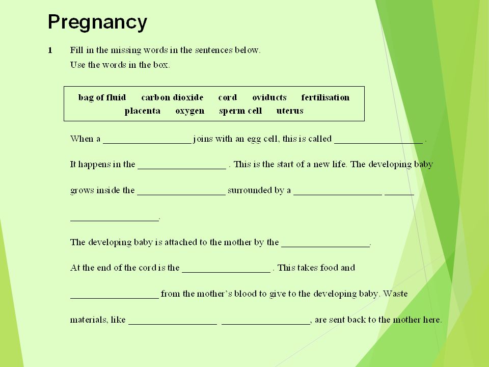
Keywords– Zygote Foetus Embryo Placenta Umbilical cord Umbilical sac Amniotic fluid

6
The placenta How does an embryo receive food and oxygen and how does it get rid of waste? An embryo forms a structure called the placenta, which attaches to the uterus wall. The umbilical cord joins the fetus to the placenta. In the placenta, food and oxygen diffuse from the mother’s blood into the blood of the fetus. Carbon dioxide and waste products diffuse from the blood of the fetus into the mother’s blood. umbilical cord

7
How does the placenta work?

8
From embryo to fetus In the earliest stages of development, a human baby is called an embryo. After the first eight weeks of pregnancy, a human embryo is then called a fetus. At this stage, the fetus has all the main human features. The fetus continues to develop and grow inside its mother’s uterus for a total of 40 weeks.

9
How is an ultrasound scan done? An ultrasound is a diagnostic or screening procedure that uses high-frequency sound waves to create a picture of internal body structures, such as a developing fetus. It may also be called an ultrasound scan, sonogram, or ultrasonography.

10
20 weeks into pregnancy. He is still a foetus.

11
The placenta

14
In the earliest stages of development, a human baby is called an embryo. After the first eight weeks of pregnancy, a human embryo is then called a fetus. At this stage the fetus has all the main human features. The fetus continues to develop and grow inside its mothers’ uterus for a total of 40 weeks. From embryo to fetus

15
What are the stages of development?

16
Fertilization Fertilization, the joining of the sperm and the egg in the fallopian tube (below) to form a unique human being, occurs.
 to form a unique human being, occurs.")
17
Only 30 hours later This is a fertilized egg only thirty hours after conception. Magnified here, it is no larger than the head of a pin. Still rapidly dividing, the developing embryo, called a zygote at this stage, floats down from the fallopian tube and towards the uterus.

18
Weeks 3-5 The embryo ’ s tiny heart begins to beat by day twenty-one. Arm and leg buds are visible and the formation of the eyes, lips, and nose has begun. The spinal cord grows faster than the rest of the body giving a tail like appearance which disappears as the embryo continues to grow. The placenta begins to provide nourishment for the embryo.

19
Week 7 Major organs have all begun to form. The embryo has developed its own blood type, unique from the mother’s. Hair follicles and knees and elbows are visible. Facial features are also observable. The eyes have a retina and lens. The major muscle system is developed and the embryo is able to move.

20
Weeks 8-12 The embryo is reactive to its environment inside the amniotic sac where it swims and moves. Hands and feet can be seen. At the end of week 8, the embryonic period is over and the foetal stage begins.

21
Weeks 13-16 The brain is fully developed and the foetus can suck, swallow, and make irregular breathing sounds. Foetus can feel pain. Foetal skin is almost transparent. Muscles tissue is lengthening and bones are becoming harder. Liver and organs produce appropriate fluids. Eyebrows and eyelashes appear and the foetus makes active movements including kicks and even somersaults.

22
Weeks 20-24 A protective waxy substance called vernix covers the skin. By birth, most of the vernix will be gone but any that is left is quickly absorbed. Foetus has a hand and footprints and fingerprints are forming. Foetus practices breathing by inhaling amniotic fluid into its developing lungs.

23
Weeks 25-28 Rapid brain development occurs during this period and the nervous system is able to control some bodily functions. The foetus’ eyelids now open and close. At 25 weeks there is a 60% chance of survival if born. There is a rapid increase in the amount of body fat the foetus has. Rhythmic breathing occurs, but the lungs are not yet mature. The foetus sleeps 90-95% of the day. At this point there the survival rate is above 95% if the baby is born Weeks 29-32

24
G Student Sheet 1Birth 3 Development: Growth data cards A B C D E F cells1 cell types1 organs0 nutrient sourcestored food survival outsideexcellent cells8 cell types1 organs0 nutrient sourcestored food survival outside very good cellsbillions cell types200 organsall finished nutrient sourcemilk survival outside very good cellsmillions cell types200 organsall formed nutrient sourcemum’s blood survival outside impossible cells Tens of thousands cell types50 organsbrain + heart nutrient sourcemum’s blood survival outside impossible cellsbillions cell types200 organsall finished nutrient sourcemum’s blood survival outside just possible Instructions: Put the cards in order to show how the baby develops. Note: The diagrams are not to scale. cells64 cell types2 organs0 nutrient sourcestored food survival outsidegood

25
The stages of pregnancy

27
PartJob Amniotic fluid Umbilical cord Placenta

28
Birth After 40 weeks of gestation, the baby is ready to be born. At this point, the head usually lies just above the cervix. After a few minutes, the placenta comes away from the uterus wall. This is pushed out as the afterbirth. Eventually the contractions cause the amniotic sac to break and the fluid escapes. The cervix then widens and dilates as the baby is pushed through the vagina. Birth begins with small contractions of the uterus wall, which gradually become stronger and more frequent. Clip

31
Learning outcomes – Describe the conditions needed for baby development [Level 4] Know how a baby develops and is born [Level 4] Understand how a baby develops and is born. [Level 5]
![Learning outcomes – Describe the conditions needed for baby development [Level 4] Know how a baby develops and is born [Level 4] Understand how a baby develops and is born.](http://images.slideplayer.com/30/9535904/slides/slide_31.jpg "[Level 5].")
Similar presentations