Download presentation
Presentation is loading. Please wait.

1
Fractures around elbow
Dr. Waleed Faris Al- Rawi

2
Supracondylar fracture
These are among the commonest fractures in children, may be the most worrisome of all pediatric upper extremity fractures because their association with neurovascualar injury.. The distal fragment displaced either posteriorly or anteriorly

3
Mechanism of injury Posterior angulation or displacement occur in 95% of all cases suggest hyper extension injury , usually due to outstretched hand, The distal fragment is pushed backwards and ( because the forearm usually in pronation ) twisted inward. The jagged end of the proximal fragment pocks into the soft tissues anteriorly, some times injuring the brachial artery or median nerve.
 twisted inward. The jagged end of the proximal fragment pocks into the soft tissues anteriorly, some times injuring the brachial artery or median nerve.")
4
Anterior displacement is rare , it is thought to be due to direct violence e.g a full on the point of the elbow with joint in flexion.

5
Classification 1- Type 1 is undisplaced fracture
2- Type 2 is an angulated fracture with the posterior cortex still in continuity, 2A being less sever and merely angulated and 2B being more sever and both angulated and malrotated. 3- Type 3 is a completely displaced fracture.

6
Clinical features Following a fall there is pain and the elbow is swollen with a posteriorly displaced fracture the S-shape deformity of the elbow is usually obvious and the bony landmarks are abnormal. It is essential to feel the pulse and check the capillary return, passive extension of the flexor muscles should be pain free. The wrist and the hand should be examined for evidence of nerve injury.

7
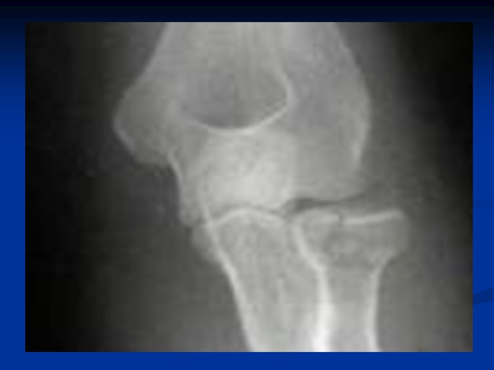
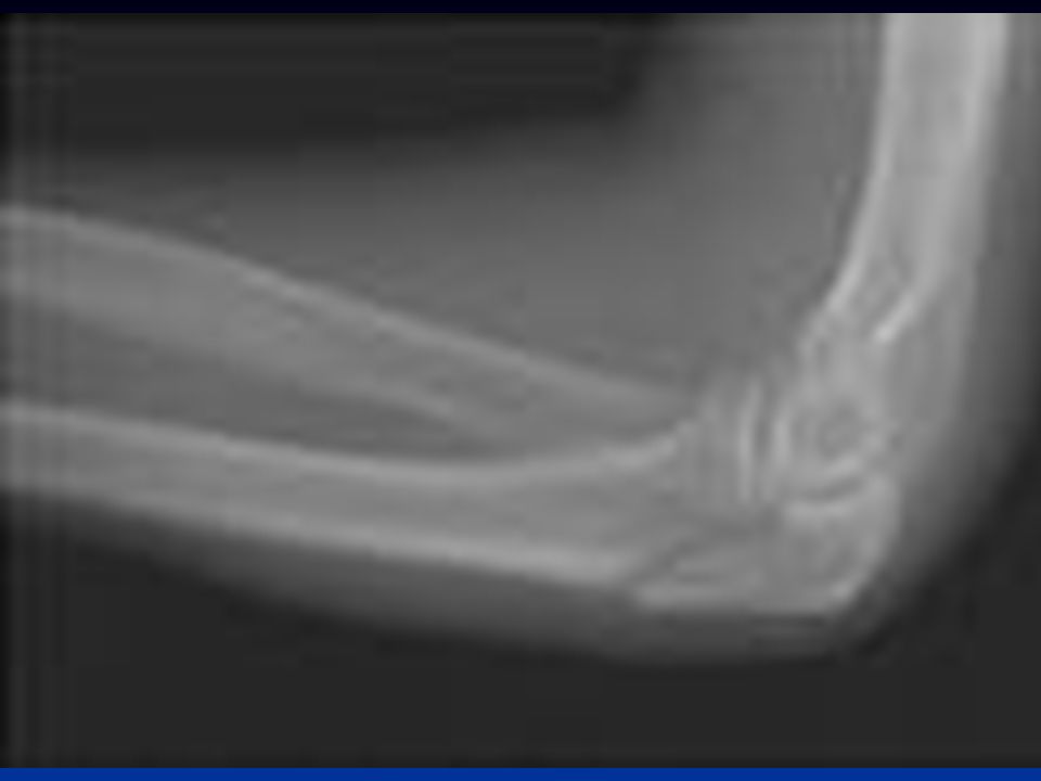
X-Ray The fracture is seen most clearly in the lateral view. In an undisplaced fracture the fat pad sign should raise suspicions, these is triangular lucency in front of distal humerus due to the fat pad being pushed forward by a haematoma.

8
In the common posteriorly displaced fracture the fracture line runs obliquely downward and forward and the distal fragment is tilted backward. In the anteriorly displaced fracture the cracks runs downward and backward and the fragment is tilted forwards.

9
The anteroposterior view is painful and may be postponed until the child has been anesthetized, it may show that the distal fragment is shifted or tilted sideways. Measurement of Baumanns angle is useful in assessing the degree of medial angulation before and after reduction.

10
Treatment The goal of management of supracondylar fracture is to restore and maintain anatomic or near anatomic aligment with a minimum of complications.

11
Treatment Type 1; undisplaced fracture
The elbow is immobilized at 90 degree and neutral rotation in cast and the arm is supported by a sling. It is essential to obtain an x-ray 5-7 days later to check that there has been no displacement. The cast is retained for 3 weeks then movement is allowed.

12
Treatment Type 2 Posteriorly angulated fracture- mild
In these cases swelling is usually not severe and the risk of vascular injury is low. If the posterior cortices are in continuity, the fracture can be reduced ander general anesthesia and during reduction pulse Is felled and check the capillary return and if distal circulation is suspected immediately relax elbow flexion until it improves.

13
Following reduction the arm held in collar and cuff and the circulation should be checked repeatedly during the first 24 hours. An x-ray is taken after 3-5 days to confirm that the fracture has not slipped. The splint is retained for 3 weeks then movement is started. If the acutely flexed position cannot be maintained without disturbing the circulation or if the reduction is unstable, the fracture should be fixed with percutaneous crossed Kirschner wires.

14
Type 2B and 3 angulated and malrotated or posteriorly displaced
These are usually associated with severe swelling, are difficult to reduced and are often unstable and there is a considerable risk of neurovascular injury or circulatory compromised due swelling. The fracture should be reduced under general anaesthesia as soon as possible and then held with percutaneous crossed Kirschnner wires and wires should be smooth to prevent physeal injury and great care should be taken not to injure ulnar and radial nerves.

15
Open reduction This is some times necessary for (1) a fracture which simply cannot be reduced closed (2) an open fracture or (3) a fracture associated with vascular damage.
 a fracture which simply cannot be reduced closed (2) an open fracture or (3) a fracture associated with vascular damage.")
16
Complications Early 1- Vascular injury; The supracondylar fracture associated with injury to brachial artery which befor introduction of percutaneous pinning was reported as occurring in over 5% which was dropped nowadays to less than 1%. Peripheral ischemia may be immediate and severe or the pulse may fail to return after reduction. More commonly the injury is complicated by forearm oedema and a mounting compartment syndrome which leads to necrosis of the muscles and nerves without causing peripheral gangrene.

17
Pain plus one positive sign ( pain on passive extension of the fingers, a tense and tender forearm, an absent pulse, blunted sensation 0r reduced capillary return on pressing the finger pulp ) demands urgent action. Flexed elbow must be extended and all dressing removed. If the circulation does not improve angiography ( on the operating table if it is saved time ) is carried out, the vessel repaired or grafted and a forearm fasciotomy performed. If angiography is not available Doppler imaging should be used. In extreme cases operative explorations would be justified on clinical criteria alone.
 is carried out, the vessel repaired or grafted and a forearm fasciotomy performed. If angiography is not available Doppler imaging should be used. In extreme cases operative explorations would be justified on clinical criteria alone..")
18
2- Nerve injury The median nerve particularly the anterior interosseous branch may be injured. Loss of function is usually temporary and recovery can be expected in 6-8 weeks. The ulnar nerve may be damage by careless pinning. If the injury is recognized and the pin removed recovery will usually follow.

19
Late 1- Malunion; it is common complication, However backwards or sideways shifts are gradually smoothed out by modeling during growth and they seldom give rise to visible deformity of the elbow. Forwards or backwards tilt may limit flexion or extension but consequent disability is slight.

20
Uncorrected sideway tilt (angulations) and rotation are much more important and may lead to varus (or rarely valgus) deformity of the elbow, this is permanent and will not improve with growth. The fracture is extraphyseal and so physeal damage should not be blamed with deformity, usually it is faulty reduction which is responsible. Cubitus is disfiguring and cubitus valgus may cause late ulnar palsy. If deformity is marked it will need correction by supracondylar osteotomy.

21
2- Elbow stiffness and myositis ossificans
Stiffness is a common complication following elbow injury. Extension is particular may take months to return. It must not be hurried. Passive movement (which includes carrying weights ) or forced movement is prohibited, this will only make matters worse and may contribute to the development of myositis ossificans.
 or forced movement is prohibited, this will only make matters worse and may contribute to the development of myositis ossificans.")
22
Fracture lateral condyle
The lateral condyle begin to ossify during first year of life and fuse with the shaft at years. Between these ages it may be sheared off or avulsed by forceful traction.

23
Mechanism of injury and pathology
The child falls on the hand with the elbow extended and forced in to varus. A large fragment which includes the lateral condyle breaks off and is pulled by the attached wrist extensors. In sever cases the elbow dislocate posterolaterally, the condyle is capsized by muscle pull and remains capsized while the elbow reduced spontaneously. The fracture is important for two reasons It may damage the growth plate It always involves the joint

24
Clinical features The elbow is swollen and deformed. There is tenderness over the lateral condyle and passive flexion of the wrist may be painfull.

25
X-Ray Two types of fractures are recognized
A fracture lateral to the trochlea, the humeroulnar joint not involved and is stable A fracture through the middle of trochlea, this injury is more common and elbow unstable and it may dislocate.

27
Treatment If there is no or minimal displacement the arm can be splinted in a back slab with the elbow flexed 90 degrees and wrist extended A displaced fracture i.e with a gap of more than 2 mm required accurate reduction and internal fixation.

28
Complications Non union and mal union Recurrent dislocation

29
Fractures of the medial condyle
Mechanism of injury The injury is usually caused by fall from height involving either a direct blow to the point of the elbow or a landing on the out stretched hand with the elbow forced in to valgus.

30
Clinical features This is an intra articular fracture resulting in considerable pain and swelling.

32
Treatment Undisplaced fracture treated by splintage, x-rays are repeated until fracture has heald. Displaced fracture are treated either by closed reduction and percutaneous pinning or by open reduction and fixation with pins.

33
Complications Early Ulnar nerve damage Late Stiffness of elbow joint
Late ulnar nerve palsy

34
Fracture neck of radius
Mechanism of injury and pathology A fall on the outstretched hand forces the elbow in to valgus and pushes the radial head against the capitulum. In the children the bone fractures through the neck of the radius, in adult the injury is more likely to fracture the radial head.

35
Clinical features Following a fall the child complains of pain in the elbow. There may be localized tenderness over the radial head and pain on rotating forearm.

36
X-Ray The fracture line is transverse. It either situated immediately distal to the physis or there is true separation of the epiphysis with a triangular fragment of shaft ( salter-Harris II injury ).
.")
38
Treatment In children up to 30 degrees of radial head tilt and up to 3 mm of transverse displacement are acceptable. The arm rested in coller and cuff and exercises are started after a week. Displacement more than 30 degrees requires reduction. If closed reduction fails open reduction are performed.

39
Pulled elbow In young children the elbow may be injured by pulling on the arm usually with the forearm pronated. It is some times called sublaxation of the radial head. A child aged 2-3 years is brought with a painful dangling arm with the forearm held in pronation and extention. A dramatic cure is achieved by forcefully supinating and then flexing the elbow.

40
Fractured head of the radius
Mechanism of injury A fall on the out stretched hand with the elbow extended and the forearm pronated causes impaction of the radial head against capitulum.The radial head is also sometimes fractured during elbow dislocation.

41
Clinical features Tenderness on pressure over the radial head and pain on pronation and supination should suggest the diagnosis.

42
X-Ray 3 types of of fractures are identified
Type I ; A vertical split in the radial head. Type II; A single fragment of the lateral portion of the head broken off and displaced distally. Type III; The head is broken into several fragments ( comminuted ).
.")
44
Treatment An undisplaced type I; The arm held in a collar and cuff for 3 weeks. Type II; if the fragment is displaced it should be reduced and held with a small screw. A comminuted fracture type III; excision of the radial head. However if there is disruption of distal radioulnar the excised head must replaced by silicone or metal prosthesis.

45
Complications Joint stiffness Myositis ossificans
Recurrent instability of the elbow joint

46
Fractures of the olecranon
Two types of injury are seen ( ) A comminuted fracture which is due to direct blow or afall on the elbow. ( ) a clean transverse break due to traction when the patient falls onto hand while triceps muscle is contracted
 A comminuted fracture which is due to direct blow or afall on the elbow. ( ) a clean transverse break due to traction when the patient falls onto hand while triceps muscle is contracted.")
48
Clinical features A bruise over the elbow suggest comminuted fracture and triceps intact and the elbow can be extended. With transverse fracture there may be a palpable gap and the patient unable to extend the elbow against resistance.

49
Treatment A comminuted fracture with triceps intact treated in arm sling for a week then patient start movement. An undisplaced fracture treated conservatively by a cast with elbow flexed 60 degrees for 2-3 weeks. Displaced fracture treated by open reduction and internal fixation by tension band wiring.

50
Complications () Stiffness of elbow joint ()Non-union
() osteoarthritis
 osteoarthritis.")
51
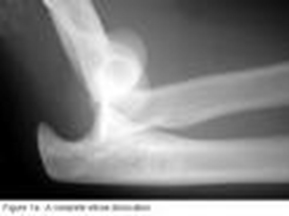
Dislocation of the elbow
Dislocation of the ulnohumeral joint is fairly common more in adult than children

53
Mechanism of injury and pathology
The cause of posterior dislocation is usually a fall on out stretched hand with the elbow in extension. Disruption of capsuloligamentous structure alone can result in posterior or posterolateral dislocation. Whoever provided there is no fractures reduction will usually stable and recurrent dislocation unlikely. The combination of ligamentous disruption and fracture of radial head, coronoid process or olecranon fracture or several fractures will render the joint unstable and unless the fractures are reduced and fixed liable to redislocate.

54
Clinical features The patient support his or her forearm with the elbow in slight flexion. Unless swelling is sever the deformity is obvios. The bony landmarks ( epicondyles and olecranon ) may be palpable and abnormally placed.
 may be palpable and abnormally placed.")
55
Treatment The surgeon pulls on the forearm while the elbow is slightly flexed, then the elbow is further flexed while the olecranon pushed forward with the thumb, then do complete flexion of the elbow joint. After reduction elbow flexed above 90 degrees by collar and cuff for 3 weeks

56
Complications () Early Vascular injury I.e Brachial artery
Nerve injury i.e Ulnar nerve or median nerve. () Late Stiffness of elbow joint. Heterotopic ossification. Unreduced dislocation Recurrent dislocation. Osteoarthritis
 Late. Stiffness of elbow joint. Heterotopic ossification. Unreduced dislocation. Recurrent dislocation. Osteoarthritis.")
57
Thank you

Similar presentations


![What am I?. What am I? Articulations of the humerus, radius, and ulna Articulations of the humerus, radius, and ulna. [ olecranon process ] Medial.](/14/4241906/big_thumb.jpg "What am I?. What am I? Articulations of the humerus, radius, and ulna Articulations of the humerus, radius, and ulna. [ olecranon process ] Medial.>")


