Download presentation
Presentation is loading. Please wait.

1
FOOD POISONING

2
Foodborne Diseases Apply to illnesses acquired by consumption of contaminated food. Sometimes incorrectly referred to as food poisoning. Includes : Foodborne intoxications Foodborne infections.

3
e.g. Salmonella Foodborne intoxication Foodborne infection
By toxins, whether biological or chemical e.g. Staphylococcus Fish poisons Heavy metals. Foodborne infection e.g. Salmonella

4
Vulnerable Groups Food poisoning is more likely to affect people with lowered resistance to disease than healthy people who might show mild symptoms or none at all. The following are particularly vulnerable to food poisoning: - Elderly or sick people Babies Young children Pregnant women

5
Symptoms of Microbiological Food Poisoning
Vomiting Abdominal pain Diarrhea Fever Others e.g. neurological; Botulism

6
Causes of Foodborne Diseases
A- Infective Bacterial Viral Protozoa B. Non infective (intoxication) Non Biological a. Chemical Poisons b. Heavy metals c. Other organic Cpds
 Non Biological. a. Chemical Poisons. b. Heavy metals. c. Other organic Cpds.")
7
Food Poisoning (contd.)
Physical contamination:- objects falling in to food – metal, glass, packaging materials etc. Chemical contamination:- Bleach, cleaning chemicals getting in to food Natural contamination: Poisonous plants and berries, undercooked red kidney beans

8
Causes of Foodborne Diseases (Continued)
Biological From: a- Mushrooms b- Harmful algal species c- Fish e.g. Ciguatera Fish poisoning Scombroid Fish poisoning associated with raised Histamine levels. Paralytic shell Fish poisoning [In Japan].

9
Causes Food Borne Disease
Bacteria Salmonella species Campylobacter jejuni Staphylococcus aureus Clostridium perfringens Clostridium botulinum Bacillus and other sporing bacilli Shigella species. E.coli Yersinia enterocolitica Vibrio parahaemolyticus, Aeromonas hydrophilia, Streptococcus species Group C, E and G . The other e.g. Listeria monocytogens

10
Causes Of Foodborne Viruses
Unlike bacteria viruses do not multiply in Foods. Acquired by: a- Primary contamination From shell Fish From Oysters From Mussels From Vegetables e.g Hepatitis A From Vegetables irrigated by untreated water. b- Secondary contamination. From Food handlers e.g. Hepatitis A

11
Foodborne Viruses (Continued)
a- Hepatitis A b- Hepatitis E c- Small rounded structured viruses(SRSV) e.g. Norovirus Caliciviruses Astroviruses Rotaviruses
 e.g. Norovirus. Caliciviruses. Astroviruses. Rotaviruses.")
12
Causes of Foodborne Disease
Protozoae 1- Giardiasis. 2- Amoebiasis 3- Cryptosporiosis 4- Isospora 5- Balantidum 6- Blastocytosis hominis 7- Microsporidia 8- Toxoplasmosis (?)
")
13
Some Diseases Which Are Transmitted By Food But Not Considered As Food Poisoning:
1- Bovine tuberculosis (milk) 2- Brucellosis (milk) 3- Toxoplasmosis (meat) 4- Listerosis (milk products) 5- Mad cow diseases etc.
 2- Brucellosis (milk) 3- Toxoplasmosis (meat) 4- Listerosis (milk products) 5- Mad cow diseases etc.")
14
Selected Clinical and Epidemiologic Characteristics of Typical Illness Caused By Common Foodborne Pathogens* Pathogen Typical Incubation Period Duration Typical Clinical Presentation Assorted Foods Bacterial Salmonella species 1-3 Days 4-7 Days Gastroenteritis Undercooked eggs or poultry, produce Campylobacter jejuni 2-5 Days 2-10 Days Undercooked poultry, unpasteurized dairy products E. coli, Enterotoxigenic 3-7 Days Many foods Shigella species 1-2 Days Produce, egg salad

15
2-6 weeks Variable 1-6 hour <24 hour 12-72 hour Days-months
Listeria monocytogenes 2-6 weeks Variable Gastroenteritis, meningitis abortion Deli meat, hotdogs, unpasteurized dairy products Bacillus cereus 1-6 hour <24 hour Vomiting, Gastroenteritis Fried rice, meats Clostridium botulinum 12-72 hour Days-months Blurred vision, paralysis Home-canned foods, fermented fish Staphylococcus aureus 1-2 Days Gastroenteritis, particularly nausea Meats, potato & pork, unpasteurized dairy products. Yersinia enterocolitica 1-3 weeks Gastroenteritis, appendicitis-like syndrome Undercooked pork, unpasteurized dairy products.

16
Viral Typical incubation period Duration Typical clinical presentation
Assorted foods Norovirus 1-2 Days 12-60 Hr Gastroenteritis Under cooked shellfish Hepatitis A virus 15-50 Days Weeks-months Hepatitis Produce, undercooked shellfish

17
Parasitic Assorted foods Typical incubation Duration
Typical clinical presentation Assorted foods Cryptos poridium parvum 2-10 Days Weeks Gastroenteritis Produce, water Cyclospora cayetanesis 1-11 Days Toxoplasma gondii 5-23 Days Months Influenza-like illness, lymphadenopathy Food contaminated by cat feces, undercooked meat Giardia lamblia 1-4 Wk Water Taenia solium Variable Asymptomatic, cysticercosis Raw pork

18
Types of bacteria Spoilage: Not particularly harmful bacteria which cause food to go off Beneficial: “Good Bacteria” which are used to make yoghurt and cheese Pathogenic: Illness causing bacteria

19
In order to grow and multiply germs need:
Warmth Time Moisture Food Remember it like this Too Many Flies Waiting

20
In ideal conditions where there is Moisture, Food and Warmth (37degrees centigrade is ideal), bacteria can double every 10 to 20 minutes. They do this by dividing in to two. This is called Binary Fission

21
These cells are beginning to divide into two

22
Bacterial growth After 10 minutes After 20 minutes After 30 minutes

23
From 0 to 1,536,000 in only 80 minutes !!!!!! Time : 9.30 Bacteria : 0
cooking chicken to a core temperature of 75°C should kill most of the bacteria Time : 9.30 Bacteria : 0 Time : 9.40 Bacteria : 12,000 Time : 9.50 Bacteria : 24,000 Knife contaminated by blood Time : Bacteria : 48,000 Time : Bacteria : 96,000 Time : Bacteria : 192,000 Time : Bacteria : 384,000 Time : Bacteria : 768,000 Time : Bacteria : 1.5 million From 0 to 1,536,000 in only 80 minutes !!!!!!

24
Bacterial Growth Curve
Numbers Of Bacteria Numbers of bacteria decrease Stationary Phase rapid multiplication Numbers of bacteria remain constant as the number produced is equal to the number dying no multiplication Log Phase Decline Phase Lag Phase Time (hours)
")
25
Spore Bacterial cell Spore forming inside cell
A resting resistant phase of some bacteria (including Clostridium Perfingens and Botulinum and Bacillus Cereus). The bacterium produces a protective coat which helps it to survive high temperatures (up to 120°C) and lack of water. When favourable conditions return, the spores split open and release the bacteria which are then able to grow and multiply Bacterial cell Spore forming inside cell
. The bacterium produces a protective coat which helps it to survive high temperatures (up to 120°C) and lack of water. When favourable conditions return, the spores split open and release the bacteria which are then able to grow and multiply. Bacterial cell. Spore forming inside cell.")
26
Spore Formation This is what happens ………….. Cell

27
Spore forms in cell

28
Cell disintegrates

29
Spore is released

30
Spore starts to germinate

31
Spore continues to germinate

32
Now see as, in suitable conditions, the cell begins to divide (binary fission)………………………….
………………………….")
41
Staphylococcus Aureus
Found in human nose and throat (also skin) Clostridium Perfingens Found in animals and birds Salmonella Found in animals, raw poultry and birds Clostridium Botulinum Found in the soil and associated with vegetables and meats Bacillus Cereus Found in soil, vegetation, cereals and spices
 Clostridium Perfingens. Found in animals and birds. Salmonella. Found in animals, raw poultry and birds. Clostridium Botulinum. Found in the soil and associated with vegetables and meats. Bacillus Cereus Found in soil, vegetation, cereals and spices.")
42
Important Microbial Causes of Food Poisoning
Toxin related A. Staphylococcus aureus Short incubation period < 6 hours An intoxication Not infection

43
Nausea, vomiting, cramps (abdominal pain) Prostration
Important Microbial Causes of Food Poisoning (Continued) Clinical Features: Abrupt, violent Nausea, vomiting, cramps (abdominal pain) Prostration Diarrhea, sub normal temperature Source infected human & food handlers Food contaminated enterotoxin produced
 Clinical Features: Abrupt, violent. Nausea, vomiting, cramps. (abdominal pain) Prostration. Diarrhea, sub normal temperature. Source infected human & food handlers. Food contaminated enterotoxin. produced.")
44
Toxin is heat stable Diagnosis: 1- Epidemiologically
2- Isolation of organism from suspected food. 3- Toxin detection in faeces of patients (rarely done)
")
45
B- Bacillus cereus (from rice meals)
Bacillus caereus is an spore forming aerobic bacillus produces two types of enterotoxins. a- Heat stable causing: emetic type of food poisoning incubation period < 6 hours vomiting, nausea b- Heat labile causing: diarrheal type incubation period 6-24 hours “BOTH TOXINS ARE PREFORMED IN FOOD”

46
Clostridium Perfringens
Causative agent: type A strains of C. perfringens (C. Welchii) Gram positive anaerobic rod spore forming characterized by sudden: Onset of colic Diarrhea Nausea
 Gram positive anaerobic rod spore forming characterized by sudden: Onset of colic. Diarrhea. Nausea.")
47
Pathogenesis Incubation period 10-16 hours
Spore-vegetate in food, when swollen they sporulate in the intestine and Produce toxin Diagnosis: Detection of spores in food >105 spores Detection of spore in faeces >106 spores

48
Canned food e.g. fish under anaerobic conditions.
Botulism: Causative agent Clostridium Botulinum Gram positive anaerobic spore forming It produces a powerful toxin The most lethal (killing toxin) 3 Kg can kill the whole population of the world. Food sources: Canned food e.g. fish under anaerobic conditions.
 3 Kg can kill the whole population of the world. Food sources: Canned food e.g. fish under anaerobic conditions.")
49
Pathogenesis (Continued)
Incubation period hours under anaerobic condition the spores germinate in the foods to produce the toxin. Classified into A – G strains they produce the responsible toxins A, B, E and F Toxin is heat labile Toxin inhibits release of acetylcholine at neurone muscular junction leading to flaccid paralysis.

50
Symptoms: Neurological (no gastrointestinal) As abnormal eye movements
 As abnormal eye movements")
51
Listeriosis Causative agent Listeria monocytogenes
Gram positive rod aerobic Resembles dipththeroids Can multiply at lower temperature 4°C

52
Sources Wide spread in dust, soil, water, sewage, vegetation, animals feeds, poultry, meat fish, vegetable

53
Diseases In pregnant women = febrile illness Neonates – meningitis
Immunocompromised patients – febrile illness

54
Food Poisoning Outbreaks:
1.Illness in a period of time - few hours, few weeks. 2.In individuals who consumed common food. 3.Many acute cases can happen 4.Proper evaluation of cases and implicated food is essential 5.Single cases are difficult to recognized unless they have a distinctive clinical syndrome e.g. Botulism

55
LABORATORY DIAGNOSIS OF FOOD POISONING OUTBREAKS
1- Type of Food 2- Incubation period 3-Isolation of the causative agent from a- Patient faeces, Vomit b- From incriminated food and related articles. 4- Investigation of Food Handlers in the same way.

56
LABORATORY INVESTIGATIONS OF FOOD HANDLERS
1- Most Important to explain the procedure 2- Don’t frightened the food handlers 3- Look for Salmonellae, Giardia, Amebae 4- Stop work until three specimens are negative

57
Salmonellosis Caused by No typhoid causing Salmonellas.
Called Food poisoning gastro intestinal Salmonellas. Cause about 85% of cases of Food poisoning or Food borne diseases (Shawrma) They are Gram –ve rods belonging to Enterobactericaee group There are more then 2500 species of Salmonellas enterica.
 They are. Gram –ve rods belonging to Enterobactericaee group. There are more then 2500 species of Salmonellas enterica.")
58
Salmonellasis (Continued)
Pathogenesis: Incubation period hours may be more depends on the dose of Bacteria Swollen.

59
CAMPYLOBACTER JEJUNI A common cause of infectious diarrhea.
Affect children. Sources: Chicken (Shwarma) Milk Person to person spread. Diarrhea extends 1- 3 days. Gram stain of faces spiral organisms Culture on selective medium at 42°C.
 Milk. Person to person spread. Diarrhea extends 1- 3 days. Gram stain of faces spiral organisms. Culture on selective medium at 42°C.")
60
10 Golden Rules For The Food Preparation
1- Choose foods processed for safety 2- Cook food thoroughly 3- Eat cooked foods immediately 4- Store cooked foods carefully 5- Reheat cooked foods thoroughly

61
10 Golden Rules For The Food Preparation (Continued)
6- Avoid contact between raw food and cooked food. 7- Wash hands repeatedly. 8- Keep all kitchen surfaces meticulously clean. 9- Protect food from insects, rodents and other animals. 10-Use safe water.

62
Are you safe? © British Nutrition Foundation 2003

63
What should you do before you cook?
What clothing should your wear? Why should you wash your hands? When is it important to wash your hands? How would you know if the ingredients are safe to use? Why should you tie back long hair? © British Nutrition Foundation 2003

64
Always wash hands before cooking and after: going to the toilet;
Washing hands Always wash hands before cooking and after: going to the toilet; handling raw food, e.g. meat; touching hair, the mouth, spots or cuts; coughing or sneezing into your hands; blowing your nose; going out of the kitchen, e.g. to put the rubbish out. © British Nutrition Foundation 2003

65
How would you know how long these foods last?
All packaged food has a date mark. A date mark: tells us by when a food is safe to eat; is in 2 formats: ‘use by’ or ‘best before’ © British Nutrition Foundation 2003

66
‘Use by’ dates are used for perishable foods, e.g. cheese, milk
Is it safe to eat? ‘Use by’ dates are used for perishable foods, e.g. cheese, milk ‘Best before’ dates are used for less perishable foods, e.g. canned baked beans, jar of jam and frozen fish fingers. © British Nutrition Foundation 2003

67
Where should these foods be stored?
Different foods are stored in a variety of ways to keep them safe to eat for longer. Dry cupboard Refrigerator Freezer © British Nutrition Foundation 2003

68
to prevent cross-contamination.
Raw and cooked foods Raw meat and poultry should be stored on the bottom shelf of the fridge to prevent cross-contamination. © British Nutrition Foundation 2003

69
Always wash fruit and vegetables before you eat or use them to cook.
This removes dirt and other and other particles. © British Nutrition Foundation 2003

70
Campylobacter enteritis
Escherichia Coli 0157 Listeriosis That’s the end of lesson 1! Coming next lesson: Foodborne diseases Typhoid and paratyphoid fever Dysentery

71
Enjoy your food! © British Nutrition Foundation 2003

72
Shigellosis

73
Epidemiology Endemic in developing countries
Poor sanitation, crowding and flies 10 to 20 percent of enteric disease 50% of the bloody diarrhea or dysentery of young children Developed countries, single-source, food or water-borne outbreaks occur sporadically under cooked food and contaminated water) Substandard sanitary facilities. Homosexual men Epidemiology Humans are the primary reservoir of Shigella species, with captive subhuman primates as accidental hosts. In developing countries with prevailing conditions of inadequate sanitation and overcrowded housing, the infection is transmitted most often by the excreta of infected individuals via direct fecal-oral contamination. Flies may contribute to spread from feces to food. The most common species, S dysenteriae and S flexneri, are also the most virulent. In developed countries, sporadic common-source outbreaks, predominantly involving S sonnei, are transmitted by uncooked food or contaminated water. The latter outbreaks usually involve semipublic water systems such as those found in camps, trailer parks, and Indian reservations. Direct fecal-oral spread can also occur in institutional environments such as child day-care centers. mental hospitals, and nursing homes. Homosexual men are also at increased risk for direct transmission of Shigella flexneri infections, and chronic, recrudescent illness complicating HIV infection has been reported.
 Substandard sanitary facilities. Homosexual men. Epidemiology. Humans are the primary reservoir of Shigella species, with captive subhuman primates as accidental hosts. In developing countries with prevailing conditions of inadequate sanitation and overcrowded housing, the infection is transmitted most often by the excreta of infected individuals via direct fecal-oral contamination. Flies may contribute to spread from feces to food. The most common species, S dysenteriae and S flexneri, are also the most virulent. In developed countries, sporadic common-source outbreaks, predominantly involving S sonnei, are transmitted by uncooked food or contaminated water. The latter outbreaks usually involve semipublic water systems such as those found in camps, trailer parks, and Indian reservations. Direct fecal-oral spread can also occur in institutional environments such as child day-care centers. mental hospitals, and nursing homes. Homosexual men are also at increased risk for direct transmission of Shigella flexneri infections, and chronic, recrudescent illness complicating HIV infection has been reported.")
74
Shigella Family Enterobacteriaceae
G-ve,facultatively anaerobic, non-spore-forming rods. Four serogroups (species) Serogroup A — S. dysenteriae Serogroup B — S flexneri Serogroup C — S. boydji Serogroup D — S sonne Structure, Classification, and Antigenic Types Shigellae are Gram-negative, nonmotile, facultatively anaerobic, non-spore-forming rods. Shigella are differentiated from the closely related Escherichia coli on the basis of pathogenicity, physiology (failure to ferment lactose or decarboxylate lysine) and serology. The genus is divided into four serogroups with multiple serotypes: A (S dysenteriae, 12 serotypes); B (S flexneri, 6 serotypes); C (S boydii, 18 serotypes); and D (S sonnei, 1 serotype
 Serogroup A — S. dysenteriae. Serogroup B — S flexneri. Serogroup C — S. boydji. Serogroup D — S sonne. Structure, Classification, and Antigenic Types. Shigellae are Gram-negative, nonmotile, facultatively anaerobic, non-spore-forming rods. Shigella are differentiated from the closely related Escherichia coli on the basis of pathogenicity, physiology (failure to ferment lactose or decarboxylate lysine) and serology. The genus is divided into four serogroups with multiple serotypes: A (S dysenteriae, 12 serotypes); B (S flexneri, 6 serotypes); C (S boydii, 18 serotypes); and D (S sonnei, 1 serotype.")
75
Developing countries (most virulant);
S. dysenteriae S flexneri Developed conteries S sonne

76
Differentiated from the closely related Escherichia coli
Pathogenicity Physiology (failure to ferment lactose or decarboxylate lysine) and serology
 and serology.")
77
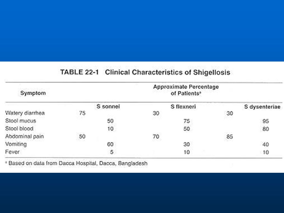
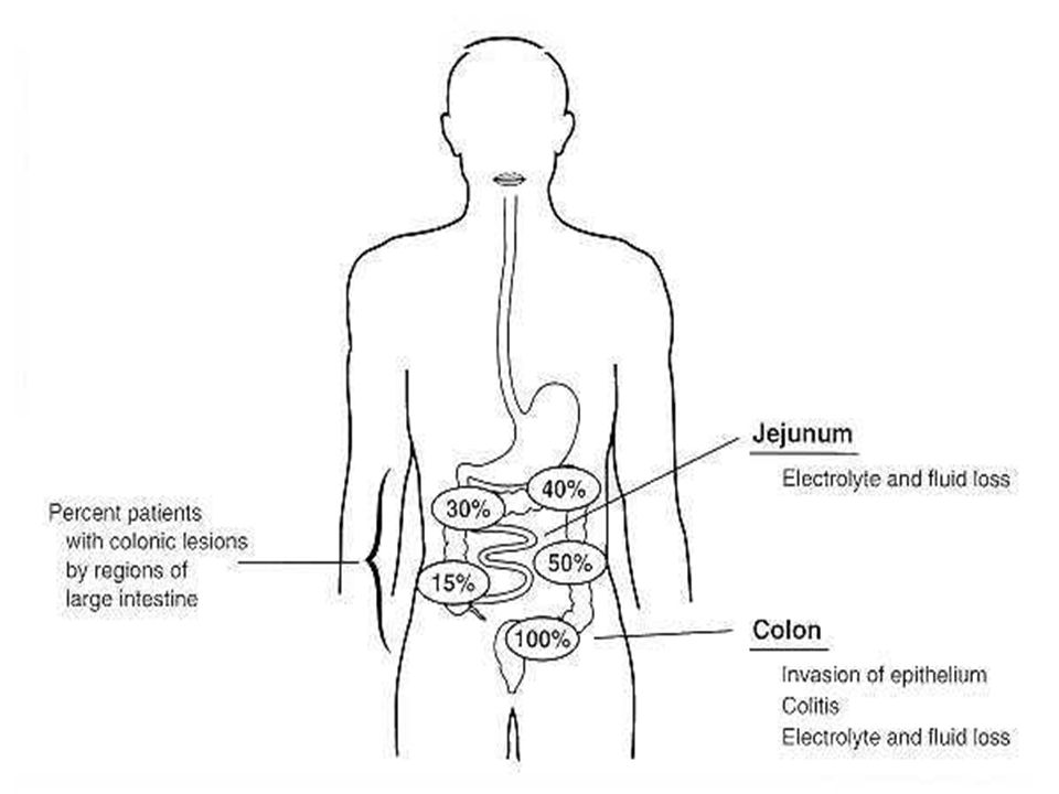
Clinical Manifestations - Shigella
Person-to-person spread Also food/water vectors IP hrs, disease 7 days & carried for 30 D Vomiting and mild to moderate dehydration Bloody diarrhea or dysentery Nonbloody diarrhea Fever, abdominal pain tenesmus with watery diarrhea or scant stools with pus, blood, mucus Clinical Manifestations Symptoms of shigellosis include abdominal pain, tenesmus, watery diarrhea, and/or dysentery (multiple scanty, bloody, mucoid stools). Other signs may include abdominal tenderness, fever, vomiting, dehydration, and convulsions.\ Shigellosis has two basic clinical presentations: watery diarrhea associated with vomiting and mild to moderate dehydration, and (2) dysentery characterized by a small volume of bloody, mucoid stools, and abdominal pain (cramps and tenesmus) (Table 22-1). Volunteer challenge studies show that shigellosis can be evoked by an extremely small inoculum ( organisms), and the time of onset of symptoms is somewhat influenced by the size of the challenge. The salient point is that shigellosis is an acute infection with onset of symptoms usually occurring within hours of ingestion of the etiologic agent. The average duration of symptoms in untreated adults is 7 days, and the organism may be cultivated from stools for 30 days or longer. The clinical features of shigellosis are summarized in Figure 22-1. Watery diarrhea occurs as a prodrome, or as the sole clinical manifestation, in a majority of patients infected with S sonnei. Diarrhea is often a prodome of the dysentery characterizing infection with other species of Shigella. Recently discovered enterotoxins secreted by S flexneri may contribute to the diarrheal phase as the etiologic agents traverse the small intestine. However, diarrhea is most common in patients who have colitis involving the transverse colon or cecum. These patients evidence net water secretion and impaired absorption in the inflamed colon. In patients experiencing dysentery, involvement is most severe in the distal colon, and the resulting inflammatory colitis is evidenced in frequent scanty stools reflecting the ileocecal fluid flow. \ Dysentery is also characterized by the daily loss of ml of serum protein into the feces. \ This loss of serum proteins results in depletion of nitrogen stores that exacerbates malnutrition and growth stunting. Depletion of immune factors also increases the risk of concurrent, unrelated infectious disease and contributes to substantial mortality
. Other signs may include abdominal tenderness, fever, vomiting, dehydration, and convulsions.\ Shigellosis has two basic clinical presentations: watery diarrhea associated with vomiting and mild to moderate dehydration, and. (2) dysentery characterized by a small volume of bloody, mucoid stools, and abdominal pain (cramps and tenesmus) (Table 22-1). Volunteer challenge studies show that shigellosis can be evoked by an extremely small inoculum ( organisms), and the time of onset of symptoms is somewhat influenced by the size of the challenge. The salient point is that shigellosis is an acute infection with onset of symptoms usually occurring within hours of ingestion of the etiologic agent. The average duration of symptoms in untreated adults is 7 days, and the organism may be cultivated from stools for 30 days or longer. The clinical features of shigellosis are summarized in Figure Watery diarrhea occurs as a prodrome, or as the sole clinical manifestation, in a majority of patients infected with S sonnei. Diarrhea is often a prodome of the dysentery characterizing infection with other species of Shigella. Recently discovered enterotoxins secreted by S flexneri may contribute to the diarrheal phase as the etiologic agents traverse the small intestine. However, diarrhea is most common in patients who have colitis involving the transverse colon or cecum. These patients evidence net water secretion and impaired absorption in the inflamed colon. In patients experiencing dysentery, involvement is most severe in the distal colon, and the resulting inflammatory colitis is evidenced in frequent scanty stools reflecting the ileocecal fluid flow. \ Dysentery is also characterized by the daily loss of ml of serum protein into the feces. \ This loss of serum proteins results in depletion of nitrogen stores that exacerbates malnutrition and growth stunting. Depletion of immune factors also increases the risk of concurrent, unrelated infectious disease and contributes to substantial mortality.")
78
FIGURE 22-1 Pathogenesis of shigellosis in humans.
Possible complications of shigellosis include bacteremia, convulsions and other neurological complications, reactive arthritis, and hemolytic-uremic syndrome. Bacteremia occasionally accompanies S dysenteriae serotype 1 infections in malnourished infants, but this complication is uncommon in otherwise healthy individuals. Convulsions have been reported in up to 25% of Shigella infections involving children under the age of 4 years. Both high fever and a family history of seizures are risk factors for a convulsive episode. Ekiri syndrome, an extremely rare, fatal encephalopathy has also been described in Japanese children with S sonnei or S flexneri infections. Reactive arthritis, a self-limiting sequela of S flexneri infection, occurs in an incidence as high as 2% in individuals expressing the HLA-B27 histocompatibility antigen. Hemolytic-uremic syndrome, characterized by a triad of microangiopathic hemolytic anemia, thrombocytopenia, and acute renal failure, is a rare complication in children infected with S dysenteriae serotype 1.The hallmarks of shigellosis are bacterial invasion of the colonic epithelium and inflammatory colitis. These are interdependent processes amplified by local release of cytokines and by the infiltration of inflammatory elements. Colitis in the rectosigmoid mucosa, with concomitant malabsorption, results in the characteristic sign of bacillary dysentery: scanty,. unformed stools tinged with blood and mucus. Host Defenses Inflammation, copious mucus secretion, and regeneration of the damaged colonic epithelium limit the spread of colitis and promote spontaneous recovery. Serotype-specific immunity is induced by a primary infection, suggesting a protective role of antibody recognizing the lipopolysaccharide (LPS) somatic antigen. Other Shigella antigens include enterotoxins, cytotoxin, and plasmid-encoded proteins that induce bacterial invasion of the epithelium. The protective role of immune responses against these antigens is unclear.
 somatic antigen. Other Shigella antigens include enterotoxins, cytotoxin, and plasmid-encoded proteins that induce bacterial invasion of the epithelium. The protective role of immune responses against these antigens is unclear.")
80
Complications Bacteremia uncommon
Convulsions 25% of Shigella infections involving children under the age of 4 years Ekiri syndrome, an extremely rare, fatal encephalopathy has also been described in Japanese children with S sonnei or S flexneri infections Reactive arthritis (Reiter’s) 2% HLA-B27 S flexneri Hemolytic-uremic syndrome S dysenteriae serotype 1 Protein loss
 2% HLA-B27 S flexneri. Hemolytic-uremic syndrome S dysenteriae serotype 1. Protein loss.")
81
Pathogenesis — Shigella
Fecal-oral Infectious dose very low (< 200 bugs) Invades mucosa with subsequent multiplication and mucosal destruction Invasiness property encoded on large 140 Mda IPA plasmid InterCellular Spread (IcsA) Shiga-toxin ( I& II) Exotoxin is an enterotoxin and cytotoxin Pathogenesis Infection is initiated by ingestion of shigellae (usually via fecal-oral contamination). An early symptom, diarrhea (possibly elicited by enterotoxins and/or cytotoxin), may occur as the organisms pass through the small intestine. The hallmarks of shigellosis are bacterial invasion of the colonic epithelium and inflammatory colitis. These are interdependent processes amplified by local release of cytokines and by the infiltration of inflammatory elements. Colitis in the rectosigmoid mucosa, with concomitant malabsorption, results in the characteristic sign of bacillary dysentery: scanty,. unformed stools tinged with blood and mucus.
 Invades mucosa with subsequent multiplication and mucosal destruction. Invasiness property encoded on large 140 Mda IPA plasmid InterCellular Spread (IcsA) Shiga-toxin ( I& II) Exotoxin is an enterotoxin and cytotoxin. Pathogenesis. Infection is initiated by ingestion of shigellae (usually via fecal-oral contamination). An early symptom, diarrhea (possibly elicited by enterotoxins and/or cytotoxin), may occur as the organisms pass through the small intestine. The hallmarks of shigellosis are bacterial invasion of the colonic epithelium and inflammatory colitis. These are interdependent processes amplified by local release of cytokines and by the infiltration of inflammatory elements. Colitis in the rectosigmoid mucosa, with concomitant malabsorption, results in the characteristic sign of bacillary dysentery: scanty,. unformed stools tinged with blood and mucus.")
82
FIGURE 22-3 Histopathology of acute colitis following peroral infection with shigellae. The organisms are initially ingested by membranous (M) cells that are associated with lymphoid microfollicles in the colon. After transcytosis through the M cell, the bacteria are deposited into the subepithelial space where they are phagocytosed by macrophages. The macrophage phagosome is subsequently degraded, and the intracellular shigellae cause release of IL-1 that evokes an influx of polymorphonuclear leukocytes (PMN). Eventually the infected macrophages undergo apoptosis (programmed cell death), and the bacteria are released onto the basolateral surface of adjacent colonic enterocytes. In addition, PMN transmigration through the epithelium disrupts tight junctions, allowing shigellae to migrate into the subepithelial space. The bacteria infect enterocytes by induced endocytosis, and the endocytic vacuoles are subsequently degraded. The intercellular shigellae attach to actin in the enterocyte junctional complex, multiply, and spread to contiguous enterocytes by induced actin polymerization. Ultimately, the infected enterocytes die, and the resulting necrosis of the epithelium, in conjunction with the continuing inflammatory response, constitutes the lesions of shigellosis. Transmigration of infiltrating PMNs through the tight junctions of local epithelial cells and into the intestinal lumen allows the reverse migration of shigellae from the lumen into the subepithelial spaces. These organisms then infect the columnar epithelial cells by inducing endocytic uptake at the basolateral surface. Immediately after infection of enterocytes, intracellular shigellae lyse endocytic vacuoles and attach to the actin cytoskeleton in the area of the junctional complex. As these organisms multiply within the enterocyte cytoplasm, occasional daughter cells induce polar nucleation of filamentous actin resulting in a "tail" that propels the shigellae into protrusions impinging on contiguous enterocytes. Plasma membranes enveloping the organisms are again lysed, and the organisms are deposited within the contiguous host cell resulting in intercellular bacterial spread. In summary, shigellosis can be characterized as an acute inflammatory bowel disease initiated by the uptake of only a few organisms into lymphoid follicles. Intracellular replication and intercellular spread leads to an amplified inflammatory cascade at the initial site of entry, and as this inflammation persists and expands, the infiltration of PMN facilitates the entry of additional bacteria into the epithelium. The inflammatory infiltrate can also cause detachment of sheets of epithelial cells in areas devoid of lymphoid structures or bacterial cells. Genetics of Virulence Shigella are exquisitely adapted for reproduction within the colonic epithelium of the human host. Many of the bacterial virulence determinants that mediate the complex interactions between these bacteria and mammalian host cells have been identified by genetic and immunological means. These virulence determinants are encoded by large extra-chromosomal elements (plasmids) that are functionally identical in all Shigella species and in EIEC. A complex of two plasmid-encoded determinants, designated Invasion Plasmid Antigens (Ipa) B and C, is recognized by antibody in the sera of convalescent patients. Ipa proteins are maximally expressed in conditions approximating the intestinal lumen (e.g., bile salts, high osmolarity, and human body temperature), and release of the IpaBC complex is triggered by contact with the mammalian host cell. This complex induces the endocytic uptake of shigellae by M cells, epithelial cells, and macrophages. IpaB also mediates lysis of endocytic vacuoles in epithelial cells or macrophages. In the latter case, Ipa proteins also cause release of the IL-1 cytokine and macrophage apoptosis. Another plasmid-encoded virulence determinant is secreted at the poles of Shigella daughter cells as these organisms multiply within the cytoplasm of infected host cells. This InterCellular Spread (IcsA) protein elicits polymerization of filamentous actin. Formation of this actin tail provides a motive force for shigellae impinging on the plasma membrane of the infected cell. The resulting protrusions deform the plasma membrane of contiguous cells. The IcsB plasmid-encoded protein then lyses the plasma membranes, resulting in intercellular bacterial spread. Biochemical characterization of the interaction between these Shigella virulence determinants and host cell components is a remaining research challenge. Characterizing and enhancing the neutralizing potential of antibody recognizing these protein virulence determinants is also an important research goal. Toxins Spent medium from S flexneri or EIEC cultures elicits fluid accumulation in rabbit ligated ileal loops and ion secretion in isolated ileal tissue. Using these assays, enterotoxins designated ShET1 and ShET2 have been identified, and the genetic loci encoding these toxins have been localized to the chromosome and plasmid, respectively. ShET1 is neutralized by convalescent sera from volunteers challenged with S flexneri 2a, suggesting that this toxic moiety is expressed by shigellae growing in the human intestine. The ShET1 locus is present on the chromosome of S flexneri 2a, but it is only occasionally found in other serotypes. In contrast, ShET2 is more widespread and detectable in 80% of shigellae representing all four species. These enterotoxins may elicit the diarrheal prodrome that often precedes bacillary dysentery; however, their role in the disease process remains to be defined by controlled challenge studies using toxin-negative mutants. S dysenteriae serotype 1 expresses Shiga toxin, an extremely potent, ricin-like, cytotoxin that inhibits protein synthesis in susceptible mammalian cells. This toxin also has enterotoxic activity in rabbit ileal loops, but its role in human diarrhea is unclear, since shigellae apparently express a number of enterotoxins. Experimental infection of rhesus monkeys with S dysenteriae 1, and with a Shiga toxin-negative mutant, suggests that this cytotoxin causes capillary destruction and focal hemorrhage that exacerbates dysentery (see Table 22-1). More importantly, Shiga toxin is associated with the hemolytic-uremic syndrome, a complication of infections with S dysenteriae 1. Closely related toxins are expressed by enterohemorrhagic E coli (EHEC) including the potentially lethal, food-borne O157-H7 serotype. Host Defense Shigellae are remarkably infectious enteric pathogenes that can cause disease after the ingestion of as few as 10 organisms. Nonetheless, shigellosis is normally an acute, self-limiting disease that exemplifies the regenerative capacity of the intestinal epithelium. Shigella virulence probably reflects both the efficient uptake by the follicle associated epithelium (M cells) and the amplifying effect of the inflammatory cascade generated by apoptic macrophages. Tenesmus and evacuation of mucus by intestinal goblet cells may effectively eliminate both extracellular shigellae and infected enterocytes from the intestinal lumen, but this defensive response, in conjunction with PMN infiltration, also constitutes the definitive sign of bacillary dysentery. In endemic areas, shigellosis is essentially a childhood disease, and the incidence decreases drastically in the indigenous population over 5 years of age. Controlled volunteer challenge studies in North American adults also indicate that prior infection with S flexneri protects against reinfection with the homologous serotype (70% efficacy). Serotype-specific immune protection against shigellosis suggests that antibody recognizing the O-polysaccharide of LPS protects against clinical symptoms. Ingested bovine colostrum containing antibody recognizing the O-polysaccharide of S flexneri 2a passively protects volunteers challenged with the homologous Shigella serotype. These observations have encouraged the development of a number of parenteral and mucosally administered O-polysaccharide vaccines that are currently in safety and/or efficacy trials. These vaccines offer the possibility of effective control of shigellosis independent of the needed improvements in the public health infrastructure of developing countries, but licensure and delivery of practical Shigella vaccines remains a distant prospect.
 cells that are associated with lymphoid microfollicles in the colon. After transcytosis through the M cell, the bacteria are deposited into the subepithelial space where they are phagocytosed by macrophages. The macrophage phagosome is subsequently degraded, and the intracellular shigellae cause release of IL-1 that evokes an influx of polymorphonuclear leukocytes (PMN). Eventually the infected macrophages undergo apoptosis (programmed cell death), and the bacteria are released onto the basolateral surface of adjacent colonic enterocytes. In addition, PMN transmigration through the epithelium disrupts tight junctions, allowing shigellae to migrate into the subepithelial space. The bacteria infect enterocytes by induced endocytosis, and the endocytic vacuoles are subsequently degraded. The intercellular shigellae attach to actin in the enterocyte junctional complex, multiply, and spread to contiguous enterocytes by induced actin polymerization. Ultimately, the infected enterocytes die, and the resulting necrosis of the epithelium, in conjunction with the continuing inflammatory response, constitutes the lesions of shigellosis. Transmigration of infiltrating PMNs through the tight junctions of local epithelial cells and into the intestinal lumen allows the reverse migration of shigellae from the lumen into the subepithelial spaces. These organisms then infect the columnar epithelial cells by inducing endocytic uptake at the basolateral surface. Immediately after infection of enterocytes, intracellular shigellae lyse endocytic vacuoles and attach to the actin cytoskeleton in the area of the junctional complex. As these organisms multiply within the enterocyte cytoplasm, occasional daughter cells induce polar nucleation of filamentous actin resulting in a tail that propels the shigellae into protrusions impinging on contiguous enterocytes. Plasma membranes enveloping the organisms are again lysed, and the organisms are deposited within the contiguous host cell resulting in intercellular bacterial spread. In summary, shigellosis can be characterized as an acute inflammatory bowel disease initiated by the uptake of only a few organisms into lymphoid follicles. Intracellular replication and intercellular spread leads to an amplified inflammatory cascade at the initial site of entry, and as this inflammation persists and expands, the infiltration of PMN facilitates the entry of additional bacteria into the epithelium. The inflammatory infiltrate can also cause detachment of sheets of epithelial cells in areas devoid of lymphoid structures or bacterial cells. Genetics of Virulence. Shigella are exquisitely adapted for reproduction within the colonic epithelium of the human host. Many of the bacterial virulence determinants that mediate the complex interactions between these bacteria and mammalian host cells have been identified by genetic and immunological means. These virulence determinants are encoded by large extra-chromosomal elements (plasmids) that are functionally identical in all Shigella species and in EIEC. A complex of two plasmid-encoded determinants, designated Invasion Plasmid Antigens (Ipa) B and C, is recognized by antibody in the sera of convalescent patients. Ipa proteins are maximally expressed in conditions approximating the intestinal lumen (e.g., bile salts, high osmolarity, and human body temperature), and release of the IpaBC complex is triggered by contact with the mammalian host cell. This complex induces the endocytic uptake of shigellae by M cells, epithelial cells, and macrophages. IpaB also mediates lysis of endocytic vacuoles in epithelial cells or macrophages. In the latter case, Ipa proteins also cause release of the IL-1 cytokine and macrophage apoptosis. Another plasmid-encoded virulence determinant is secreted at the poles of Shigella daughter cells as these organisms multiply within the cytoplasm of infected host cells. This InterCellular Spread (IcsA) protein elicits polymerization of filamentous actin. Formation of this actin tail provides a motive force for shigellae impinging on the plasma membrane of the infected cell. The resulting protrusions deform the plasma membrane of contiguous cells. The IcsB plasmid-encoded protein then lyses the plasma membranes, resulting in intercellular bacterial spread. Biochemical characterization of the interaction between these Shigella virulence determinants and host cell components is a remaining research challenge. Characterizing and enhancing the neutralizing potential of antibody recognizing these protein virulence determinants is also an important research goal. Toxins. Spent medium from S flexneri or EIEC cultures elicits fluid accumulation in rabbit ligated ileal loops and ion secretion in isolated ileal tissue. Using these assays, enterotoxins designated ShET1 and ShET2 have been identified, and the genetic loci encoding these toxins have been localized to the chromosome and plasmid, respectively. ShET1 is neutralized by convalescent sera from volunteers challenged with S flexneri 2a, suggesting that this toxic moiety is expressed by shigellae growing in the human intestine. The ShET1 locus is present on the chromosome of S flexneri 2a, but it is only occasionally found in other serotypes. In contrast, ShET2 is more widespread and detectable in 80% of shigellae representing all four species. These enterotoxins may elicit the diarrheal prodrome that often precedes bacillary dysentery; however, their role in the disease process remains to be defined by controlled challenge studies using toxin-negative mutants. S dysenteriae serotype 1 expresses Shiga toxin, an extremely potent, ricin-like, cytotoxin that inhibits protein synthesis in susceptible mammalian cells. This toxin also has enterotoxic activity in rabbit ileal loops, but its role in human diarrhea is unclear, since shigellae apparently express a number of enterotoxins. Experimental infection of rhesus monkeys with S dysenteriae 1, and with a Shiga toxin-negative mutant, suggests that this cytotoxin causes capillary destruction and focal hemorrhage that exacerbates dysentery (see Table 22-1). More importantly, Shiga toxin is associated with the hemolytic-uremic syndrome, a complication of infections with S dysenteriae 1. Closely related toxins are expressed by enterohemorrhagic E coli (EHEC) including the potentially lethal, food-borne O157-H7 serotype. Host Defense. Shigellae are remarkably infectious enteric pathogenes that can cause disease after the ingestion of as few as 10 organisms. Nonetheless, shigellosis is normally an acute, self-limiting disease that exemplifies the regenerative capacity of the intestinal epithelium. Shigella virulence probably reflects both the efficient uptake by the follicle associated epithelium (M cells) and the amplifying effect of the inflammatory cascade generated by apoptic macrophages. Tenesmus and evacuation of mucus by intestinal goblet cells may effectively eliminate both extracellular shigellae and infected enterocytes from the intestinal lumen, but this defensive response, in conjunction with PMN infiltration, also constitutes the definitive sign of bacillary dysentery. In endemic areas, shigellosis is essentially a childhood disease, and the incidence decreases drastically in the indigenous population over 5 years of age. Controlled volunteer challenge studies in North American adults also indicate that prior infection with S flexneri protects against reinfection with the homologous serotype (70% efficacy). Serotype-specific immune protection against shigellosis suggests that antibody recognizing the O-polysaccharide of LPS protects against clinical symptoms. Ingested bovine colostrum containing antibody recognizing the O-polysaccharide of S flexneri 2a passively protects volunteers challenged with the homologous Shigella serotype. These observations have encouraged the development of a number of parenteral and mucosally administered O-polysaccharide vaccines that are currently in safety and/or efficacy trials. These vaccines offer the possibility of effective control of shigellosis independent of the needed improvements in the public health infrastructure of developing countries, but licensure and delivery of practical Shigella vaccines remains a distant prospect.")
84
Diagnosis Shigellosis on the basis of fresh blood in the stool.
Neutrophils in fecal smears is also a strongly suggestive sign. Watery, mucoid diarrhea may be the only symptom of many S sonnei infections DDx EIEC, Salmonella enteritidis, Yersinia enterocolitica, Campylobacter species, and Entamoeba histolytica Clinical Patients presenting with watery diarrhea and fever should be suspected of having shigellosis. The diarrheal stage of the infection cannot be distinguished clinically from other bacterial, viral, and protozoan infections. Nausea and vomiting can accompany Shigella diarrhea, but these symptoms are also observed during infections with nontyphoidal salmonellae and enterotoxigenic E coli. Bloody, mucoid stools are highly indicative of shigellosis, but the differential diagnosis should include EIEC, Salmonella enteritidis, Yersinia enterocolitica, Campylobacter species, and Entamoeba histolytica. Although blood is common in the stools of patients with amebiasis, it is usually dark brown rather than bright red, as in Shigella infections. Microscopic examination of stool smears from patients with amebiasis should reveal erythrophagocytic trophozoites in the absence of PMN, whereas bacillary dysentery is characterized by sheets of PMN. Sigmoidoscopic examination of a shigellosis patient reveals a diffusely erythematous mucosal surface with small ulcers, whereas amebiasis is characterized by discrete ulcers in the absence of generalized inflammation.

85
Isolation Procedures - Shigella
Enrichment media GN broth & Selenite broth Plate media Low selectivity —MAC Med selectivity — XLD High selectivity — DCA, HEK Caution — some Shigella strains are inhibited by SS agar Laboratory Although clinical signs may evoke the suspicion of shigellosis, diagnosis is dependent upon the isolation and identification of Shigella from the feces. Positive cultures are most often obtained from blood-tinged plugs of mucus in freshly passed stool specimens obtained during the acute phase of disease. Rectal swabs may also be used to culture shigellae if the specimen is processed rapidly or is deposited in a buffered glycerol saline holding solution. Isolation of shigellae in the clinical laboratory typically involves an initial streaking for isolation on differential/selective media with aerobic incubation to inhibit the growth of the anaerobic normal flora. Commonly used primary isolation media include MacConkey, Hektoen Enteric Agar, and Salmonella-Shigella (SS) Agar. These media contain bile salts to inhibit the growth of other Gram-negative bacteria and pH indicators to differentiate lactose fermenters (Coliforms) from non-lactose fermenters such as shigellae. A liquid enrichment medium (Hajna Gram-negative broth) may also be inoculated with the stool specimen and subcultured onto the selective/differential agarose media after a short growth period. Following overnight incubation of primary isolation media at 37° C, colorless, non-lactose-fermenting colonies are streaked and stabbed into tubed slants of Kligler's Iron Agar or Triple Sugar Iron Agar. In these differential media, Shigella species produce an alkaline slant and an acid butt with no bubbles of gas in the agar. This reaction gives a presumptive identification, and slide agglutination tests with antisera for serogroup and serotype confirm the identification. Some E coli biotypes of the normal intestinal flora closely resemble Shigella species (i.e. they are nonmotile, delayed lactose fermenters). These coliforms can usually be differentiated from shigellae by the ability to decarboxylate lysine. However, some coliforms cause enteroinvasive disease because they carry the Shigella-like virulence plasmid, and these pathogens are conventionally identified by laborious serological screening for EIEC serotypes. Sensitive and rapid methodology for identification of both EIEC and Shigella species utilizes DNA probes that hybridize with common virulence plasmid genes or DNA primers that amplify plasmid genes by polymerase chain reaction (PCR). Enzyme-linked immunosorbent assay (ELISA) using antiserum or monoclonal antibody recognizing Ipa proteins can also be used to screen stools for enteroinvasive pathogens. These experimental diagnostic techniques are useful for epidemiological studies of enteroinvasive infections, but they are probably too specialized for routine use in the clinical laboratory.
 Agar. These media contain bile salts to inhibit the growth of other Gram-negative bacteria and pH indicators to differentiate lactose fermenters (Coliforms) from non-lactose fermenters such as shigellae. A liquid enrichment medium (Hajna Gram-negative broth) may also be inoculated with the stool specimen and subcultured onto the selective/differential agarose media after a short growth period. Following overnight incubation of primary isolation media at 37° C, colorless, non-lactose-fermenting colonies are streaked and stabbed into tubed slants of Kligler s Iron Agar or Triple Sugar Iron Agar. In these differential media, Shigella species produce an alkaline slant and an acid butt with no bubbles of gas in the agar. This reaction gives a presumptive identification, and slide agglutination tests with antisera for serogroup and serotype confirm the identification. Some E coli biotypes of the normal intestinal flora closely resemble Shigella species (i.e. they are nonmotile, delayed lactose fermenters). These coliforms can usually be differentiated from shigellae by the ability to decarboxylate lysine. However, some coliforms cause enteroinvasive disease because they carry the Shigella-like virulence plasmid, and these pathogens are conventionally identified by laborious serological screening for EIEC serotypes. Sensitive and rapid methodology for identification of both EIEC and Shigella species utilizes DNA probes that hybridize with common virulence plasmid genes or DNA primers that amplify plasmid genes by polymerase chain reaction (PCR). Enzyme-linked immunosorbent assay (ELISA) using antiserum or monoclonal antibody recognizing Ipa proteins can also be used to screen stools for enteroinvasive pathogens. These experimental diagnostic techniques are useful for epidemiological studies of enteroinvasive infections, but they are probably too specialized for routine use in the clinical laboratory.")
86
Screening Procedures - Shigella
Lactose or xylose NON-fermenting colonies Screen suspect colonies biochemically TSI — K/A I no gas / no H25 Some sonnei and fiexneri produce gas Nonmotile, urease negative Screen typical biochemically ID colonies with grouping antisera

87
Antimicrobial Susceptibility Testing
Shigella Susceptibility testing should be done Empiric treatment when susceptibility is not known is TMP-SMX Use Quinolone if resistance to TMP-SMX is suspected Treatment Although severe dehydration is uncommon in shigellosis, the first consideration in treating any diarrheal disease is correction of abnormalities that result from isotonic dehydration, metabolic acidosis, and significant potassium loss. The oral rehydration treatment developed by the World Health Organization has proven effective and safe in the treatment of acute diarrhea, provided that the patient is not vomiting or in shock from severe dehydration. In the latter case, intravenous fluid replacement is required until initial fluid and electrolyte losses are corrected. With proper hydration, shigellosis is generally a self-limiting disease, and the decision to prescribe antibiotics is predicated on the severity of disease, the age of the patient, and the likelihood of further transmission of the infection. Effective antibiotic treatment reduces the average duration of illness from approximately 5-7 days to approximately 3 days and also reduces the period of Shigella excretion after symptoms subside. Absorbable drugs such as ampicillin (2 g/day for 5 days) are likely to be effective when the isolate is sensitive. Trimethoprim (8 mg/kg/day) and sulfamethoxazole (40 mg/kg/day) will eradicate sensitive organisms quickly from the intestine, but resistance to this agent is increasing. Ciprofloxacin (1 g/day for 3 days) is effective against multiple drug resistant strains, but this antibiotic is not approved by the United States Food and Drug Administration for use in children less than 17 years of age because there is a theoretical risk of cartilage damage. Opiates, such as paregoric, induce intestinal stasis and may promote bacterial invasion, prolonging the febrile state.
 are likely to be effective when the isolate is sensitive. Trimethoprim (8 mg/kg/day) and sulfamethoxazole (40 mg/kg/day) will eradicate sensitive organisms quickly from the intestine, but resistance to this agent is increasing. Ciprofloxacin (1 g/day for 3 days) is effective against multiple drug resistant strains, but this antibiotic is not approved by the United States Food and Drug Administration for use in children less than 17 years of age because there is a theoretical risk of cartilage damage. Opiates, such as paregoric, induce intestinal stasis and may promote bacterial invasion, prolonging the febrile state.")
88
Control Prevention of fecal-oral transmission
Vaccines are not currently available, but some promising candidates are being developed Control As is the case with other intestinal infections, the most effective methods for controlling shigellosis are provision of safe and abundant water and effective feces disposal. These public health measures are, at best, long range strategies for control of enteric infections in developing countries. The estimated five million deaths annually attributed to diarrheal disease in these countries, in addition to the malabsorption and growth stunting among survivors, require more immediate and practical approaches. The most effective intervention strategy to minimize morbidity and mortality would involve comprehensive media and personal outreach programs consisting of the following components: (1) education of all residents to actively avoid fecal contamination of food and water and to encourage hand washing after defecation; (2) encourage mothers to breast-feed infants; (3) promote the use of oral rehydration therapy to offset the effects of acute diarrhea; (4) encourage mothers to provide convalescent nutritional care in the form of extra food for children recovering from diarrhea or dysentery.
 education of all residents to actively avoid fecal contamination of food and water and to encourage hand washing after defecation; (2) encourage mothers to breast-feed infants; (3) promote the use of oral rehydration therapy to offset the effects of acute diarrhea; (4) encourage mothers to provide convalescent nutritional care in the form of extra food for children recovering from diarrhea or dysentery.")
Similar presentations