Download presentation
Presentation is loading. Please wait.

1
Lab #2

3
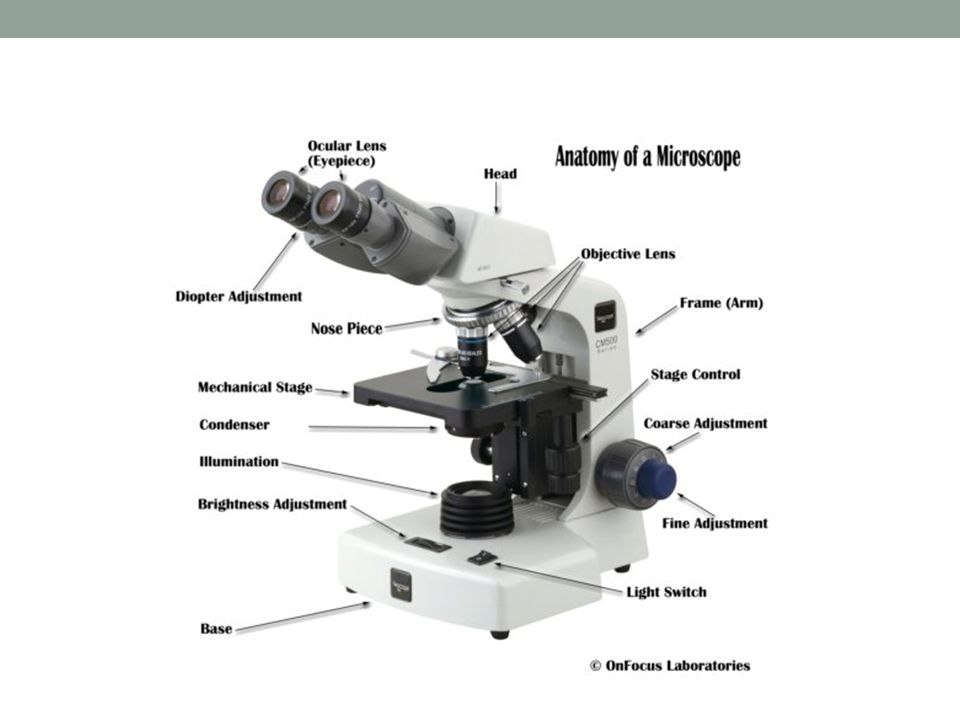
Review of Lab #1 Properly putting away the microscope: Light off
Wrap the cord Stage all the way down On lowest objective (4X) Iris diaphragm open Clean any oil on 100X using LENS paper. Also, clean other objectives with lens paper Clean ocular lenses! DO NOT ROTATE THE HEAD WITH THE OCULAR LENSES! Show bibulous paper/ blotting paper vs lens paper
 Iris diaphragm open. Clean any oil on 100X using LENS paper. Also, clean other objectives with lens paper. Clean ocular lenses! DO NOT ROTATE THE HEAD WITH THE OCULAR LENSES! Show bibulous paper/ blotting paper vs lens paper.")
4
Review of Lab #1

5
Review of Lab #1 Bacillus, chains Bacillus, singles

6
Review of Lab #1 Cocci, clusters Cocci, chains

7
Review of Lab #1 Cocci, tetrads

8
Review of Lab #1 Prokaryotes: no nucleus (bacteria and archae)
Eukaryotes: nucleus and membrane bound organelles Protozoa: Giardia, Amoeba, Trypanosoma Unicellular Fungi: Pencillium Unicellular algae: Chlorella Viruses Giardia Show bibulous paper/ blotting paper vs lens paper Amoeba Trypanosoma

9
Lab #2 – 3. Ubiquity of Microbes
Microbes are found in every environment Each environment has a unique collection of residents Your petri plate Observe 3 different colonies on your petri plate Describe each colony fill out table on pg. 17 Use pg. 19 for reference Are microbes ubiquitous? Are they diverse? Each colony is a population of clones derived from a single dividing cell Microbiologists study isolated colonies Colonies are described on the basis of size, shape, margin, elevation, texture, and pigment A lot of diversity in microbes

10
Colony morphology

11
Lab #2: 4A. Aseptic Technique
Aseptic technique: a technique utilized by microbiologists to prevent contamination – only the desired organism is inoculated in the growth medium Includes: Washing hands Tying back your hair Disinfecting your work station Turning on the flame Sterilizing your tools (loop, needle, etc)
")
12
Lab #2: 4A. Aseptic Technique
Growth media contains all nutrients: CHNOPS, minerals, and vitamins Many different types of growth media: Liquid media: Broth Solid media: prepared by adding a solidifying agent called agar Petri plate Slant Tall/ deep Microbes can be transferred from any one form of media to another Media is sterilized using an autoclave or a filter before use

13
Types of growth media Broth Slant Petri plate Tall/ deep

14
Autoclave

15
Lab #2: Aseptic Technique
Tools (pg. 22): Loop – broth, slants, petri plates Needle – Talls/ deeps Swab – petri plates Today you will learn the proper use of a loop and a needle Each student: Choose one of the following organisms: B. subtilis E. coli K. pneumoniae S. epidermidis Inoculate organism onto a Tryptic Soy Agar slant (use loop), Motility Media (use needle), and thioglycollate broth (use loop)
: Loop – broth, slants, petri plates. Needle – Talls/ deeps. Swab – petri plates. Today you will learn the proper use of a loop and a needle. Each student: Choose one of the following organisms: B. subtilis. E. coli. K. pneumoniae. S. epidermidis. Inoculate organism onto a Tryptic Soy Agar slant (use loop), Motility Media (use needle), and thioglycollate broth (use loop)")
16
Lab #2: Gram staining Type of a differential stain separate major groups of microorganisms based on retention or loss of the initial dye Grams stain: divides bacteria into 2 major groups: Gram positive Gram negative One group retains the initial dye of the specific gram staining procedure and the second group loses the initial dye in the staining procedure Discovered by a Danish biologist, Hans Christian Joachim Gram.

17
How does Gram staining work?
Gram positive cells have a thicker layer of peptidoglycan in their cell wall as compared to gram negative cell wall The staining dye crystal violet forms a large complex with iodine this complex stays in the thick cell wall of G+ bacteria cells don’t decolorize Gram negative cells are decolorized when treated with Gram’s alcohol the counter stain, safranin, is then taken up by gram negative cells

18
Preparation of a smear Smear (pg 31 & 32):
Suspension of cells in water which is dried on a slide Should be light and uniform in appearance for best results! Madigan 10th edition, Figure 4.3, pg 58

19
Gram Staining: Procedure (Pg. 34)
1 min 30-45 sec 5-10 sec You may save your slides after preparing and/or viewing Madigan, 10th edition, figure 4.4, page 59

20
Gram positive cocci in cluster (masses) Gram positive bacilli
Gram negative rods in singles Mix of gram positve and gram negative organisms

21
Lab #2: Gram staining Read pages 31 – 32 – know common terms
Each student gram stain two organisms: B. subtilis & K. pneumoniae E. coli & S. epidermidis Go around class to view the other two organisms Record results on Pg. 35 Use a separate slide for each gram stain

Similar presentations


 –algae.>")


 in microbiology. Functions : To allow us to study.>")
