Download presentation
Presentation is loading. Please wait.

1
Electrocardiography for Healthcare Professionals
Chapter 2: The Cardiovascular System

2
Learning Outcomes 2.1 Describe circulation as it relates to the ECG. 2.2 Recall the structures of the heart. 2.3 Differentiate between the pulmonary, systemic and coronary circulation. 2.4 Explain the cardiac cycle, and relate the difference between systole and diastole.

3
Learning Outcomes (Cont’d)
2.5a Describe the parts and function of the conduction system. 2.5b Recall the unique qualities of the heart and their relationship to the cardiac conduction system. 2.5c Explain the conduction system as it relates to the ECG.

4
Learning Outcomes (Cont’d)
2.6a Identify each part of the ECG waveform. 2.6b Describe the heart activity that produces the ECG waveform.

5
2.1 Circulation and the ECG
Transports blood Muscular pump Electrical activity LO 2.1: Describe circulation as it relates to the ECG. ----- Function of heart – pump blood to and from all the cells and tissues Blood supplies nutrients & O2 and removes CO2 and waste products Process of transporting blood is known as circulation Circulation dependent upon the heart and its ability to contract or beat The electrical activity of the heart causes the heart to contract Each electrical activity of heart is recorded on the ECG Knowledge of the heart, its functions, and what produces the ECG tracing will provide you with a clear understanding of the tasks you will be performing as an ECG healthcare professional

6
2.1 Apply Your Knowledge What is the function of the heart?

7
2.1 Apply Your Knowledge What is the function of the heart?
Checkpoint questions What is circulation? The process of transporting blood to and from the body tissues What is recorded on the ECG strip? The electrical activity of the heart ANSWER: To pump blood to and from body tissues

8
2.2 Anatomy of the Heart Lies in center of chest Under sternum
Between the lungs The size of your fist Weighs 10.6 oz or 300 grams LO 2.2: Anatomy of the Heart 2/3 of heart lies left of sternum

9
2.2 Heart Statistics Average beats per minute = 72
Total output = 5 liters per minute LO 2.2: Anatomy of the Heart powerful muscular pump 140 ml of blood per beat Each day 1800 gallons enough to fill an average size bathtub about 36 times

10
2.2 Heart Anatomy Pericardium Pericardial space Epicardium Myocardium
Endocardium LO 2.2: Anatomy of the Heart -----photo on next slide Pericardium -Sack of tissue surrounding the heart Two layers Parietal layer is the tough outer layer Visceral or epicardium layer is the inner layer and adheres closely to the heart Purpose is to protect the heart from infection and trauma Pericardial space Between parietal and visceral layers of the pericardium Contains approximately 10 to 20 mL of fluid Serves to cushion the heart against trauma Heart consists of three layers: Epicardium – outer layer Also known as the pericardium Thin and contains the coronary arteries Myocardium – middle, muscular layer, contracts the heart Endocardium – inner layer Lines the inner surfaces of the heart chambers and valves Location of the Purkinje fibers is just below this layer

11
Click to Return to Last Screen Viewed
2.2 Heart Layers LO 2.2: Anatomy of the Heart Click to Return to Last Screen Viewed

12
2.2 Heart Chambers Left Atrium Right Atrium Left Ventricle
LO 2.2: Anatomy of the Heart Interventricular Septum Varies in thickness Thin in the atria Thick in the Right ventricle Thickest in the left ventricle Thicker = stronger the muscular contraction of that chamber Left ventricle sometimes know as the “workhorse of the heart” Right Ventricle

13
2.2 Heart Valves Tricuspid valve Mitral (bicuspid) valve
Semilunar valves LO 2.2: Anatomy of the Heart ----- Tricuspid valve – located between the right atrium and right ventricle Mitral (bicuspid) valve – located between left atrium and left ventricle Mitral and tricuspid valves also known as Atrioventricular (AV) valves prevent backflow of blood from the ventricles to the atria The pulmonary artery and the aorta each have a semilunar valve Half (semi) Lunar (moon) Semilunar valves prevent the backflow of blood into the ventricles Flaps or cusps open to allow the blood to flow then close AV valves open = semilunar valves closed
 valve – located between left atrium and left ventricle. Mitral and tricuspid valves also known as Atrioventricular (AV) valves prevent backflow of blood from the ventricles to the atria. The pulmonary artery and the aorta each have a semilunar valve. Half (semi) Lunar (moon) Semilunar valves prevent the backflow of blood into the ventricles. Flaps or cusps open to allow the blood to flow then close. AV valves open = semilunar valves closed.")
14
2.2 Heart Vessels Vena cava Pulmonary artery Pulmonary veins Aorta
Coronary arteries LO 2.2: Anatomy of the Heart ----- Vena cava (largest vein in the body) Superior vena cava returns blood from upper body to the heart Inferior vena cava returns blood from lower body to the heart Pulmonary artery Carries deoxygenated blood from the right ventricle to the lungs Pulmonary vein Carries oxygenated blood from the lungs to the left atrium Aorta Carries blood from the left ventricle to the body First branches from aorta are the coronary arteries Coronary arteries Supply blood to the heart muscle
 Superior vena cava returns blood from upper body to the heart. Inferior vena cava returns blood from lower body to the heart. Pulmonary artery. Carries deoxygenated blood from the right ventricle to the lungs. Pulmonary vein. Carries oxygenated blood from the lungs to the left atrium. Aorta. Carries blood from the left ventricle to the body. First branches from aorta are the coronary arteries. Coronary arteries. Supply blood to the heart muscle.")
15
2.2 Heart Valves and Vessels
Aorta Tricuspid Valve Vena Cava Bicuspid Valve Aortic Valve Pulmonary Artery Pulmonary Valve Pulmonary Veins LO 2.2: Anatomy of the Heart Click to Return to Last Screen Viewed Using the on-screen pen, draw a line from the label to its location.

17
2.2 Apply Your Knowledge Which valve of the heart lies between the left atrium and the left ventricle?

18
2.2 Apply Your Knowledge Which valve of the heart lies between the left atrium and the left ventricle? ANSWER: The mitral or bicuspid valve

19
2.2 Apply Your Knowledge Name the three layers of the heart.

20
2.2 Apply Your Knowledge Name the three layers of the heart.
Checkpoint Questions LO 2.2 What is the name of the middle layer of the heart (the muscular layer)? Myocardium Which valve is located between the left atrium and left ventricle? Bicuspid or mitral valve ANSWER: The endocardium, myocardium, and epicardium
 Myocardium. Which valve is located between the left atrium and left ventricle Bicuspid or mitral valve. ANSWER: The endocardium, myocardium, and epicardium.")
21
2.3 Principles of Circulation
Pulmonary Systemic Coronary L.O 2.3: Principles of Circulation

22
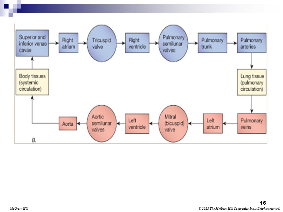
2.3 Pulmonary Circulation
Enters right atrium Blood passes through the tricuspid valve to the right ventricle Right ventricle pumps to the lungs Blood returns into the left atrium L.O. 2.3: Principles of Circulation ----- Pulmonary Circulation: Deoxygenated blood enters the right atrium from the superior and inferior venae cavae. Blood travels through the tricuspid valve into the right ventricle. The right ventricle pumps the blood through the pulmonary semilunar valve into the pulmonary artery, then into the lungs. In the lungs, the blood is oxygenated. The blood returns to the heart through the pulmonary veins into the left atrium. The left atrium is the last step of pulmonary circulation.

23
2.3 Systemic Circulation Enters the left atrium and passes through into the left ventricle The left ventricle pumps to the aorta From the aorta, blood circulates throughout the body Deoxygenated blood returns to the heart L.O 2.3: Principles of Circulation ----- Oxygenated blood enters the left atrium and travels through the mitral valve into the left ventricle. The left ventricle pumps the blood through the aortic semilunar valve into the aorta. After traveling through the body, the deoxygenated blood returns to the heart through the superior and inferior venae cavae.

24
2.3 Coronary Circulation A
L.O. 2.3: Principles of Circulation

25
2.3 Coronary Circulation B
L.O. 2.3: Principles of Circulation

26
2.3 Coronary Circulation Oxygenated blood travels from left ventricle to the coronary arteries Coronary arteries supply entire heart Deoxygenated blood returns to the right atrium L.O. 2.3: Principles of Circulation ----- Oxygenated blood from the left ventricle travels through the aorta to the coronary arteries. Two main coronary arteries-left main and right main. These arteries branch to supply oxygenated blood to the entire heart. The left main artery has more branches because the left side of the heart is more muscular and requires more blood supply. The deoxygenated blood travels through the coronary veins and is collected in the coronary sinus, which empties the blood directly into the right atrium. The Heart as a Pump Each ventricle pumps 70 mL of blood during contraction – this volume of blood that is ejected with each contraction is referred to as stroke volume This amount varies depending on gender, level of fitness, disease state, or genetics The volume of blood pumped each minute is referred to as cardiac output Average cardiac output is approx. 5 liters/min. Volume of blood decreases = heart rate decreases Contractile force decreases = cardiac output decreases Decreased output causes pallor, confusion, low blood pressure, nausea, and dizziness. Cardiac output is estimated by multiplying heart rate by stroke volume HR x SV = CO

27
2.3 Apply Your Knowledge Great!
Which vessels transport blood from the lungs to the left atrium? Great!

28
2.3 Apply Your Knowledge Great!
Which vessels transport blood from the lungs to the left atrium? ANSWER: The pulmonary veins Checkpoint questions(2.3): What are the first vessels to branch off the aorta known as? Coronary arteries Describe the three types of circulation. The pathways for pumping blood to and from the lungs are known as pulmonary circulation. The pathways for pumping blood throughout the body and back to the heart are known as systemic circulation. The circulation of blood to and from the heart muscle is known as coronary circulation. What is cardiac output? The volume pumped each minute is referred to as cardiac output. Great!
: What are the first vessels to branch off the aorta known as Coronary arteries. Describe the three types of circulation. The pathways for pumping blood to and from the lungs are known as pulmonary circulation. The pathways for pumping blood throughout the body and back to the heart are known as systemic circulation. The circulation of blood to and from the heart muscle is known as coronary circulation. What is cardiac output The volume pumped each minute is referred to as cardiac output. Great!")
29
2.4 The Cardiac Cycle L.O 2.4: The Cardiac Cycle
Each beat has two phases – contraction and relaxation which together makeup the cardiac cycle Relaxation = diastole – expanding and refilling Contraction = systole – squeezing blood out

30
2.4 The Cardiac Cycle (Cont’d)
L.O 2.4: The Cardiac Cycle

31
2.4 Diastole – Relaxation Phase
Blood returns to the heart via the superior and inferior vena cava Blood flows from the right atrium into the right ventricle Blood from the pulmonary veins flows from the left atrium into the left ventricle L.O. 2.4: The Cardiac Cycle ----- Blood from the upper body returns via the superior vena cava. Blood from the lower body returns via the inferior vena cava. The right atrium fills with blood, pushes open the tricuspid valve, and into the right ventricle. Blood returns from the lungs via the pulmonary veins to the left atrium, then forces the mitral valve to open to allow blood flow to the left ventricle.

32
2.4 Systole – Contraction Phase
Contraction creates pressure, opening the pulmonary and aortic valves Blood from the right ventricle flows to the lungs Blood from the left ventricle flows through the aorta to the body L.O. 2.4: The Cardiac Cycle ----- Blood is pushed into the lungs to exchange oxygen and carbon dioxide by the right ventricle. Blood from the left ventricle is pushed through the aorta to be distributed through the body to provide oxygen for tissues and to remove carbon dioxide. Listening with a stethoscope you will hear two sounds: “lubb” and “dupp” which are made by the opening and closing of the valves caused by the contraction of the heart “Lubb” sound made during the systolic phase by the contraction of the ventricles and the closing of the mitral and tricuspid valves “Dupp” sound is made during the diastolic phase. It is shorter and occurs during the beginning of ventricular relaxation.

33
2.4 Apply Your Knowledge The relaxation phase of the cardiac cycle is known as:

34
2.4 Apply Your Knowledge The relaxation phase of the cardiac cycle is known as: ANSWER: Diastole Checkpoint Questions (LO2.4) What is the cardiac cycle? The contraction and relaxation of the heart together make up the cardiac cycle. How are diastole and systole the same? They are both parts of the cardiac cycle.

35
2.5 Unique Qualities of the Heart
Automaticity Conductivity Contractivity Excitability L.O. 2.5 a., b., c.: Conduction System of the Heart Pumping cycle is controlled by electrical impulses – the impulses stimulate contraction of the heart muscle and absence of impulses allows the heart muscle to relax Impulses are initiated by specialized pacemaker cells in the heart. Impulses are transferred through the heart by the conduction system The working cells respond by shortening, causing cardiac contraction and blood to flow ----- Automaticity – heart’s ability to initiate an electrical impulse Conductivity – ability of myocardial cells to receive and conduct (transmit) electrical impulses Contractility – ability of the heart muscle to shorten in response to an electrical impulse Excitability – ability of the heart to respond to an impulse or stimulus Without these qualities the heart would not beat and would not be rhythmical
 electrical impulses. Contractility – ability of the heart muscle to shorten in response to an electrical impulse. Excitability – ability of the heart to respond to an impulse or stimulus. Without these qualities the heart would not beat and would not be rhythmical.")
36
2.5 Regulation of the Heart
Autonomic Nervous System Speeds up or slows down the heart rate Sympathetic branch can increase the heart rate Parasympathetic branch can decrease the heart rate L.O. 2.5 a., b., c.: Conduction System of the Heart ----- In addition to automaticity, the heartbeat is controlled by the ANS, which is involuntary Sympathetic branch can increase the heart rate in response to norepinephrine. Happens when you are under stress or frightened Normal rate of heart is 60 to 100 beats a minute When sympathetic branch of ANS stimulated the heartbeat speeds up Parasympathetic branch can decrease the heart rate via the vagus nerve. Acts like the brake of the heart Other factors can also affect the heart Exercise, stress, or fever can increase the rate The cardiac control center, in the brain, sends impulses to decrease the heart rate when the BP rises Levels of potassium and calcium Low potassium ions in the blood, heart rate decreases High potassium ions causes abnormal heart rate or rhythm Low calcium depresses heart actions High calcium causes contractions that are longer then normal heart contractions

37
2.5 Pathways for Conduction
SA node AV node Bundle of His Bundle branches Purkinje fibers L.O. 2.5 a., b., c.: Conduction System of the Heart

38
2.5 Sinoatrial (SA) Node Located in upper right portion of right atrium Initiates the heartbeat Pacemaker of the heart ( beats per minute) Normal conduction begins in SA node L.O. 2.5 a., b., c.: Conduction System of the Heart SA & AV nodes are small, round structures that consist of many specialized cardiac cells. Automaticity of the fibers in the SA node produces the contraction of the right and left atria

39
2.5 Atrioventricular (AV) Node
Located on the floor of the right atrium Causes delay in the electrical impulse, allowing for blood to travel to ventricles Can act as pacemaker if SA node is not working (40-60 bpm) L.O. 2.5 a., b., c.: Conduction System of the Heart
 L.O. 2.5 a., b., c.: Conduction System of the Heart.")
40
2.5 Bundle of His (AV bundle)
Located next to the AV node Transfers electrical impulses from the atria to the ventricles via bundle branches L.O. 2.5 a., b., c.: Conduction System of the Heart ----- The bundle branches are located along the left and right side of the interventricular septum.

41
2.5 Bundle Branches Split the electrical impulse down the right and left side From interventricular septum, the impulse activates myocardial tissue, causing contraction Contractions occur in left-to-right pattern L.O. 2.5 a., b., c.: Conduction System of the Heart

42
2.5 Purkinje Fibers Electrical pathway for each cardiac cell
Impulse activates left and right ventricles simultaneously Produce an electrical wave L.O. 2.5 a., b., c.: Conduction System of the Heart

43
2.5 Conduction System A. Identify each part of the conduction system (A to H), then view the next slide for the answers. B. L.O. 2.5 a., b., c.: Conduction System of the Heart F. C. D. G. H. E.

44
2.5 Conduction System A. B. C. D. E. H. G. F. SA node AV node
AV bundle Purkinje fibers Interventricular septum Left bundle branch Right bundle branch Apex Would recommend using Figure 2-9 on page 39 instead of current picture Answers to Slide #44 above.

45
2.5 Apply Your Knowledge What is the ability of the heart to generate an electrical impulse called?

46
2.5 Apply Your Knowledge What is the ability of the heart to generate an electrical impulse called? ANSWER: Automaticity

47
Apply Your Knowledge Which part of the conduction system is known as the pacemaker of the heart? \

48
Apply Your Knowledge Which part of the conduction system is known as the pacemaker of the heart? Checkpoint Questions (LO 2.5a,b,c) List the parts of the conduction system in the order the electrical impulse travels. Sinoatrial (SA) node (pacemaker), atrioventricular (AV) node, bundle of His (AV bundle), bundle branches, Purkinje fibers (network) The ability of the heart muscle cell to respond to a stimulus is called Excitability Where does the electrical impulse get delayed to allow all the blood to leave the atria before the ventricles contract? AV node ANSWER: The sinoatrial or SA node
 node (pacemaker), atrioventricular (AV) node, bundle of His (AV bundle), bundle branches, Purkinje fibers (network) The ability of the heart muscle cell to respond to a stimulus is called Excitability. Where does the electrical impulse get delayed to allow all the blood to leave the atria before the ventricles contract AV node. ANSWER: The sinoatrial or SA node.")
49
2.6 Electrical Stimulation
Depolarization State of stimulation, preceding contraction Electrical activation of heart cells Causes the heart to contract Most important electrical event L.O. 2.6 a., b., c.: Electrical Stimulation and the ECG Waveform Polarization is the state during which the heart cells are at their peak resting energy. During this portion of the cycle, the cells are electrically polarized. This means the inside of the cell is negatively charge in relation to the outside of the cell Depolarization is a state of cellular stimulation which precedes contraction. It is the electrical activation of the cells of the heart when the electrical charge is reversed across the cell membrane so the interior becomes positively charged. This rapid change in polarization is known as action potential Most important electrical event in the heart—it causes the heart to contract and pump blood to the body

50
2.6 Electrical Stimulation (Cont’d)
Repolarization State of cellular recovery, following contraction Cell returns to a resting state Heart relaxes, allowing for refilling of the chambers L.O. 2.6 a., b., c.: Electrical Stimulation and the ECG Waveform

51
2.6 ECG Waveform Recorded activity of depolarization and repolarization Isoelectric line or baseline Labeled P,Q,R,S,T L.O. 2.6 a., b., c.: Electrical Stimulation and the ECG Waveform ----- Isoelectric line or baseline is when no electrical activity occurs. Discovered by Einthoven. Deflections, which appear as waves on the ECG tracing, indicate electrical activity in the heart Up = positive down = negative U wave was added later. Each of the waves indicates specific activity in the heart An ECG waveform contains intervals, segments, and complexes in addition to the waves – QRS complex, ST segment, the PR interval, and the QT interval

52
2.6 ECG Waveform (Cont’d) P wave First positive deflection
Occurs when the atria depolarize Small compared to other ECG waves L.O. 2.6 a., b., c.: Electrical Stimulation and the ECG Waveform During the delay of conduction that occurs at the AV node, a small baseline segment is seen on the waveform. There is no electrical activity occurring (depolarization or repolarization). It is during this time that atrial kick occurs. Atrial kick occurs when the blood is ejected into the ventricles by the atria.
. It is during this time that atrial kick occurs. Atrial kick occurs when the blood is ejected into the ventricles by the atria.")
53
2.6 ECG Waveform (Cont’d) QRS complex Q wave
Represents conduction of impulse down the interventricular septum First negative deflection before the R wave Not always visualized on the ECG Less than 1/4 the height of the R wave L.O. 2.6 a., b., c.: Electrical Stimulation and the ECG Waveform ----- The next three waves occur together as the QRS complex QRS complex represents ventricular depolarization.

54
2.6 ECG Waveform (Cont’d) QRS complex R wave
First positive wave of the QRS complex Represents conduction of electrical impulse to the left ventricle Usually easiest to find L.O. 2.6 a., b., c.: Electrical Stimulation and the ECG Waveform

55
2.6 ECG Waveform (Cont’d) QRS complex S wave
First negative deflection after the R wave Represents conduction of electrical impulse through both ventricles L.O. 2.6 a., b., c.: Electrical Stimulation and the ECG Waveform

56
2.6 ECG Waveform (Cont’d) QRS complex
Represents complete ventricular depolarization Reflects the time required for impulses to activate the ventricular myocardium to contract L.O. 2.6 a., b., c.: Electrical Stimulation and the ECG Waveform QRS complex represents ventricular depolarization and is reflective of the time it takes for the impulses to depolarize the interventricular septum down through the ventricular myocardium causing the ventricles to contract

57
2.6 ECG Waveform (Cont’d) ST segment
Measured from end of the S wave to the beginning of T wave Indicates end of ventricular depolarization and beginning of ventricular repolarization Elevated ST segment indicates myocardial damage L.O. 2.6 a., b., c.: Electrical Stimulation and the ECG Waveform ----- Normally on the isoelectric line. The reason this segment is studied in a 12-lead ECG recording is to determine whether there is any ischemia or myocardial (heart) damage. Ischemia , which is lack of blood causing reduced oxygen to the heart muscle and either an elevation or depression of the segment depending upon the extent of the ischemia and the amount of damage to the cardiac cells.
 damage. Ischemia , which is lack of blood causing reduced oxygen to the heart muscle and either an elevation or depression of the segment depending upon the extent of the ischemia and the amount of damage to the cardiac cells.")
58
2.6 ECG Waveform (Cont’d) T wave Represents ventricular repolarization
Normal T wave is in same direction as R wave and P wave L.O. 2.6 a., b., c.: Electrical Stimulation and the ECG Waveform As repolarization occurs the ventricular muscles relax The T wave look like a small mountain with one sloping side

59
2.6 ECG Waveform (Cont’d) U wave Follows the T wave
Represents repolarization of Bundle of His and Purkinje fibers Not on all ECGs Presence can indicate an electrolyte imbalance L.O. 2.6 a., b., c.: Electrical Stimulation and the ECG Waveform

60
2.6 ECG Waveform (Cont’d) PR interval
Measured from beginning of P wave to beginning of QRS complex Normal length of time is second Interval should be consistent L.O. 2.6 a., b., c.: Electrical Stimulation and the ECG Waveform ----- This time interval represents the time the electrical impulse is initiated until the ventricles are stimulated by the impulse to start the contraction.

61
2.6 ECG Waveform (Cont’d) QT interval
Time required for ventricular depolarization and repolarization to occur Starts at beginning of QRS complex and ends at end of T wave Includes QRS complex, ST segment, and T wave L.O. 2.6 a., b., c.: Electrical Stimulation and the ECG Waveform Many variables such as the heart rate, coronary artery disease, electrolyte imbalance, and antidysrhythmic medications affect QT intervals Longer than normal QT interval may indicate that the patient is at an increased risk for certain ventricular dysrhythmias and sudden cardiac death If the heart rate exceeds 100 beats/min. the QT interval is of little clinical significance due to the intervals closing as a result of the increased heart rate.

62
2.6 ECG Waveform (Cont’d) R to R interval
Measurement of time from start of one QRS complex to start of next QRS complex Used to calculate heart rate Readily seen on ECG L.O. 2.6 a., b., c.: Electrical Stimulation and the ECG Waveform R-R can only be used to calculate the heart rate if the rhythm is regular

63
2.6 ECG Waveform (Cont’d) J point
Junction of the QRS interval and the ST interval Represents end of the QRS complex and ventricular depolarization Important when measuring the length of the QRS complex L.O. 2.6 a., b., c.: Electrical Stimulation and the ECG Waveform ----- The J point is important when measuring the length of the QRS complex and interpreting the ECG tracing. A normal QRS complex is 0.06 to 0.1 second.

64
2.6 ECG Waveform (Cont’d) L.O. 2.6 a., b., c.: Electrical Stimulation and the ECG Waveform
 L.O. 2.6 a., b., c.: Electrical Stimulation and the ECG Waveform")
65
2.6 Apply Your Knowledge Which wave on the ECG waveform represents ventricular repolarization?

66
2.6 Apply Your Knowledge Which wave on the ECG waveform represents ventricular repolarization? ANSWER: T wave

67
2.6 Apply Your Knowledge What is the normal length of time for the PR interval?

68
2.6 Apply Your Knowledge What is the normal length of time for the PR interval? ANSWER: 0.12 to 0.20 second Checkpoint Questions (LO 2.6a, b, c) Which electrical event is represented by the cardiac contraction of the heart? Depolarization Which wave represents the atrial depolarization? Ventricle repolarization? P wave (atrial), T wave (ventricular) What is ischemia? Lack of blood causing reduced oxygen to the heart muscle; can result in a change in the ST segment.
 Which electrical event is represented by the cardiac contraction of the heart Depolarization. Which wave represents the atrial depolarization Ventricle repolarization P wave (atrial), T wave (ventricular) What is ischemia Lack of blood causing reduced oxygen to the heart muscle; can result in a change in the ST segment.")
69
Chapter Summary The heart consists of four chambers and four valves
Blood travels through the pulmonary and systemic circulation Coronary circulation supplies blood to the coronary arteries

70
Chapter Summary (Cont’d)
The cardiac cycle consists of a contraction phase and a relaxation phase Diastole is known as the relaxation phase and systole is the contraction phase

71
Chapter Summary (Cont’d)
The conduction system of the heart together maintains electrical activity The conduction system and conducting tissue have qualities that control the heartbeat The conduction system creates the electrical activity and thus the ECG waveform

72
Chapter Summary (Cont’d)
Depolarization causes the heart to contract, repolarization allows for cellular recovery The ECG waveform includes the P wave, QRS complex, T wave, U wave, PR interval, QT interval, and ST segment

73
Chapter Summary (Cont’d)
The ECG waveform is created by the various activities of the conduction system

Similar presentations


>")