Download presentation
Presentation is loading. Please wait.

1
Copyright © The McGraw-Hill Companies, Inc. Permission required for reproduction or display. Chapter 19 Lecture Slides

2
Functions 1. Production of gametes 2. Fertilization 3. Development and nourishment of new individuals 4. Production of sex hormones

3
Formation of Sex Cells Gametes: - sex cells - sperm in males - oocytes (eggs) in females Meiosis: special type of cell division that leads to formation of sex cells Each sperm cell and each oocyte contains 23 chromosomes 3
 in females Meiosis: special type of cell division that leads to formation of sex cells Each sperm cell and each oocyte contains 23 chromosomes 3")
5
5 Fertilization: union of sperm and oocyte Zygote: - what develops after fertilization - develops into an embryo 3-14 days after fertilization Embryo: 14-56 days after fertilization Fetus: 56 days after fertilization

6
6 Functions of Male Reproductive System Produce sperm cells (sex cells) Produce male sex hormones Transfer sperm cells to female
 Produce male sex hormones Transfer sperm cells to female")
7
7 Male Reproductive Organs Scrotum: - contains testes - contains dartos muscle that moves scrotum and testes close to and away from body depending on temp. - sperm must develop at temp. less than body temp.

8
8 Testes: - primary male reproductive organ - produces sperm - in scrotum - contain seminiferous tubules: where sperm is produced - contain interstitial cells: secrete testosterone - contain germ cells: begin of sperm cell - contain Sustentacular cells: nourish germ cells and produce hormones

9
Epididymis: - thread-like tubules on side of each testis - where seminiferous tubules empty new sperm - where sperm continue to mature develop ability to swim and bind to oocytes 9

10
10 Ductus deferens: - “vas deferens” - extends from epididymis and joins seminal vesicle - cut during a vasectomy

11
Clinical Focus 19Bf

12
12 Urethra: - extends from urinary bladder to end of penis - passageway for urine and male reproductive fluids (not at same time)
")
13
Penis: - corporus cavernosum, corpus spongiosum, spongy urethra: 3 columns of erectile tissue which fill with blood for erection - transfer sperm from male to female - excrete urine 13

14
14 Glands Seminal Vesicles: next to ductus deferens help form ejaculatory duct Prostate gland: - surrounds urethra - size of a walnut Bulbourethral gland: small mucus secreting glands near base of prostate gland

15
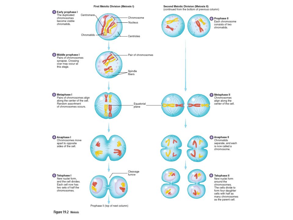
Figure 19.2

16
Figure 19.5a

17
17 Secretions Semen: - mixture of sperm and secretions from glands - provides a transport medium and nutrients that protect and activate sperm - 60% of fluid is from seminal vesicles - 30% of fluid is from prostate gland - 5% of fluid is from bulbourethral gland - 5% of fluid is from testes

18
18 Seminal vesicles: - provide fructose - contain prostaglandins which decrease mucus thickness around cervix and uterine tubes and help sperm move through female repro. tract - contains coagulants that help deliver semen into female

19
Prostate gland: - contains enzymes to liquefy semen after it is inside female - neutralizes acidity of vagina Bulbourethral gland: neutralize acidity of male urethra and female vagina 19

20
20 Testicular secretions: include sperm and small amount of fluid 2-5 ml of semen is ejaculated each time 1 ml of semen contains 100 million sperm Sperm can live for 72 hours once inside female

21
21 Path of Sperm 1.Sperm develop in seminiferous tubules (testes) 2.Epididymis (mature) 3.Ductus deferens 4.Receive secretions from seminal vesicles, prostate gland, and bulbourethral gland 5.Urethra where semen (sperm) exit body
 2.Epididymis (mature) 3.Ductus deferens 4.Receive secretions from seminal vesicles, prostate gland, and bulbourethral gland 5.Urethra where semen (sperm) exit body")
22
22 Spermatogenesis What is it? - formation of sperm cells - begins at puberty - interstitial cells (in semin. tubules) increase in number and size - seminiferous tubules enlarge - seminiferous tubules produce germ cells and Sustentacular cells
 increase in number and size - seminiferous tubules enlarge - seminiferous tubules produce germ cells and Sustentacular cells.")
23
23 Production of Sperm Cells 1.Germ cells 2.Spermatogonia 3.Primary spermatocytes 4.Secondary spermatocytes 5.Spermatids 6.Sperm cells

25
25 Sperm Cells Structure Head: contain a nucleus and DNA Midpiece: contain mitochondria Tail: flagellum for movement

26
Figure 19.3

27
27 Male Sex Hormones HormoneSourceFunction Gonadotropin-hypothalamusstimulates secretion of LH releasing hormoneand FSH LH (luteinizing)anterior pituitarystimulates secretion of gland testosterone FSH anterior pituitaryprompts spermatogenesis (follicle-stimulating)gland Testosteroneinterstitial cellsinvolved in development and in testesmaintenance of reproductive organs
anterior pituitarystimulates secretion of gland testosterone FSH anterior pituitaryprompts spermatogenesis (follicle-stimulating)gland Testosteroneinterstitial cellsinvolved in development and in testesmaintenance of reproductive organs")
29
29 Male Puberty What is it? - sequence of events in which a boy begins to produce male hormones and sperm cells - begins at 12-14 and ends around 18 - testosterone is major male hormone - secondary sexual characteristics develop: Ex. Skin texture, fat distribution, hair growth, skeletal muscle growth, and larynx changes

30
30 Functions of Female Reproductive System Produce female oocytes (sex cells) Produce female sex hormones Receive sperm from males Develop and nourish embryos
 Produce female sex hormones Receive sperm from males Develop and nourish embryos")
31
31 External Female Genitalia Vulva: - external female sex organs - mons pubis, labia majora and minora, clitoris, and vestibule Mons pubis: fatty layer of skin covering pubic symphysis

32
Labia majora: - larger, outer folds of skin - equivalent to male scrotum Labia minora: thin, inner folds of skin 32

33
33 Clitoris: - small erectile structure located in vestibule - equivalent to male penis Prepuce: where 2 labia minora unite over clitoris Vestibule: space in which vagina and urethra are located

35
Figure 19.7

36
36 Female Reproductive Organs Ovaries: - primary female reproductive organ - produces oocytes and sex hormones - one on either side of uterus - ovarian ligaments: anchor ovaries to uterus - suspensory ligaments: anchor ovaries to pelvic cavity - ovarian follicle: cells in ovaries that contain oocytes

37
37 Uterine (Fallopian) Tubes: - part of uterus which extends toward ovaries and receive oocytes - fimbriae: fringe-like structures around opening of uterine tubes that help sweep oocyte into uterine tubes - tubal ligation (sterilization of female) - ectopic pregnancy: if fertilized oocyte (zygote) implants somewhere beside uterus (usually in uterine tube)
 Tubes: - part of uterus which extends toward ovaries and receive oocytes - fimbriae: fringe-like structures around opening of uterine tubes that help sweep oocyte into uterine tubes - tubal ligation (sterilization of female) - ectopic pregnancy: if fertilized oocyte (zygote) implants somewhere beside uterus (usually in uterine tube)")
38
Clinical Focus 19Bg

39
39 Uterus: - pear sized structure located in pelvic cavity - functions: receive, retain, and provide nourishment for fertilized oocyte, where embryo resides and develops - body: main part - cervix: narrow region that leads to vagina

40
40 Uterus layers: - perimetrium (serous): outermost layer - myometrium (muscular): middle layer composed of smooth muscle - endometrium: innermost layer that is sloughed off during menstruation
: outermost layer - myometrium (muscular): middle layer composed of smooth muscle - endometrium: innermost layer that is sloughed off during menstruation")
41
41 Vagina: - extends from uterus to outside of body - female copulation organ that receives penis during intercourse - allows menstrual flow - involved in childbirth - contains very muscular walls and a mucous membrane - very acidic to keep bacteria out

42
Figure 19.8

43
43 Follicle and Oocyte Development OocyteFollicle Fetus oogonium primordial follicle primary oocyteprimordial follicle Puberty primary oocyteprimary follicle To primary oocytesecondary follicle Menopause primary oocytemature follicle secondary oocytemature follicle

45
45 Ovulation What is it? - when a mature follicle ruptures forcing oocyte into peritoneal (pelvic) cavity - due to LH (anterior pit. gland) Corpus luteum: - mature follicle after ovulation - degenerates if egg is not fertilized
 cavity - due to LH (anterior pit. gland) Corpus luteum: - mature follicle after ovulation - degenerates if egg is not fertilized.")
46
Figure 19.9

47
47 Other Female Reproductive Facts Females are born with all of their oogonia (2 million), unlike males that only begin to produce sperm during puberty. At puberty about 300,000-400,000 oogonia are left. Puberty to menopause, FSH stimulates several follicles to begin developing during each menstrual cycle but only 1 follicle should be ovulated.

48
48 Oocytes are swept into one of uterine tubes by fimbriae. If sperm is present in uterine tube during ovulation oocyte could be fertilized. If fertilization occurs then zygote implants in uterus. Oocyte only lives for 24 hours, so if no sperm is present at ovulation no zygote develops, and oocyte dies.

49
49 Female Puberty Begins between 11-13 and is usually completed by 16 Menarche first episode of menstrual bleeding Vagina, uterus, uterine tubes, and external genitalia to enlarge and fat is deposited in breast and hips Elevated levels of estrogen and progesterone are secreted by ovaries

50
Mammary Glands Organs of milk production in breasts Modified sweat glands Female breasts begin to enlarge during puberty Consists of lobes covered by adipose Lobes, ducts, lobules are altered during lactation to expel milk

52
52 Female Sex Hormones HormoneSourceFunction Gonadotropin-hypothalamusstimulates secretion of LH releasing hormoneand FSH LH (luteinizing)anterior pituitarycauses ovulation gland FSH anterior pituitarysignals the follicle in ovaries (follicle-stimulating) glandto being development Estrogenfollicles of ovariesaffects endometrial lining of uterus, breasts, regulates secretions of LH and FSH Progesteroneovariesaffects endometrial lining of uterus, secretions, breasts, affects LH and FSH, secondary sexual charac.
anterior pituitarycauses ovulation gland FSH anterior pituitarysignals the follicle in ovaries (follicle-stimulating) glandto being development Estrogenfollicles of ovariesaffects endometrial lining of uterus, breasts, regulates secretions of LH and FSH Progesteroneovariesaffects endometrial lining of uterus, secretions, breasts, affects LH and FSH, secondary sexual charac.")
53
53 Menstrual Cycle What is it? series of changes that occur in sexually mature, nonpregnant females Menses: time when endometrium is shed from uterus Average is 28 days and results from cyclical changes that occur in endometrium

54
54 Stages of Menstrual Cycle Days 1-5 Menses (shedding of endometrium) - menstrual bleeding (menses) - estrogen and progesterone levels are low - follicle begins to mature Days 6-13 Proliferative (between end of menses and ovulation) - endometrium rebuilds - estrogen levels begin to increase - progesterone levels remain low - follicle matures
 - menstrual bleeding (menses) - estrogen and progesterone levels are low - follicle begins to mature Days 6-13 Proliferative (between end of menses and ovulation) - endometrium rebuilds - estrogen levels begin to increase - progesterone levels remain low - follicle matures")
55
55 Day 14 Ovulation - oocyte is released due to LH - estrogen levels high - progesterone levels are increasing - cervical mucus thins Days 15-28 Secretory (between ovulation and next menses) - endometrium is preparing for implantation - estrogen levels decrease (low) - progesterone levels high - cervical mucus thickens
 - endometrium is preparing for implantation - estrogen levels decrease (low) - progesterone levels high - cervical mucus thickens")
56
Figure 19.14

57
57 Menopause What is it? - time when ovaries secrete less hormones and number of follicles in ovaries is low - menstrual cycle and ovulation are less regular - hot flashes, fatigue, irritability may occur - estrogen replacement therapy may be used to decreases side effects

Similar presentations