Download presentation
Presentation is loading. Please wait.

1
The Reproductive System
Copyright © 2003 Pearson Education, Inc. publishing as Benjamin Cummings

2
The Reproductive System
Gonads – primary sex organs Testes in males Ovaries in females Gonads produce gametes (sex cells) and secrete hormones Sperm – male gametes Eggs (ova) – female gametes Slide 16.1 Copyright © 2003 Pearson Education, Inc. publishing as Benjamin Cummings
 and secrete hormones. Sperm – male gametes. Eggs (ova) – female gametes. Slide Copyright © 2003 Pearson Education, Inc. publishing as Benjamin Cummings.")
3
Male Reproductive System
Accessory organs Seminal vesicle Prostate gland Bulbourethral gland External genitalia Penis Scrotum Slide 16.2b Copyright © 2003 Pearson Education, Inc. publishing as Benjamin Cummings

4
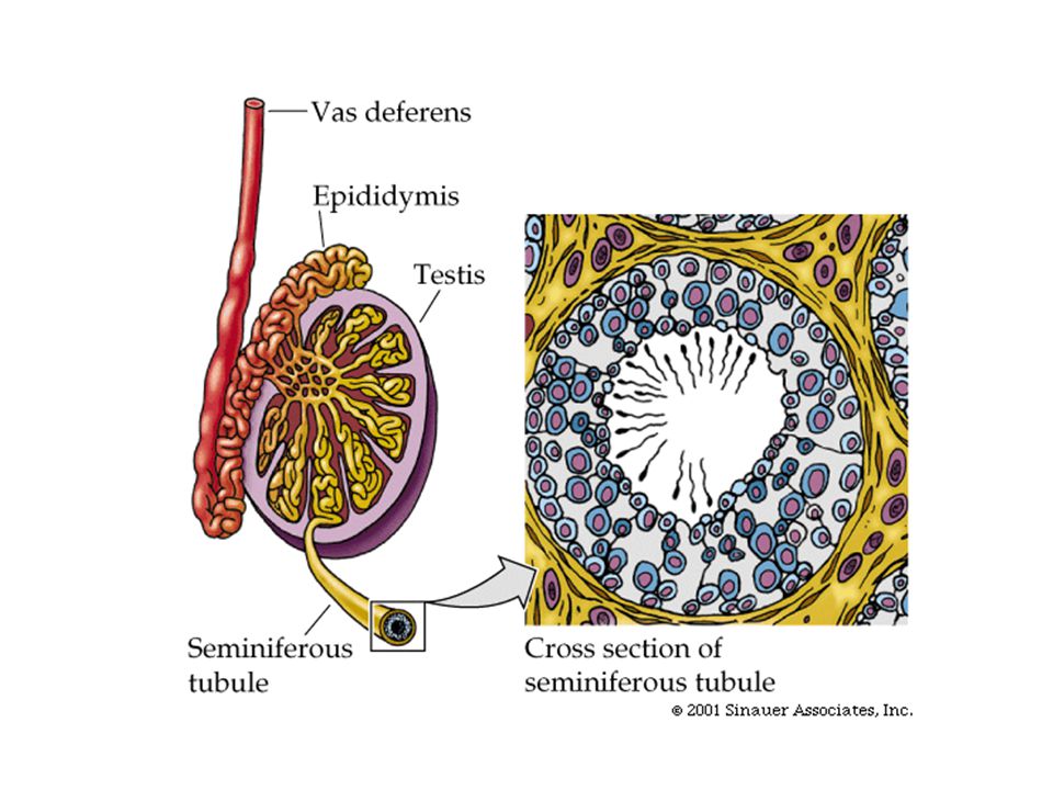
Testes Testes contain structures called seminiferous tubules
Tightly coiled structures Function as sperm-forming factories Empty sperm into the epididymis Once sperm have been formed they travel to the epididymis Testes produce hormones such as testosterone Slide 16.4 Copyright © 2003 Pearson Education, Inc. publishing as Benjamin Cummings

5
Male Reproductive System
Figure 16.2 Slide 16.2c Copyright © 2003 Pearson Education, Inc. publishing as Benjamin Cummings

6
Testes Figure 16.1 Slide 16.3a Copyright © 2003 Pearson Education, Inc. publishing as Benjamin Cummings

8
Epididymis Comma-shaped, tightly coiled tube
Functions to mature and store sperm cells (at least 20 days) Expels sperm with the contraction of muscles in the epididymis walls to the vas deferens Slide 16.5 Copyright © 2003 Pearson Education, Inc. publishing as Benjamin Cummings
 Expels sperm with the contraction of muscles in the epididymis walls to the vas deferens. Slide Copyright © 2003 Pearson Education, Inc. publishing as Benjamin Cummings.")
9
Male Reproductive System
Figure 16.2 Slide 16.2c Copyright © 2003 Pearson Education, Inc. publishing as Benjamin Cummings

10
Vas Deferens Carries sperm from the epididymis to the ejaculatory duct
Moves sperm by contraction The ejaculatory duct connects with the urethra (exit out of the penis) Slide 16.6a Copyright © 2003 Pearson Education, Inc. publishing as Benjamin Cummings
 Slide 16.6a. Copyright © 2003 Pearson Education, Inc. publishing as Benjamin Cummings.")
11
Male Reproductive System
Figure 16.2 Slide 16.2c Copyright © 2003 Pearson Education, Inc. publishing as Benjamin Cummings

12
Urethra Extends from the base of the urinary bladder to the tip of the penis Carries both urine and sperm Sperm enters from the ejaculatory duct Slide 16.7a Copyright © 2003 Pearson Education, Inc. publishing as Benjamin Cummings

13
Male Reproductive System
Figure 16.2 Slide 16.2c Copyright © 2003 Pearson Education, Inc. publishing as Benjamin Cummings

14
Seminal Vesicles Located at the base of the bladder
Produces a thick, yellowish secretion (60% of semen) Fructose (sugar) Vitamin C Other substances that nourish and activate sperm Slide 16.8 Copyright © 2003 Pearson Education, Inc. publishing as Benjamin Cummings
 Fructose (sugar) Vitamin C. Other substances that nourish and activate sperm. Slide Copyright © 2003 Pearson Education, Inc. publishing as Benjamin Cummings.")
15
Male Reproductive System
Figure 16.2 Slide 16.2c Copyright © 2003 Pearson Education, Inc. publishing as Benjamin Cummings

16
Prostate Gland Encircles the upper part of the urethra
Secretes a milky fluid Helps to activate sperm Enters the urethra through several small ducts Slide 16.9 Copyright © 2003 Pearson Education, Inc. publishing as Benjamin Cummings

17
Male Reproductive System
Figure 16.2 Slide 16.2c Copyright © 2003 Pearson Education, Inc. publishing as Benjamin Cummings

18
Bulbourethral Glands Also known as the Cowper’s Gland
Pea-sized gland under the prostate Produces a thick, clear mucus Cleanses the urethra of acidic urine Serves as a lubricant during sexual intercourse Secreted into the urethra Slide 16.10 Copyright © 2003 Pearson Education, Inc. publishing as Benjamin Cummings

19
Male Reproductive System
Figure 16.2 Slide 16.2c Copyright © 2003 Pearson Education, Inc. publishing as Benjamin Cummings

20
Semen Mixture of sperm and accessory gland secretions
Advantages of accessory gland secretions Fructose provides energy for sperm cells Alkalinity of semen helps neutralize the acidic environment of vagina Semen inhibits bacterial multiplication Elements of semen enhance sperm motility Slide 16.11 Copyright © 2003 Pearson Education, Inc. publishing as Benjamin Cummings

21
External Genitalia Scrotum Divided sac of skin outside the abdomen
Maintains testes at 3°C lower than normal body temperature to protect sperm Slide 16.12 Copyright © 2003 Pearson Education, Inc. publishing as Benjamin Cummings

22
Male Reproductive System
Figure 16.2 Slide 16.2c Copyright © 2003 Pearson Education, Inc. publishing as Benjamin Cummings

23
External Genitalia Penis
Delivers sperm into the female reproductive tract Regions of the penis Shaft Glans penis (enlarged tip) Prepuce (foreskin) Folded cuff of skin around proximal end Often removed by circumcision Slide 16.13a Copyright © 2003 Pearson Education, Inc. publishing as Benjamin Cummings
 Prepuce (foreskin) Folded cuff of skin around proximal end. Often removed by circumcision. Slide 16.13a. Copyright © 2003 Pearson Education, Inc. publishing as Benjamin Cummings.")
24
Male Reproductive System
Figure 16.2 Slide 16.2c Copyright © 2003 Pearson Education, Inc. publishing as Benjamin Cummings

25
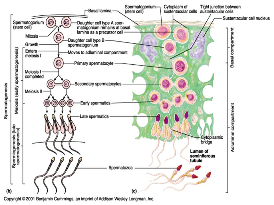
Spermatogenesis Production of sperm cells
Begins at puberty and continues throughout life Occurs in the seminiferous tubules (in testes) Slide 16.14 Copyright © 2003 Pearson Education, Inc. publishing as Benjamin Cummings
 Slide Copyright © 2003 Pearson Education, Inc. publishing as Benjamin Cummings.")
27
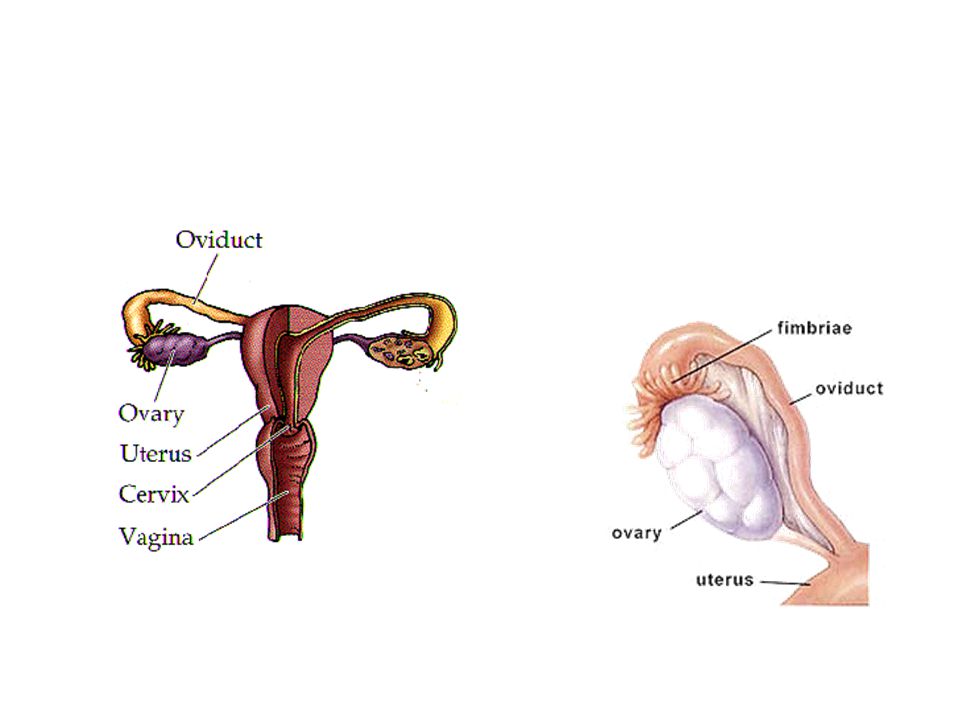
Female Reproductive System
Ovaries Fallopian tubes Uterus Vagina External genitalia Slide 16.21a Copyright © 2003 Pearson Education, Inc. publishing as Benjamin Cummings

28
Female Reproductive System
Figure 16.8a Slide 16.21b Copyright © 2003 Pearson Education, Inc. publishing as Benjamin Cummings

29
Composed of ovarian follicles (sac-like structures)
Ovaries Composed of ovarian follicles (sac-like structures) Produce eggs Figure 16.7 Slide 16.22 Copyright © 2003 Pearson Education, Inc. publishing as Benjamin Cummings
 Produce eggs. Figure Slide Copyright © 2003 Pearson Education, Inc. publishing as Benjamin Cummings.")
31
Ovarian Follicle Stages
Ovulation – when the egg is mature the follicle ruptures Occurs about every 28 days Slide 16.23 Copyright © 2003 Pearson Education, Inc. publishing as Benjamin Cummings

32
Fallopian Tubes/Oviducts
Receive the ovulated egg (oocyte) Provide a site for fertilization Attaches to the uterus Does not physically attach to the ovary Slide 16.25 Copyright © 2003 Pearson Education, Inc. publishing as Benjamin Cummings
 Provide a site for fertilization. Attaches to the uterus. Does not physically attach to the ovary. Slide Copyright © 2003 Pearson Education, Inc. publishing as Benjamin Cummings.")
33
Female Reproductive System
Figure 16.8a Slide 16.21b Copyright © 2003 Pearson Education, Inc. publishing as Benjamin Cummings

34
Fallopian Tube Function
Fimbriae – finger-like projections at the end of the fallopian tube that help move the egg from the ovary to the fallopian tube Cilia inside the fallopian tube slowly move the egg towards the uterus (takes 3–4 days) Fertilization occurs inside the fallopian tube Slide 16.26 Copyright © 2003 Pearson Education, Inc. publishing as Benjamin Cummings
 Fertilization occurs inside the fallopian tube. Slide Copyright © 2003 Pearson Education, Inc. publishing as Benjamin Cummings.")
35
Fallopian tube, cont’d If fertilized egg implants into oviduct, an ectopic pregnancy occurs (very dangerous)
")
36
Uterus Located between the urinary bladder and rectum Hollow organ
Functions of the uterus Receives a fertilized egg Nourishes the fertilized egg as it develops into a fetus Slide 16.27 Copyright © 2003 Pearson Education, Inc. publishing as Benjamin Cummings

37
Female Reproductive System
Figure 16.8a Slide 16.21b Copyright © 2003 Pearson Education, Inc. publishing as Benjamin Cummings

38
Walls of the Uterus Inner layer of the uterus is called the endometrium Fertilized egg implants in the endometrium Released if no pregnancy occurs (menstration) Slide 16.30 Copyright © 2003 Pearson Education, Inc. publishing as Benjamin Cummings
 Slide Copyright © 2003 Pearson Education, Inc. publishing as Benjamin Cummings.")
39
Cervix The lower entrance into the uterus. The opening lies between the uterus and the vagina.

40
Female Reproductive System
Figure 16.8a Slide 16.21b Copyright © 2003 Pearson Education, Inc. publishing as Benjamin Cummings

41
Vagina Extends from cervix to exterior of body
Behind bladder and in front of rectum Serves as the birth canal Receives the penis during sexual intercourse Slide 16.31 Copyright © 2003 Pearson Education, Inc. publishing as Benjamin Cummings

42
Female Reproductive System
Figure 16.8a Slide 16.21b Copyright © 2003 Pearson Education, Inc. publishing as Benjamin Cummings

43
Oogenesis The total supply of eggs are present at birth
Ability to release eggs begins at puberty Reproductive ability ends at menopause Oocytes (eggs) are matured in follicles inside the ovaries Slide 16.34 Copyright © 2003 Pearson Education, Inc. publishing as Benjamin Cummings
 are matured in follicles inside the ovaries. Slide Copyright © 2003 Pearson Education, Inc. publishing as Benjamin Cummings.")
44
Oogenesis Slide 16.37 Figure 16.10
Copyright © 2003 Pearson Education, Inc. publishing as Benjamin Cummings

45
Menstrual Cycle Regulated by hormones such as estrogen and progesterone Happens approximately every 28 days if a fertilized egg has not been implanted in the uterine wall. Blood and tissue from the endometrium are shed. Slide 16.38 Copyright © 2003 Pearson Education, Inc. publishing as Benjamin Cummings

46
Hormonal Control of the Ovarian and Uterine Cycles
Figure 16.12a, b Slide 16.39a Copyright © 2003 Pearson Education, Inc. publishing as Benjamin Cummings

47
Hormonal Control of the Ovarian and Uterine Cycles
Figure 16.12c, d Slide 16.39b Copyright © 2003 Pearson Education, Inc. publishing as Benjamin Cummings

48
Mammary Glands Present in both sexes, but only function in females
Modified sweat glands Function is to produce milk Stimulated by sex hormones (mostly estrogen) to increase in size Slide 16.42 Copyright © 2003 Pearson Education, Inc. publishing as Benjamin Cummings
 to increase in size. Slide Copyright © 2003 Pearson Education, Inc. publishing as Benjamin Cummings.")
49
Developmental Aspects of the Reproductive System
Reproductive system organs do not function until puberty Puberty usually begins between ages 10 and 15 The first menstruation usually occurs about two years after the start of puberty Most women reach peak reproductive ability in their late 20s Slide 16.65 Copyright © 2003 Pearson Education, Inc. publishing as Benjamin Cummings

50
Developmental Aspects of the Reproductive System
Menopause occurs when ovulation and menstruation stop There is a no equivalent of menopause in males, but there is a steady decline in testosterone as males age Slide 16.66 Copyright © 2003 Pearson Education, Inc. publishing as Benjamin Cummings

Similar presentations