Download presentation
Presentation is loading. Please wait.

1
© 2012 Pearson Education, Inc. PowerPoint ® Lecture Slides Prepared by Patty Bostwick-Taylor, Florence-Darlington Technical College C H A P T E R 3 Cells and Tissues

2
© 2012 Pearson Education, Inc. Crash Course Intro to TISSUES https://www.youtube.com/watch?v=i5tR3csC WYohttps://www.youtube.com/watch?v=i5tR3csC WYo

3
© 2012 Pearson Education, Inc. Body Tissues HISTOLOGY = the study of tissues Tissues Groups of cells with similar structure and function Four primary types Epithelial tissue (epithelium) Connective tissue Muscle tissue Nervous tissue
 Connective tissue Muscle tissue Nervous tissue.")
4
© 2012 Pearson Education, Inc. Crash Course Epithelial tissue https://www.youtube.com/watch?v=lUe_RI_m- Vghttps://www.youtube.com/watch?v=lUe_RI_m- Vg

5
© 2012 Pearson Education, Inc. Epithelial Tissues Locations Body coverings Body linings Glandular tissue Functions Protection Absorption Filtration Secretion

6
© 2012 Pearson Education, Inc. Epithelium Characteristics Cells fit closely together and often form sheets The apical surface is the free surface of the tissue The lower surface of the epithelium rests on a basement membrane Avascular (no blood supply) Regenerate easily if well nourished
 Regenerate easily if well nourished.")
7
© 2012 Pearson Education, Inc. Apical surface Basal surface Simple Apical surface Basal surface Stratified (a) Classification based on number of cell layers Figure 3.17a
 Classification based on number of cell layers Figure 3.17a.")
8
© 2012 Pearson Education, Inc. Classification of Epithelia Number of cell layers Simple—one layer Stratified—more than one layer

9
© 2012 Pearson Education, Inc. Apical surface Basal surface Simple Apical surface Basal surface Stratified (a) Classification based on number of cell layers Figure 3.17a DRAW THIS IN YOUR NOTES
 Classification based on number of cell layers Figure 3.17a DRAW THIS IN YOUR NOTES.")
10
© 2012 Pearson Education, Inc. Crash Course Epithelial tissue https://www.youtube.com/watch?v=lUe_RI_m- Vghttps://www.youtube.com/watch?v=lUe_RI_m- Vg

11
© 2012 Pearson Education, Inc. Classification of Epithelia Shape of cells Squamous flattened Cuboidal cube-shaped Columnar column-like

12
© 2012 Pearson Education, Inc. Simple Epithelia Simple squamous Single layer of flat cells Location - usually forms membranes Lines body cavities Lines lungs and capillaries Functions in diffusion, filtration, or secretion in membranes

13
© 2012 Pearson Education, Inc. Figure 3.18a Nucleus of squamous epithelial cell Basement membrane (a) Diagram: Simple squamous Photomicrograph: Simple squamous epithelium forming part of the alveolar (air sac) walls (185×). Nuclei of squamous epithelial cells Air sacs of lungs
 Diagram: Simple squamous Photomicrograph: Simple squamous epithelium forming part of the alveolar (air sac) walls (185×). Nuclei of squamous epithelial cells Air sacs of lungs.")
14
© 2012 Pearson Education, Inc. Simple Epithelia Simple cuboidal Single layer of cube-like cells Locations Common in glands and their ducts Forms walls of kidney tubules Covers the ovaries Functions in secretion and absorption; ciliated types propel mucus or reproductive cells

15
© 2012 Pearson Education, Inc. Figure 3.18b (b) Diagram: Simple cuboidal Nucleus of simple cuboidal epithelial cell Photomicrograph: Simple cuboidal epithelium in kidney tubules (250 × ). Basement membrane Connective tissue Basement membrane Simple cuboidal epithelial cells
 Diagram: Simple cuboidal Nucleus of simple cuboidal epithelial cell Photomicrograph: Simple cuboidal epithelium in kidney tubules (250 × ). Basement membrane Connective tissue Basement membrane Simple cuboidal epithelial cells.")
16
© 2012 Pearson Education, Inc. Simple Epithelia Simple columnar Single layer of tall cells Often includes mucus-producing goblet cells Location - lines digestive tract Functions in secretion and absorption; ciliated types propel mucus or reproductive cells

17
© 2012 Pearson Education, Inc. Figure 3.18c Nucleus of simple columnar epithelial cell Connective tissue Photomicrograph: Simple columnar epithelium of the small intestine (430×). Basement membrane (c) Diagram: Simple columnar Basement membrane Goblet cell Simple columnar epithelial cell
. Basement membrane (c) Diagram: Simple columnar Basement membrane Goblet cell Simple columnar epithelial cell.")
18
© 2012 Pearson Education, Inc. Simple Epithelia Pseudostratified columnar Single layer, but some cells are shorter than others Often looks like a double layer of cells but all cells rest on the basement membrane Location - respiratory tract, where it is ciliated Functions in absorption or secretion

19
© 2012 Pearson Education, Inc. Figure 3.18d Pseudo- stratified epithelial layer Basement membrane (d) Diagram: Pseudostratified (ciliated) columnar Photomicrograph: Pseudostratified ciliated columnar epithelium lining the human trachea (430×). Pseudo- stratified epithelial layer Basement membrane Connective tissue Cilia
 Diagram: Pseudostratified (ciliated) columnar Photomicrograph: Pseudostratified ciliated columnar epithelium lining the human trachea (430×). Pseudo- stratified epithelial layer Basement membrane Connective tissue Cilia.")
20
© 2012 Pearson Education, Inc. Stratified Epithelia Stratified squamous Cells at the apical surface are flattened Functions as a protective covering where friction is common Locations - lining of the: Skin Mouth Esophagus

21
© 2012 Pearson Education, Inc. Figure 3.18e Stratified squamous epithelium Basement membrane (e) Diagram: Stratified squamous Photomicrograph: Stratified squamous epithelium lining of the esophagus (140×). Connective tissue Stratified squamous epithelium Nuclei Basement membrane
 Diagram: Stratified squamous Photomicrograph: Stratified squamous epithelium lining of the esophagus (140×). Connective tissue Stratified squamous epithelium Nuclei Basement membrane.")
22
© 2012 Pearson Education, Inc. Stratified Epithelia Stratified cuboidal — two layers of cuboidal cells; functions in protection Stratified columnar — surface cells are columnar, cells underneath vary in size and shape; functions in protection Stratified cuboidal and columnar Rare in human body Found mainly in ducts of large glands

23
© 2012 Pearson Education, Inc. Stratified Epithelia Transitional epithelium Composed of modified stratified squamous epithelium Shape of cells depends upon the amount of stretching Functions in stretching and the ability to return to normal shape Location - lines organs of the urinary system

24
© 2012 Pearson Education, Inc. Figure 3.18f Transi- tional epithelium Basement membrane Photomicrograph: Transitional epithelium lining of the bladder, relaxed state (215×); surface rounded cells flatten and elongate when the bladder fills with urine. (f) Diagram: Transitional Connective tissue Transitional epithelium Basement membrane
; surface rounded cells flatten and elongate when the bladder fills with urine. (f) Diagram: Transitional Connective tissue Transitional epithelium Basement membrane.")
25
© 2012 Pearson Education, Inc. Glandular Epithelium Gland One or more cells responsible for secreting a particular product Secretions contain protein molecules in an aqueous (water-based) fluid
 fluid.")
26
© 2012 Pearson Education, Inc. Glandular Epithelium Two major gland types Endocrine gland Ductless since secretions diffuse into blood vessels All secretions are hormones Exocrine gland Secretions empty through ducts to the epithelial surface Include sweat and oil glands LAST EPITHELIAL SLIDE

27
© 2012 Pearson Education, Inc. CONNECTIVE TISSUE Crash Course video #3 https://www.youtube.com/watch?v=D- SzmURNBH0https://www.youtube.com/watch?v=D- SzmURNBH0

28
© 2012 Pearson Education, Inc. Connective Tissue Found everywhere in the body Includes the most abundant and widely distributed tissues Functions Binds body tissues together Supports the body Provides protection

29
© 2012 Pearson Education, Inc. Connective Tissue Characteristics Variations in blood supply Some tissue types are well vascularized Some have a poor blood supply or are avascular Extracellular matrix Non-living material that surrounds living cells

30
© 2012 Pearson Education, Inc. Extracellular Matrix Two main elements Ground substance—mostly water along with adhesion proteins and polysaccharide molecules Fibers Produced by the cells Three types Collagen (white) fibers Elastic (yellow) fibers Reticular fibers
 fibers Elastic (yellow) fibers Reticular fibers.")
31
© 2012 Pearson Education, Inc. Connective Tissue TYPES Crash Course #5 https://www.youtube.com/watch?v=Jvtb0a2RX aYhttps://www.youtube.com/watch?v=Jvtb0a2RX aY

32
© 2012 Pearson Education, Inc. Connective Tissue Types Bone (osseous tissue) Composed of Bone cells in lacunae (cavities) Hard matrix of calcium salts Large numbers of collagen fibers Functions to protect and support the body
 Composed of Bone cells in lacunae (cavities) Hard matrix of calcium salts Large numbers of collagen fibers Functions to protect and support the body.")
33
© 2012 Pearson Education, Inc. Figure 3.19a Bone cells in lacunae (a) Diagram: Bone Photomicrograph: Cross-sectional view of ground bone (300×). Lamella Lacunae Central canal
 Diagram: Bone Photomicrograph: Cross-sectional view of ground bone (300×). Lamella Lacunae Central canal.")
34
© 2012 Pearson Education, Inc. Connective Tissue Types Hyaline cartilage Most common type of cartilage Composed of Abundant collagen fibers Rubbery matrix Locations Larynx Entire fetal skeleton prior to birth Functions as a more flexible skeletal element than bone

35
© 2012 Pearson Education, Inc. Figure 3.19b Chondrocyte (Cartilage cell) Lacunae (b) Diagram: Hyaline cartilage Photomicrograph: Hyaline cartilage from the trachea (500×). Matrix Chondrocyte in lacuna
 Lacunae (b) Diagram: Hyaline cartilage Photomicrograph: Hyaline cartilage from the trachea (500×). Matrix Chondrocyte in lacuna.")
36
© 2012 Pearson Education, Inc. Connective Tissue Types Elastic cartilage Provides elasticity Location Supports the external ear Fibrocartilage Highly compressible Location Forms cushion-like discs between vertebrae

37
© 2012 Pearson Education, Inc. Figure 3.19c Chondro- cites in lacunae Collagen fibers (c) Diagram: Fibrocartilage Photomicrograph: Fibrocartilage of an intervertebral disc (110×). Collagen fiber Chondrocytes in lacunae
 Diagram: Fibrocartilage Photomicrograph: Fibrocartilage of an intervertebral disc (110×). Collagen fiber Chondrocytes in lacunae.")
38
© 2012 Pearson Education, Inc. Connective Tissue Types Dense connective tissue (dense fibrous tissue) Main matrix element is collagen fiber Fibroblasts are cells that make fibers Locations Tendons— attach skeletal muscle to bone Ligaments— attach bone to bone at joints Dermis— lower layers of the skin
 Main matrix element is collagen fiber Fibroblasts are cells that make fibers Locations Tendons— attach skeletal muscle to bone Ligaments— attach bone to bone at joints Dermis— lower layers of the skin.")
39
© 2012 Pearson Education, Inc. Figure 3.19d Ligament Tendon Collagen fibers Nuclei of fibroblasts (d) Diagram: Dense fibrous Photomicrograph: Dense fibrous connective tissue from a tendon (500×). Nuclei of fibroblasts Collagen fibers
 Diagram: Dense fibrous Photomicrograph: Dense fibrous connective tissue from a tendon (500×). Nuclei of fibroblasts Collagen fibers.")
40
© 2012 Pearson Education, Inc. Connective Tissue Types Loose connective tissue types Areolar tissue Most widely distributed connective tissue Soft, pliable tissue like “cobwebs” Functions as a packing tissue Contains all fiber types Can soak up excess fluid (causes edema)
.")
41
© 2012 Pearson Education, Inc. Figure 3.19e Mucosa epithelium Lamina propria Fibers of matrix Nuclei of fibroblasts (e) Diagram: Areolar Photomicrograph: Areolar connective tissue, a soft packaging tissue of the body (300×). Fibroblast nuclei Collagen fibers Elastic fibers
 Diagram: Areolar Photomicrograph: Areolar connective tissue, a soft packaging tissue of the body (300×). Fibroblast nuclei Collagen fibers Elastic fibers.")
42
© 2012 Pearson Education, Inc. Connective Tissue Types Loose connective tissue types Adipose tissue Matrix is an areolar tissue in which fat globules predominate Many cells contain large lipid deposits Functions Insulates the body Protects some organs Serves as a site of fuel storage

43
© 2012 Pearson Education, Inc. Figure 3.19f Nuclei of fat cells Vacuole containing fat droplet (f) Diagram: Adipose Photomicrograph: Adipose tissue from the subcutaneous layer beneath the skin (430×). Vacuole containing fat droplet Nuclei of fat cells
 Diagram: Adipose Photomicrograph: Adipose tissue from the subcutaneous layer beneath the skin (430×). Vacuole containing fat droplet Nuclei of fat cells.")
44
© 2012 Pearson Education, Inc. Connective Tissue Types Loose connective tissue types Reticular connective tissue Delicate network of interwoven fibers Locations Forms stroma (internal supporting network) of lymphoid organs Lymph nodes Spleen Bone marrow
 of lymphoid organs Lymph nodes Spleen Bone marrow.")
45
© 2012 Pearson Education, Inc. Figure 3.19g Spleen Reticular cell Reticular fibers Blood cell (g) Diagram: Reticular Photomicrograph: Dark-staining network of reticular connective tissue (430×). White blood cell (lymphocyte) Reticular fibers
 Diagram: Reticular Photomicrograph: Dark-staining network of reticular connective tissue (430×). White blood cell (lymphocyte) Reticular fibers.")
46
© 2012 Pearson Education, Inc. Connective Tissue Types Blood (vascular tissue) Blood cells surrounded by fluid matrix called blood plasma Fibers are visible during clotting Functions as the transport vehicle for materials
 Blood cells surrounded by fluid matrix called blood plasma Fibers are visible during clotting Functions as the transport vehicle for materials.")
47
© 2012 Pearson Education, Inc. Figure 3.19h Neutrophil (white blood cell) Red blood cells Monocyte (white blood cell) Photomicrograph: Smear of human blood (1300×)(h) Diagram: Blood White blood cell Red blood cells Blood cells in capillary LAST CONNECTIVE SLIDE
 Red blood cells Monocyte (white blood cell) Photomicrograph: Smear of human blood (1300×)(h) Diagram: Blood White blood cell Red blood cells Blood cells in capillary LAST CONNECTIVE SLIDE.")
48
© 2012 Pearson Education, Inc. Muscle Tissue Function is to produce movement Three types Skeletal muscle Cardiac muscle Smooth muscle https://www.youtube.com/watch?v=MK1sf8W H0CY

49
© 2012 Pearson Education, Inc. Muscle Tissue Types Skeletal muscle Under voluntary control Contracts to pull on bones or skin Produces gross body movements or facial expressions Characteristics of skeletal muscle cells Striated Multinucleate (more than one nucleus) Long, cylindrical cells
 Long, cylindrical cells.")
50
© 2012 Pearson Education, Inc. Figure 3.20a Nuclei Part of muscle fiber (a) Diagram: Skeletal musclePhotomicrograph: Skeletal muscle (approx. 300×).
 Diagram: Skeletal musclePhotomicrograph: Skeletal muscle (approx. 300×)..")
51
© 2012 Pearson Education, Inc. Muscle Tissue Types Cardiac muscle Under involuntary control Found only in the heart Function is to pump blood Characteristics of cardiac muscle cells Striated One nucleus per cell Cells are attached to other cardiac muscle cells at intercalated disks

52
© 2012 Pearson Education, Inc. Figure 3.20b Intercalated discs Nucleus (b) Diagram: Cardiac musclePhotomicrograph: Cardiac muscle (430×).
 Diagram: Cardiac musclePhotomicrograph: Cardiac muscle (430×)..")
53
© 2012 Pearson Education, Inc. Muscle Tissue Types Smooth muscle Under involuntary muscle Found in walls of hollow organs such as stomach, uterus, and blood vessels Characteristics of smooth muscle cells No visible striations One nucleus per cell Spindle-shaped cells

54
© 2012 Pearson Education, Inc. Figure 3.20c Smooth muscle cell Nuclei (c) Diagram: Smooth musclePhotomicrograph: Sheet of smooth muscle (approx. 300×). LAST MUSCLE SLIDE
 Diagram: Smooth musclePhotomicrograph: Sheet of smooth muscle (approx. 300×). LAST MUSCLE SLIDE.")
55
© 2012 Pearson Education, Inc. Nervous Tissue

56
© 2012 Pearson Education, Inc. Nervous Tissue Composed of neurons and nerve support cells Function is to send impulses to other areas of the body Irritability Conductivity Support cells called neuroglia insulate, protect, and support neurons

57
© 2012 Pearson Education, Inc. Figure 3.21 Brain Spinal cord Nuclei of supporting cells Cell body of neuron Neuron processes Diagram: Nervous tissue Photomicrograph: Neurons (150×) Nuclei of supporting cells Cell body of neuron Neuron processes
 Nuclei of supporting cells Cell body of neuron Neuron processes.")
58
© 2012 Pearson Education, Inc. Brain, spinal cord, and nerves Nervous tissue: Internal communication Muscle tissue: Contracts to cause movement Muscles attached to bones (skeletal) Muscles of heart (cardiac) Muscles of walls of hollow organs (smooth) Epithelial tissue: Forms boundaries between different environments, protects, secretes, absorbs, filters Lining of GI tract organs and other hollow organs Skin surface (epidermis) Connective tissue: Supports, protects, binds other tissues together Bones Tendons Fat and other soft padding tissue Figure 3.22 LAST TISSUE TYPE SLIDE
 Muscles of heart (cardiac) Muscles of walls of hollow organs (smooth) Epithelial tissue: Forms boundaries between different environments, protects, secretes, absorbs, filters Lining of GI tract organs and other hollow organs Skin surface (epidermis) Connective tissue: Supports, protects, binds other tissues together Bones Tendons Fat and other soft padding tissue Figure 3.22 LAST TISSUE TYPE SLIDE.")
59
© 2012 Pearson Education, Inc. Tissue Repair (Wound Healing) Regeneration Replacement of destroyed tissue by the same kind of cells Fibrosis Repair by dense (fibrous) connective tissue (scar tissue) Whether regeneration or fibrosis occurs depends on: Type of tissue damaged Severity of the injury
 Regeneration Replacement of destroyed tissue by the same kind of cells Fibrosis Repair by dense (fibrous) connective tissue (scar tissue) Whether regeneration or fibrosis occurs depends on: Type of tissue damaged Severity of the injury.")
60
© 2012 Pearson Education, Inc. Events in Tissue Repair Inflammation Capillaries become very permeable Clotting proteins migrate into the area from the blood stream A clot walls off the injured area Granulation tissue forms Growth of new capillaries Rebuild collagen fibers Regeneration of surface epithelium Scab detaches

61
© 2012 Pearson Education, Inc. Regeneration of Tissues Tissues that regenerate easily Epithelial tissue (skin and mucous membranes) Fibrous connective tissues and bone Tissues that regenerate poorly Skeletal muscle Tissues that are replaced largely with scar tissue Cardiac muscle Nervous tissue within the brain and spinal cord
 Fibrous connective tissues and bone Tissues that regenerate poorly Skeletal muscle Tissues that are replaced largely with scar tissue Cardiac muscle Nervous tissue within the brain and spinal cord.")
62
© 2012 Pearson Education, Inc. Developmental Aspects of Tissue Epithelial tissue arises from all three primary germ layers Muscle and connective tissue arise from the mesoderm Nervous tissue arises from the ectoderm With old age, there is a decrease in mass and viability in most tissues

63
© 2012 Pearson Education, Inc. Notebook P AGE 1 = C OVER = Ch.3: Human Body Tissues 2 FRONT =Epithelial 2 BACK = Connective 3 FRONT = Muscle 3 BACK = Nervous 4 FRONT (& BACK IF NEEDED ) = Tissue Repair 5 FRONT (& BACK IF NEEDED ) = Developmental Aspects… Each page must include: Heading 3 characteristics corresponding tissue illustrations. DRAW nervous tissue!
 = Tissue Repair 5 FRONT (& BACK IF NEEDED ) = Developmental Aspects… Each page must include: Heading 3 characteristics corresponding tissue illustrations. DRAW nervous tissue!.")
64
© 2012 Pearson Education, Inc. Notebook P AGE 6 = SUMMARY Assume you are teaching a fellow student (not enrolled in this class) about HUMAN BODY TISSUES. Summarize all you have learned about tissues. Organize your thoughts and ideas. Use entire page (and back if necessary). Skip lines Write in pen clearly.
 about HUMAN BODY TISSUES. Summarize all you have learned about tissues. Organize your thoughts and ideas. Use entire page (and back if necessary). Skip lines Write in pen clearly..")
65
© 2012 Pearson Education, Inc. Short Answer questions for test. 1.Describe the structure of epithelial tissue. (include apical surface and basement membrane) 2.Compare endocrine glands to exocrine glands (similarities & differences). 3.Compare areolar tissue to hyaline cartilage in terms of structure, location, and function. 4.Compare blood tissue to osseous tissue in terms of structure, location and function. 5.Compare the 3 types of muscle tissue to each other in terms of structure, location and function. 6.Summarize the process by which tissue is repaired. BE PREPARED TO ANSWER 3 OF THESE.
 2.Compare endocrine glands to exocrine glands (similarities & differences). 3.Compare areolar tissue to hyaline cartilage in terms of structure, location, and function. 4.Compare blood tissue to osseous tissue in terms of structure, location and function. 5.Compare the 3 types of muscle tissue to each other in terms of structure, location and function. 6.Summarize the process by which tissue is repaired. BE PREPARED TO ANSWER 3 OF THESE..")
66
© 2012 Pearson Education, Inc. TISSUE REVIEW

67
© 2012 Pearson Education, Inc.

Similar presentations
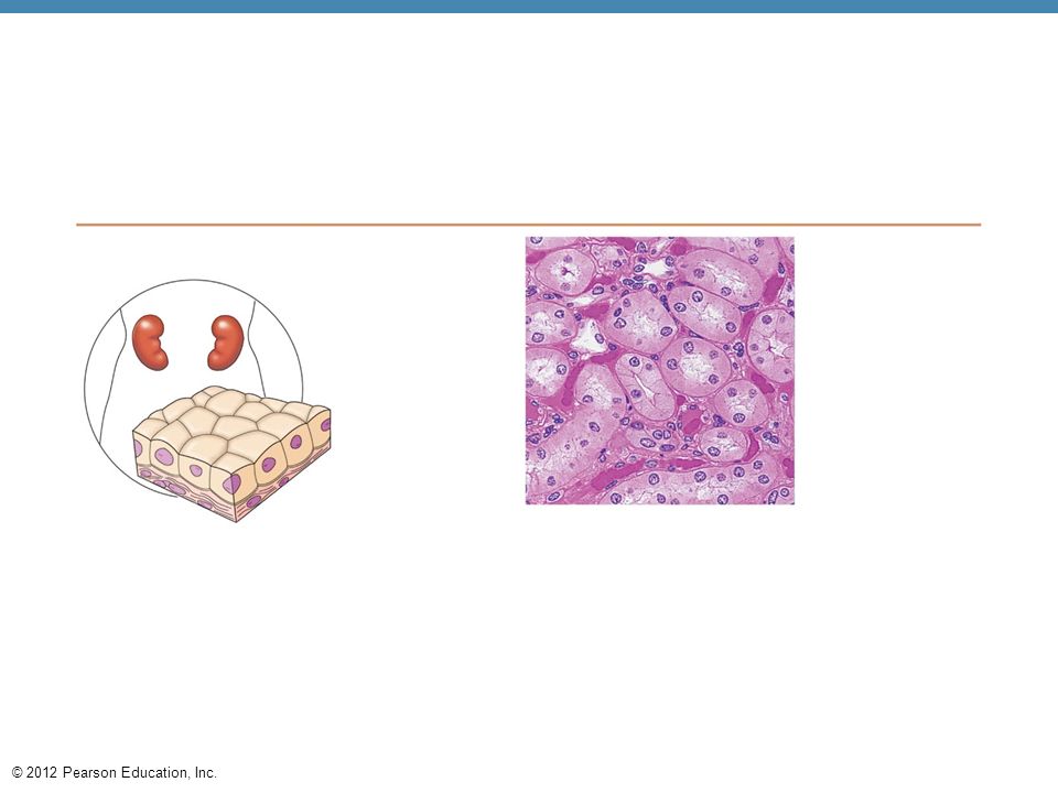
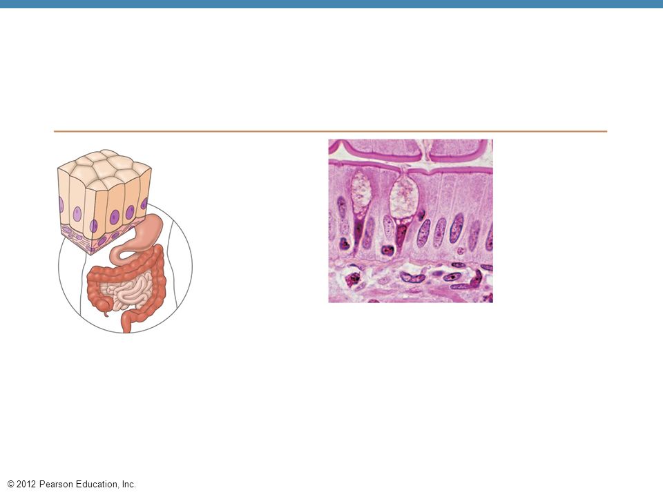
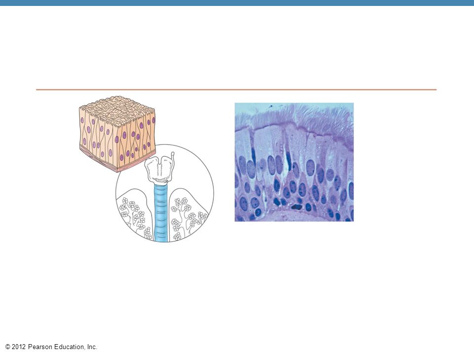

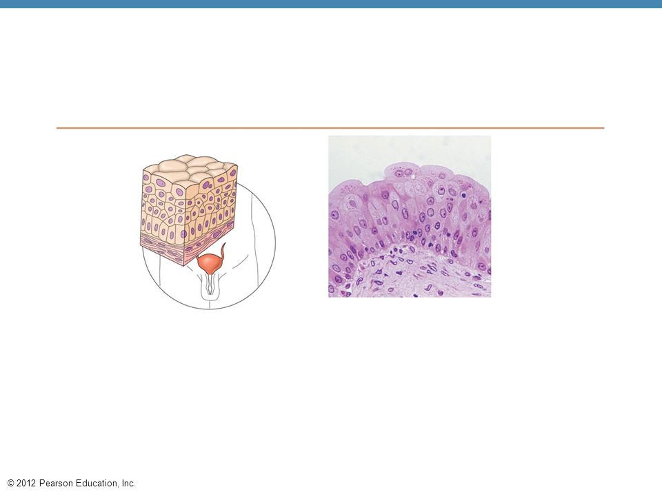
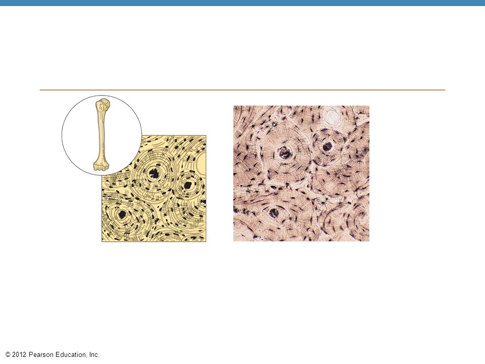

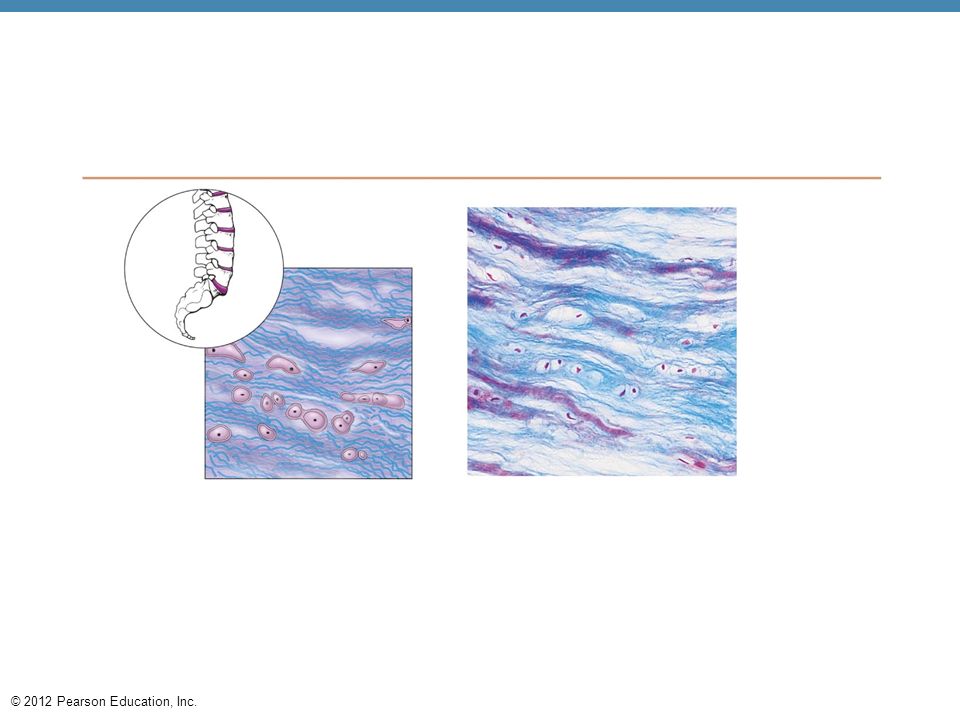
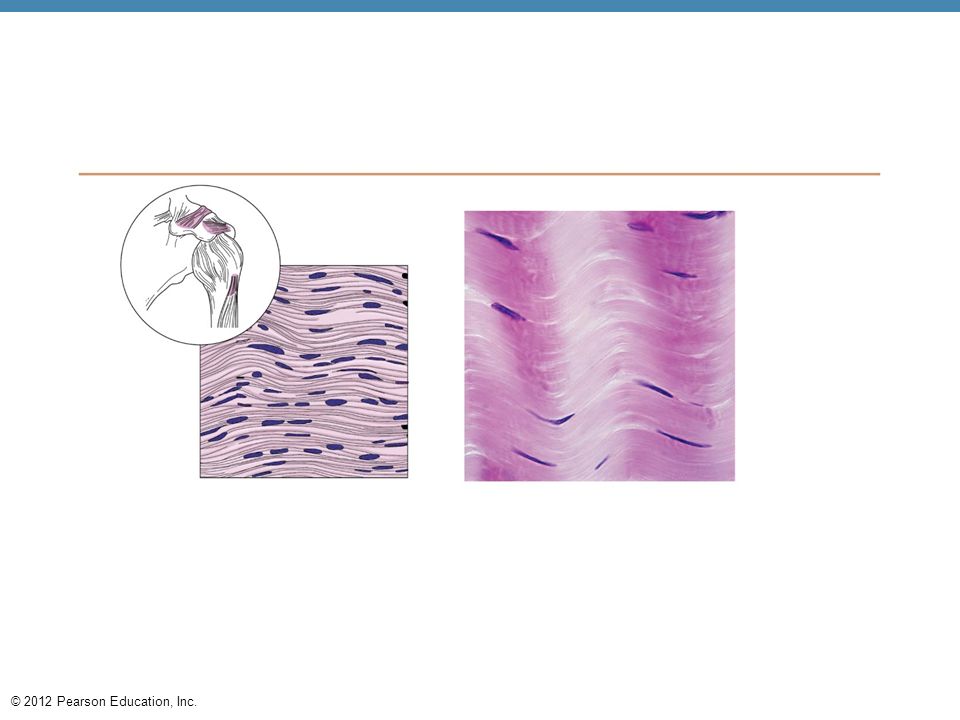

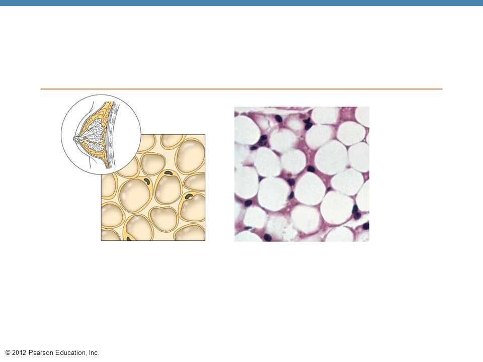
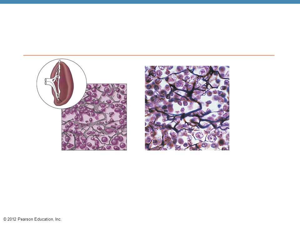


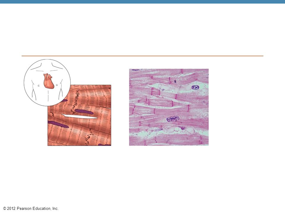

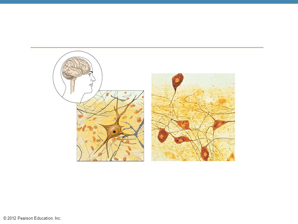
 Diagram: Simple squamous Photomicrograph: Simple.>")


 Connective tissue Muscle tissue.>")


 Connective tissue Muscle tissue.>")


