Download presentation
Presentation is loading. Please wait.

1
Lower limb : Bone, Muscles, Nerves and vessels
Lecturer: Dr. M. Samsam University of Central Florida, Orlando, Pictures from Platzer atlas and textbook of human anatomy and K. Moore anatomy And Netter atlas of human body

2
Hip bone: Study the bones from book and lab manual.
*Bone marrow biopsy and Transplantation. *Bone grafting from iliac crest

3
Pelvic ligaments: 8- iliolumbar ligament 11- sacrotuberous ligament
12- sacrospinous ligament 13- obturator membrane 19- greater sciatic foramen 20- lesser sciatic foramen 24- inguinal ligament 27- lacunar ligament 30- iliopectineal arch

4
male and a female pelvis:
Differences between a male and a female pelvis:

5
Types of pelvis and Pelvic Diameters: Pelvic inlet:
1- transverse diameter ( cm) 2- Oblique diameter I ( cm) 3- Oblique diameter II ( cm) 4- Anatomical conjugate (12 cm) 5- True conjugate (11.5 cm) 6- Diagonal conjugate (13 cm) Pelvic outlet: 7- Straight conjugate ( cm) 8- median conjugate (11.5 cm) *interspinous diameter (not less than 9.5 cm) Others: 10- intercristal distance 29 cm External conjugate (20 cm)
 2- Oblique diameter I ( cm) 3- Oblique diameter II ( cm) 4- Anatomical conjugate (12 cm) 5- True conjugate (11.5 cm) 6- Diagonal conjugate (13 cm) Pelvic outlet: 7- Straight conjugate ( cm) 8- median conjugate (11.5 cm) *interspinous diameter. (not less than 9.5 cm) Others: 10- intercristal distance 29 cm. External conjugate (20 cm)")
6
Femur:

7
*Angle of Inclination *Rickets and Osteomalacia *Hip fracture due to osteoporosis in elderly (fracture causes the person to fall). *Congenital dislocation of the hip: Common birth defect, more in female infants. Either the acetabulum fails to form completely or the ligaments of the hip joint are loose. Treatment: splint or harness of straps to hold femur in its proper position

8
Fibula Used sometimes for bone grafting Mid-shaft fractures of tibia +
fibula are common distal fractures in Skiers

9
Metatarsals and phalangeal bones: Metatarsal stress fractures:
As a result of repetitive stress on foot. 2nd and 3rd metatarsals are mostly affected Treatment: rest and wearing stiff or well cushioned shoes.

10
Lumbosacral plexus Sacral plexus: Sciatic nerve (roots): L4 L5 S1 S2 S3 *Sciatic nerve is the thickest nerve of body. *It is composed of Common Peroneal and Tibial nerves. *Com. Peroneal: composed of dorsal rami Tibial: composed of ventral rami *L4+L5= Lumbosacral trunk 3- Lumbosacral trunk 4- Sciatic nerve 5- common peroneal N. 6- tibial N. 12- posterior femoral cutaneous nerve 13- pudendal nerve 14- superior gluteal nerve

11
Dorsal Hip muscles: 1- Tensor fascia lata:
Origin: anterior superior iliac spine (2), extends to greater trochanter , into iliotibial tract (3) which has >2cm width (It is thickened deep fascia of the thigh). Insertion: lateral tibial condyle Innervation: superior gluteal nerve (L4-L5). Function: Abduction, medial rotation and flexion of the thigh, protects the knee joint. Gluteal region has 3 important muscles: Gluteus Maximus, Medius and minimus. Gluteus Maximus (4) Origin: superficial and deep: Iliac crest (5), post. sup. Iliac spine (6), sacrum (7), coccyx, ilium, posterior to posterior iliac line (9) and sacrotuberous ligament (10). Insertion: mostly to iliotibial tract and only about 25% to gluteal tuberosity (11) of femur. Innervation: Inferior gluteal N. (L5- S2) Function: powerful extensor of hip joint, lateral rotator, active in rising, sitting, climbing
, extends. to greater trochanter , into iliotibial tract (3) which has >2cm width (It is thickened deep. fascia of the thigh). Insertion: lateral tibial condyle. Innervation: superior gluteal nerve (L4-L5). Function: Abduction, medial rotation and flexion. of the thigh, protects the knee joint. Gluteal region has 3 important muscles: Gluteus Maximus, Medius and minimus. Gluteus Maximus (4) Origin: superficial and deep: Iliac crest (5), post. sup. Iliac spine (6), sacrum (7), coccyx, ilium, posterior to posterior iliac line (9) and. sacrotuberous ligament (10). Insertion: mostly to iliotibial tract and only about. 25% to gluteal tuberosity (11) of femur. Innervation: Inferior gluteal N. (L5- S2) Function: powerful extensor of hip joint, lateral rotator, active in rising, sitting, climbing.")
12
Dorsal Hip muscles: (gluteal region) 13- Gluteus Medius:
Origin: external surface of ilium (ala), between anterior and posterior gluteal lines (14). Insertion: greater trochanter of femur (16). Innervation: superior gluteal N. (L5-S1) Function: Abduction, medial rotation of thigh, It keeps pelvis level when opposite leg is raised. 17- Gluteus minimus: Origin: ala of ilium between anterior and inferior Gluteal lines (18). Insertion: greater trochanter of femur (19). same Innervation and function as the Gluteus medius. *Superior gluteal artery gives the blood supply of these 2 muscles while the inf. Gluteal artery gives the blood supply of gluteus maximus. *Positive Trendelenberg sign. *Waddling gate *Lurching gate (one sided defect)
, between. anterior and posterior gluteal lines (14). Insertion: greater trochanter of femur (16). Innervation: superior gluteal N. (L5-S1) Function: Abduction, medial rotation of thigh, It keeps pelvis level when opposite leg is raised. 17- Gluteus minimus: Origin: ala of ilium between anterior and inferior. Gluteal lines (18). Insertion: greater trochanter of femur (19). same Innervation and function as the. Gluteus medius. *Superior gluteal artery gives the blood supply. of these 2 muscles while the inf. Gluteal artery. gives the blood supply of gluteus maximus. *Positive Trendelenberg sign. *Waddling gate. *Lurching gate (one sided defect)")
14
Dorsal Hip muscles: (gluteal region continued)
Deep muscles in the gluteal region: There are a few muscles deep to gluteus Max. These are: Piriformis, obturator internus, superior and inferior gemellus and quadratus femoris. Piriformis Muscle (20): Origin: as several slips from anterior surface of sacrum (21) and sacro-tuberous ligament. Insertion: greater trochanter of femur (22). Innervation: nerve to piriformis (sacral plexus) S1-S2. Function: lateral rotator and abductor of the thigh, keeps femur head in acetabulum. *The muscle may partially or totally be absent.
: Origin: as several slips from anterior surface. of sacrum (21) and sacro-tuberous ligament. Insertion: greater trochanter of femur (22). Innervation: nerve to piriformis (sacral plexus) S1-S2. Function: lateral rotator and abductor of the. thigh, keeps femur head in acetabulum. *The muscle may partially or totally be absent.")
15
Ventral hip muscles: Function as lateral rotators, are stronger than
medial rotators and control the balance***. 1- Obturator internus Origin: inner surface of hip bone, around the Obturator foramen. It passes through the LESSER Sciatic Foramen. Insertion: Trochanteric fossa (2). Innervation: N. to obturator internus (L5-S1) 3- Superior gemellus, arises from ischial spine (4) and inferior gemellus (5) from ischial tuberosity (6). Both insert to trochanteric fossa. Innerv, Sup. Gemellus: N. to Obturator int. Innerv, inf. Gemellus: N. to quadratus femoris. 7- Quadratus femoris: from ischial tuberosity to Intertochanteric crest. Innervation: Nerve to quadratus femoris All function: lateral rotator of the thigh when It is extended and, abductor of the thigh when flexed.
. Innervation: N. to obturator internus (L5-S1) 3- Superior gemellus, arises from ischial. spine (4) and inferior gemellus (5) from. ischial tuberosity (6). Both insert to trochanteric fossa. Innerv, Sup. Gemellus: N. to Obturator int. Innerv, inf. Gemellus: N. to quadratus femoris. 7- Quadratus femoris: from ischial. tuberosity to Intertochanteric crest. Innervation: Nerve to quadratus femoris. All function: lateral rotator of the thigh when. It is extended and, abductor of the thigh when. flexed.")
16
Posterior thigh muscles:
Hamstring muscles: Long head of biceps, Semitendinous, Semimembranous, Adductor mag (ischial part). All innervated by tibial branch of sciatic nerve. Charactristic of hamstring muscles: 1- originate from ischial tuberosity 2- innervated by tibial nerve 3- Flexors (strong) of the knee joint 4- Extensors (weak) of hip joint 5- insert around bones of the knee joint
. All innervated by tibial branch of sciatic nerve. Charactristic of hamstring muscles: 1- originate from ischial tuberosity. 2- innervated by tibial nerve. 3- Flexors (strong) of the knee joint. 4- Extensors (weak) of hip joint. 5- insert around bones of the knee joint.")
17
Posterior thigh muscles:
Hamstring muscles: 1- Biceps femoris: Long head origin: Ischial tuberosity (3) Short head origin: linea aspera (lateral lip) Insertion: head of fibula (encircles collateral lig) Innervation: long head: tibial N. (L5-S2) short head: common peroneal nerve (S1-S2). Function: Long head: extension at hip. It is a flexor and lateral rotator at knee joint. *Short head may be absent. 4- Semitendineous: Origin: ischial tuberos (3) Insertion: shaft of tibia medially (upper part) Pes Anserinus Superficialis (8), +gracilis (9), + Sartorius (10). Function: extension at hip, flexion and medial rotation at knee. Innervation: tibial Nerve (L5-S2). 11- Semimembraneous: Origin: Ischial tuberosity (3). Insertion: near semi/ Tendineous (Pes Anserinus Profundus) It’s extension is the oblique popliteal ligament Innervation: tibial N., Function: hip extensor, knee flexor, medial rotator
 Short head origin: linea aspera (lateral lip) Insertion: head of fibula (encircles collateral lig) Innervation: long head: tibial N. (L5-S2) short head: common peroneal nerve (S1-S2). Function: Long head: extension at hip. It is a flexor and lateral rotator at knee joint. *Short head may be absent. 4- Semitendineous: Origin: ischial tuberos (3) Insertion: shaft of tibia medially (upper part) Pes Anserinus Superficialis (8), +gracilis (9), + Sartorius (10). Function: extension at hip, flexion and medial. rotation at knee. Innervation: tibial Nerve (L5-S2). 11- Semimembraneous: Origin: Ischial tuberosity (3). Insertion: near semi/ Tendineous (Pes Anserinus Profundus) It’s extension is the oblique popliteal ligament. Innervation: tibial N., Function: hip extensor, knee flexor, medial rotator.")
18
Adductor Magnus (4): Origin: inferior ramus of pubis (6), Inferior ramus of ischium (7) and ischial tuberosity (8) Insertion: Linea Aspera (medial lip) 10. Adductor tubercle (12) at medial epicondyle. Function: powerful adductor, lateral rotator (part inserted to linea aspera), medial rotator at knee joint. Extensor at hip joint, 13- Hiatus tendineus Innervation: Tibial nerve to the part inserted to linea aspera. obturator nerve to the part inserted to adductor tubercle (puberty and conception). *Perforating arteries (3-4) usually pierce this muscle from deep femoral artery in anterior femoral region to posterior thigh region to give blood to the dorsal muscles. Adductor hiatus may be considered as the 5th hiatus, its content: Popliteal artery and vein
 10. Adductor tubercle (12) at medial epicondyle. Function: powerful adductor, lateral rotator. (part inserted to linea aspera), medial rotator. at knee joint. Extensor at hip joint, 13- Hiatus tendineus. Innervation: Tibial nerve to the part inserted. to linea aspera. obturator nerve to the part inserted to adductor. tubercle (puberty and conception). *Perforating arteries (3-4) usually pierce this. muscle from deep femoral artery in anterior. femoral region to posterior thigh region to. give blood to the dorsal muscles. Adductor hiatus may be considered as the 5th. hiatus, its content: Popliteal artery and vein.")
19
Posterior leg muscles, Superficial layer: The Triceps Surae, consisting of Soleus (1), Gastrocnemius (2) and Plantaris (3) muscles. 1- Soleus muscle: Origin: head and upper fibula (4), soleal line of Tibia (5). Insertion: into tuber calcanei (8), Achilles T (7). Active in hard and sustain motion (red fiber type). Gastrocnemius (2) muscle: Origin: by 2 heads (9, 11) proximal to femoral Condyles (10). Insertion: tuber calcanei (8). Active for fast movements (white fiber types) Plantaris muscle (3): slight and delicate, long Tendon. Originates from lateral femoral condyle Insertion is to the tuber calcanei (8). May disappear by evolution. Innervation: All 3 muscles, tibial nerve (S1-S2). Function: best plantar flexors, active in Walking. Achilles tendon (calcaneal tendon) is the most powerful tendon and important in walking.
, soleal line of. Tibia (5). Insertion: into tuber calcanei (8), Achilles T (7). Active in hard and sustain motion (red fiber type). Gastrocnemius (2) muscle: Origin: by 2 heads (9, 11) proximal to femoral. Condyles (10). Insertion: tuber calcanei (8). Active for fast movements (white fiber types) Plantaris muscle (3): slight and delicate, long. Tendon. Originates from lateral femoral condyle. Insertion is to the tuber calcanei (8). May disappear by evolution. Innervation: All 3 muscles, tibial nerve (S1-S2). Function: best plantar flexors, active in Walking. Achilles tendon (calcaneal tendon) is the. most powerful tendon and important in walking.")
20
Poaterior leg muscles Deep group, Tibialis posterior (1) muscle: Origin: tibia (3), fibula (4) and interosseous membrane (2). Insertion: navicular bone (7) and 3 cuneiform bones (8). May extend to 2,3,4 metatarsal bones. Innervation: tibial nerve (L4-L5). Flexor Hallusis longus (9): Origin: fibula (10), interosseous membrane (11). Insertion: base of terminal phalanx,1st digit (14). Innervation: tibial nerve (S1-S2). Flexor digitorum Longus (15): Origin: tibia (16) Insertion: by 4 tendons to base of terminal Phalanx of 2nd to 5th digits (18). Innervation: Tibial nerve (S1-S3). *The tendon of all 3 muscle pass beneath the Flexor retinaculum (13). Function: all 3 are active in plantar flexion and Supination of the foot.
. Insertion: navicular bone (7) and 3 cuneiform. bones (8). May extend to 2,3,4 metatarsal bones. Innervation: tibial nerve (L4-L5). Flexor Hallusis longus (9): Origin: fibula (10), interosseous membrane (11). Insertion: base of terminal phalanx,1st digit (14). Innervation: tibial nerve (S1-S2). Flexor digitorum Longus (15): Origin: tibia (16) Insertion: by 4 tendons to base of terminal. Phalanx of 2nd to 5th digits (18). Innervation: Tibial nerve (S1-S3). *The tendon of all 3 muscle pass beneath. the Flexor retinaculum (13). Function: all 3 are active in plantar flexion. and Supination of the foot.")
21
Poaterior leg muscles Deep group, Popliteus (19) muscle: Origin: lateral femoral epicondyle (20). Insertion: tibia (21), above soleal line. Function: Flexion of the knee, unlocking of the Knee joint, protection of lateral meniscus. Innervation: tibial nerve (L4- S1).
, above soleal line. Function: Flexion of the knee, unlocking. of the Knee joint, protection of lateral. meniscus. Innervation: tibial nerve (L4- S1).")
22
Peroneal group of muscles
1-Peroneus longus: has a long tendon. Origin: head and proximal parts of fibula (2). *The tendon runs behind the lateral malleolus Passing in tendon groove of cuboid bone (8). Insertion: tuberosity of 1st metatarsal (6)and the medial cuneiform bone (7). Peroneus brevis (3): short tendon Origin: lateral surface of fibula (9). Its tendon pass beneath the superior (4) and Inferior (5) peroneal retinaculum with the tendon Peroneus longus. Insertion: tuberosity of 5th metatarsal bone (10). Function: they are strongest pronator of the foot. That is Eversion at the subtalar and talocalca- -neonavicular joints (standing on medial margin of the foot while lateral margin is up). Innervation of both muscles: Superficial peroneal nerve (L5-S1).
. *The tendon runs behind the lateral malleolus. Passing in tendon groove of cuboid bone (8). Insertion: tuberosity of 1st metatarsal (6)and. the medial cuneiform bone (7). Peroneus brevis (3): short tendon. Origin: lateral surface of fibula (9). Its tendon pass beneath the superior (4) and. Inferior (5) peroneal retinaculum with the. tendon Peroneus longus. Insertion: tuberosity of 5th metatarsal bone (10). Function: they are strongest pronator of the. foot. That is Eversion at the subtalar and talocalca- -neonavicular joints (standing on medial margin. of the foot while lateral margin is up). Innervation of both muscles: Superficial peroneal nerve (L5-S1).")
23
Deep gluteal region: Greater sciatic foramen Lesser sciatic foramen is divided by Piriformis muscle (2). Suprapiriformis hiatus: sup. Gluteal vessels (3, 4, 5) Infrapiriformis hiatus: Inferior Gluteal vessels (8,9) Internal pudendal artery and vein (10) Pudendal nerve (11) Posterior Cutaneous N. of the thigh (14) Sciatic nerve (15) Nerve to obturator internus (not shown) [18- inf. Clunial N., and 19- perineal N. are branches of posterior cutaneous N. of the thigh.] Int. pudendal artery, pudendal nerve and nerve to Obturator int. reenter the pelvis through lesser Sciatic foramen. Lesser Sciatic Foramen: Int. pudendal vessels. Pudendal nerve. Nerve to Obturator int. and tendon of Obturator internus.
 Infrapiriformis hiatus: Inferior Gluteal vessels (8,9) Internal pudendal artery and vein (10) Pudendal nerve (11) Posterior Cutaneous N. of the thigh (14) Sciatic nerve (15) Nerve to obturator internus (not shown) [18- inf. Clunial N., and 19- perineal N. are. branches of posterior cutaneous N. of the thigh.] Int. pudendal artery, pudendal nerve and nerve to. Obturator int. reenter the pelvis through lesser. Sciatic foramen. Lesser Sciatic Foramen: Int. pudendal vessels. Pudendal nerve. Nerve to. Obturator int. and tendon of Obturator internus.")
24
Posterior femoral region
Division of sciatic nerve Perforating vessels (artery and vein)
")
25
Popliteal region (superficial)
1- greater Saphenous vein (medially) 2- Saphenous nerve (medially) 3- Small saphenous vein 4- medial sural cutaneous nerve 5- branches of post. Femoral cutaneous N.
 2- Saphenous nerve (medially) 3- Small saphenous vein. 4- medial sural cutaneous nerve. 5- branches of post. Femoral cutaneous N.")
26
Popliteal fossa: Diamond shape. Walls: Inferiorly: Gastrocnemius (medial and lateral heads) M. Superiorly: Semitendinosus and semimembranosus (medial) Biceps femoris (lateral) Its floor is composed of: Popliteal surface of femur, knee joint and upper Tibial bone, oblique popliteal ligament and Popliteal muscle with its covering fascia. Content of popliteal fossa: Popliteal artery Popliteal vein Tibial N. Common peroneal N. Genicular arteries and veins
 Biceps femoris (lateral) Its floor is composed of: Popliteal surface of femur, knee joint and upper. Tibial bone, oblique popliteal ligament and. Popliteal muscle with its covering fascia. Content of popliteal fossa: Popliteal artery. Popliteal vein. Tibial N. Common peroneal N. Genicular arteries and veins.")
27
Posterior region of the leg
1- triceps surae m. 2- gastrocnemius 3- soleus 4- calcaneal tendon 5- saphenous nerve 6- great saphenous vein 7- small saphenous vein 9- medial sural nerve 10- communicating branch (lateral sural N.) 11- sural nerve 12- lateral dorsal cutaneous nerve 15- common peroneal nerve 16- posterior tibial artery 17- peroneal (fibular artery 18- popliteal artery 19- anterior tibial artery 20- perforating branch of fibular artery
 11- sural nerve. 12- lateral dorsal cutaneous nerve. 15- common peroneal nerve. 16- posterior tibial artery. 17- peroneal (fibular artery. 18- popliteal artery. 19- anterior tibial artery. 20- perforating branch of fibular artery.")
28
Medial retromalleolar region:
1-2, the Flexor retinaculum 3- greater saphenous vein 5- saphenous nerve Structures passing beneath the Flexor retinaculum (from medial to lateral): Tibialis posterior tendon (7) Flexor digitorum longus (8) Posterior tibial artery and veins (10, 11) Tibial nerve (12) Flexor Hallucis longus (9)
: Tibialis posterior tendon (7) Flexor digitorum longus (8) Posterior tibial artery and veins (10, 11) Tibial nerve (12) Flexor Hallucis longus (9)")
29
Arteries of the leg 4-5: lateral and medial superior genicular aa. 6-7:lateral and medial inferior genicular aa. 8- medial (middle) genicular artery (piercing the oblique popliteal ligament to reach inside the knee joint). 1- Anterior tibial artery 2- posterior tibial artery 20- fibular artery 12- Dorsalis pedis artery
 genicular artery (piercing. the oblique popliteal ligament to reach. inside the knee joint). 1- Anterior tibial artery. 2- posterior tibial artery. 20- fibular artery. 12- Dorsalis pedis artery.")
30
Lumbosacral plexus Lumbar plexus (T12- L4): 1- Obturator nerve (L2-L3-L4) 2- Femoral nerve (L2-L3-L4) 3- Lumbosacral trunk (L4-L5) 7- subcostal nerve (T12) 8- iliohypogastric N. (T12-L1) 9- ilioinguinal N. (L1) 10- genitofamoral N. (L1-L2) 11- lateral cutaneous N. of the thigh (L2-L3)
 7- subcostal nerve (T12) 8- iliohypogastric N. (T12-L1) 9- ilioinguinal N. (L1) 10- genitofamoral N. (L1-L2) 11- lateral cutaneous N. of the thigh (L2-L3)")
31
Diaphragm and posterior abdominal wall:
The psoas major and minor muscles, the quadratus lumborum muscle. The lumbar plexus and its related nerves.

32
Adductors of the thigh:
All are innervated by Obturator nerve except the pectineus muscle. 1- Gracilis muscle: Origin: inferior ramus of pubis (2). Insertion: medial tibial condyle (pes anserinus). Function: adductor of the thigh, flexor at hip and flexor at knee joints 5- Pectineus muscle Origin: pecten of pubis (6), iliopubic eminence Insertion: Pectineal line of femur (8) Function: adductor of thigh and flexor of hip joint. Innervation: Femoral nerve (L2-L3) and Obturator nerve 10- Adductor brevis muscle: Origin: inferior ramus of pubis (11) Insertion: upper part of linea aspera (9) Function: adductor of thigh and flexor of hip joint. Innervation: Obturator nerve (L2-L4).
. Insertion: medial tibial condyle (pes anserinus). Function: adductor of the thigh, flexor at hip. and flexor at knee joints. 5- Pectineus muscle. Origin: pecten of pubis (6), iliopubic eminence. Insertion: Pectineal line of femur (8) Function: adductor of thigh and flexor of. hip joint. Innervation: Femoral nerve (L2-L3) and Obturator nerve. 10- Adductor brevis muscle: Origin: inferior ramus of pubis (11) Insertion: upper part of linea aspera (9) Function: adductor of thigh and flexor of hip joint. Innervation: Obturator nerve (L2-L4).")
33
Adductor Longus (1): Origin: superior ramus of pubis (2), Insertion: middle 1/3 of linea aspera (3). It lies on the adductor magnus (4). Adductor brevis (5) is interposed between them. Function: Adduction of thigh, hip flexion NN: Obturator nerve
. Adductor brevis (5) is interposed between them. Function: Adduction of thigh, hip flexion. NN: Obturator nerve.")
34
Adductor Magnus (4): Origin: inferior ramus of pubis (6), Inferior ramus of ischium (7) and ischial tubrosity (8) Insertion: Linea Aspera (medial lip) 10. Adductor tubercle (12) at medial epicondyle. Function: powerful adductor, lateral rotator (part inserted to linea aspera), medial rotator at knee joint. Extensor at hip joint, 13- Hiatus tendineus Innervation: Tibial nerve to the part inserted to linea aspera. Obturator nerve to the part inserted to adductor tubercle (puberty and conception). Adductor minimus (14): Incompletely separated division of adductor Magnus. Origin: inferior ramus of pubis (6) Insertion: upper part of linea aspera Function and innervation: same as adduct. Magnus. 13- Adductor hiatus 16- Vasto-adductor membrane
 10. Adductor tubercle (12) at medial epicondyle. Function: powerful adductor, lateral rotator. (part inserted to linea aspera), medial rotator. at knee joint. Extensor at hip joint, 13- Hiatus tendineus. Innervation: Tibial nerve to the part inserted. to linea aspera. Obturator nerve to the part inserted to adductor. tubercle (puberty and conception). Adductor minimus (14): Incompletely separated division of adductor. Magnus. Origin: inferior ramus of pubis (6) Insertion: upper part of linea aspera. Function and innervation: same as adduct. Magnus. 13- Adductor hiatus. 16- Vasto-adductor membrane.")
35
Anterior Thigh region:
Iliopsoas muscle: Composed of Psoas major (1) and Iliacus (4) muscles. Psoas major originates from body (2) and Transverse processes (3) of 1st-4th lumbar Vertebrae and The Iliacus originates from ala of the ilium (7). Insertion: lesser trochanter of femur (6). Function: chief flexor of the hip Innervation: lumbar plexus to psoas (L1-L3) and femoral nerve to iliacus (L2-L4)
 and Iliacus (4) muscles. Psoas major originates from body (2) and. Transverse processes (3) of 1st-4th lumbar. Vertebrae and. The Iliacus originates from ala of the ilium (7). Insertion: lesser trochanter of femur (6). Function: chief flexor of the hip. Innervation: lumbar plexus to psoas (L1-L3) and femoral nerve to iliacus (L2-L4)")
36
Anterior thigh muscles:
Quadriceps femoris muscle: Consist of 4 parts: Rectus femoris (1): originates from ant inf Iliac spine (2) Vastus intermedius (3), vastus medialis (5) and vastus lateralis (7), all originating from femur. Insertion: by a common tendon (tendon of the quadriceps femoris) to patella (9). Inferior to patella, the tendon continues as the patellar ligament (10) to insert on tibial tuberosity (11). Function: Chief extensor of the knee joint Rectus femoris also flexes the hip joint. Innervation: femoral nerve (L2-L4). Sartorius muscle (12): Origin: anterior superior iliac spine (13) Insertion: pes anserinus superficialis (14) Function: acts on 2 joints: flexes at both hip and knee joints.
: originates from ant inf. Iliac spine (2) Vastus intermedius (3), vastus medialis (5) and vastus lateralis (7), all originating from. femur. Insertion: by a common tendon (tendon of. the quadriceps femoris) to patella (9). Inferior to patella, the tendon continues as. the patellar ligament (10) to insert on tibial. tuberosity (11). Function: Chief extensor of the knee joint. Rectus femoris also flexes the hip joint. Innervation: femoral nerve (L2-L4). Sartorius muscle (12): Origin: anterior superior iliac spine (13) Insertion: pes anserinus superficialis (14) Function: acts on 2 joints: flexes at both hip. and knee joints.")
37
Anterior thigh region (deep) The femoral triangle
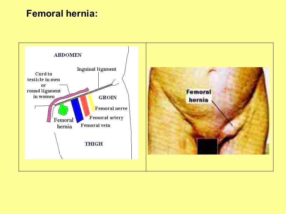
Borders: Sartorius laterally Adductor longus, medially and also the floor Ilioinguinal ligament superiorly. Floor: iliopsoas m., pectineus m., and adductor longus. Content: A- lateral femoral cutaneous nerve B- Femoral nerve C- Structures inside the femoral sheath *Content of Femoral Ring: Femoral artery and genitofemoral N. (fem) Femoral vein Lymph nodes and areolar tissue (femoral Canal), the Rosenmuller node (they drain the Glans penis and clitoris) *Femoral hernia: painful, more in female, below and lateral to pubic tubercle.
 Femoral vein. Lymph nodes and areolar tissue (femoral. Canal), the Rosenmuller node (they drain the. Glans penis and clitoris) *Femoral hernia: painful, more in female, below and lateral to pubic tubercle.")
38
Femoral hernia:
39
Arteries to the pelvis The internal iliac artery and it’s branches

40
Arteries of the pelvis and the thigh
5- abdominal aorta 4- common iliac artery 1- internal iliac artery 15- external iliac artery 19- femoral artery 24- deep femoral artery 25- medial circumflex femoral artery 27- lateral circumflex femoral artery 30- terminal branches of deep femoral artery, the perforating arteries. 33- descending genicular artery

41
Subinguinal region 9- external pudendal vessels 10- superficial epigastric vessels 11- superficial circumflex iliac vessels

42
Saphenous hiatus

43
Anterior thigh region (deep)
Subsartorius (adductor, Hunter’s) canal: *Starts distal to femoral triangle. Content: Femoral artery and vein, saphenous nerve, nerve to Vastus medialis, small branches of Obturator nerve and great saphenous vein
 canal: *Starts distal to femoral triangle. Content: Femoral artery and vein, saphenous nerve, nerve to Vastus medialis, small branches of. Obturator nerve and great saphenous. vein.")
44
Anterior leg: Extensor group 1-2: Tibialis anterior
Origin: tibia and interosseous memb (2) Its tendon passes beneath the extesor RT (3) Insertion: medial cuneiform (5) and 1st metatarsal (6). Function: dorsiflexion and supination; with Peron. L., keep the transverse arch of the foot Innervation: deep peroneal N. (L4-L5). 7-8: extensor digitorum longus Origin: tibial condyle, fibula, interosseous M. Insertion: Base of distal phalanx (2nd-5th). Function: Dorsiflexion of the foot Innervation: deep peroneal N. (L5-S1). Peroneus Tertius: Extensor digitorum’s additional part which may extend to the base of 5th metatarsal (9). It may even be a muscle and have a separate origin. NN: Deep peronael nerve 10-11: Extensor Hallucis longus Origin: fibula, inteosseous membrane (11) Insertion: terminal phalanx of the big toe (12) Function: first toe dorsiflexion Innervation: deep peroneal N. (L4- S1).
 Its tendon passes beneath the extesor RT (3) Insertion: medial cuneiform (5) and. 1st metatarsal (6). Function: dorsiflexion and supination; with. Peron. L., keep the transverse arch of the foot. Innervation: deep peroneal N. (L4-L5). 7-8: extensor digitorum longus. Origin: tibial condyle, fibula, interosseous M. Insertion: Base of distal phalanx (2nd-5th). Function: Dorsiflexion of the foot. Innervation: deep peroneal N. (L5-S1). Peroneus Tertius: Extensor digitorum’s. additional part which may extend to the. base of 5th metatarsal (9). It may even be a. muscle and have a separate origin. NN: Deep peronael nerve : Extensor Hallucis longus. Origin: fibula, inteosseous membrane (11) Insertion: terminal phalanx of the big toe (12) Function: first toe dorsiflexion. Innervation: deep peroneal N. (L4- S1).")
45
Muscles of the dorsum of the foot:
Tendon of the long extensors of the foot, lie superficial to these muscles and they form a dorsal aponeurosis into which the short Extensors of the digits, plantar and dorsal Interosseous muscles radiate. Extensor digitorum brevis (6): Origin: Calcaneus (7) Insertion: with 3 tendons to dorsal aponeurosis (8). Function: dorsiflexion of these digits Innervation: Deep peroneal nerve (S1-S2). Extensor Hallucis brevis (9): Origin: Calcaneus Insertion: Dorsal aponeurosis of 1st digit Function: Dorsiflexion of 1st digit 10- tibialis anterior tendon 11- Peroneus tertius
: Origin: Calcaneus (7) Insertion: with 3 tendons to dorsal. aponeurosis (8). Function: dorsiflexion of these digits. Innervation: Deep peroneal nerve (S1-S2). Extensor Hallucis brevis (9): Origin: Calcaneus. Insertion: Dorsal aponeurosis of 1st digit. Function: Dorsiflexion of 1st digit. 10- tibialis anterior tendon. 11- Peroneus tertius.")
46
Muscles of the sole of the foot:
1- Plantar Aponeurosis Consist of longitudinal and transverse fibers. It maintains the longitudinal arch of the foot and protects the vessels and nerves there. 5- Abductor Hallucis: Origin: Tuber Calcanei (6), plantar aponeurosis (7). Insertion: medial sesomoid bone (8) and base of proximal phalanx of 1st toe (9). Innervation: Medial plantar Nerve (L5-S1). 10- Fexor Hallucis Brevis: Origin: medial cuneiform bone (11) It has 2 heads. A medial head (12) which extends to medial sesamoid bone (13) and Its lateral head (15) extend to lateral sesamoid bone (16) and inserted on proximal phalanx of 1st toe. Innervation: Medial Plantar Nerve (L5-S1).
, plantar aponeurosis (7). Insertion: medial sesomoid bone (8) and base. of proximal phalanx of 1st toe (9). Innervation: Medial plantar Nerve (L5-S1). 10- Fexor Hallucis Brevis: Origin: medial cuneiform bone (11) It has 2 heads. A medial head (12) which. extends to medial sesamoid bone (13) and. Its lateral head (15) extend to lateral sesamoid. bone (16) and inserted on proximal phalanx. of 1st toe. Innervation: Medial Plantar Nerve (L5-S1).")
47
Muscles of the sole of the foot:
11- Flexor digitorum Brevis: Origin: tuber calcanei Insertion: middle phalax of 2nd-4th digits Innervation: Medial Plantar N (L5-S1). 1- Lumbriclas (4 ones) Tiny muscles originating from tendon (2) of the flexor digitorum longus (medial side). Insertion: Dorsal aponeurosis of 2nd-5th digit. Function: plantar flexion of these digits Innervation: Medial Plantar N to 1, and Lat Plantar N to 2, 3 and 4 (S2 and S3) 3- Quadratus Plantae: Origin: by 2 heads from calcaneus Insertion: lateral border of the tendon of the Flexor digitorum longus. Innervation: Lateral Plantar N. (S1-S2).
. 1- Lumbriclas (4 ones) Tiny muscles originating from tendon (2) of the flexor digitorum longus (medial side). Insertion: Dorsal aponeurosis of 2nd-5th digit. Function: plantar flexion of these digits. Innervation: Medial Plantar N to 1, and. Lat Plantar N to 2, 3 and 4 (S2 and S3) 3- Quadratus Plantae: Origin: by 2 heads from calcaneus. Insertion: lateral border of the tendon of the. Flexor digitorum longus. Innervation: Lateral Plantar N. (S1-S2).")
48
Muscles of the sole of the foot:
Plantar interossei MM (3), Blue They have single head, Number 7 Originate from medial side of 3rd-5th metatarsals bones. Insertion: medial side of 3rd-5th digits. Function: Adductors of the digits Innervation: Lateral Plantar N (S2-S3). Dorsal interossei MM (4), Red They have 2 heads Number 9 Originate from opposing surface of all metatarsals Insertion: to base of 2nd-4th digits Function: Abductors of the digits
, Blue. They have single head, Number 7. Originate from medial side of 3rd-5th. metatarsals bones. Insertion: medial side of 3rd-5th digits. Function: Adductors of the digits. Innervation: Lateral Plantar N (S2-S3). Dorsal interossei MM (4), Red. They have 2 heads Number 9. Originate from opposing surface of all. metatarsals. Insertion: to base of 2nd-4th digits. Function: Abductors of the digits.")
49
Muscles of the sole of the foot:
1- Adductor Hallucis: Has 2 heads: oblique head (3) and the transverse head (9). Innervation: Lateral Plantar N (S1-S2) 7- Long Plantar Ligament 12-Opponens digiti minimi, Innervation: Lat Plantar N (S1-S2) 15-16: Flexor Digiti Minimi: Innervation: Lateral Plantar N (S1-S2) 18- Abductor Digiti Minimi Origin: calcaneus(20) and 5th metatarsal (21) Insertion: Base of proximal phalanx of the 5th digit (22). Innervation: Lateral Plantar N (S1-S2). 23- Quadratus Plantae
 and the transverse head (9). Innervation: Lateral Plantar N (S1-S2) 7- Long Plantar Ligament. 12-Opponens digiti minimi, Innervation: Lat Plantar N (S1-S2) 15-16: Flexor Digiti Minimi: Innervation: Lateral Plantar N (S1-S2) 18- Abductor Digiti Minimi. Origin: calcaneus(20) and 5th metatarsal (21) Insertion: Base of proximal phalanx of the. 5th digit (22). Innervation: Lateral Plantar N (S1-S2). 23- Quadratus Plantae.")
50
Arteries of the leg 4-5: lateral and medial superior genicular aa. 6-7:lateral and medial inferior genicular aa. 8- medial (middle) genicular artery (piercing the oblique popliteal ligament to reach inside the knee joint). 1- Anterior tibial artery 2- posterior tibial artery 20- fibular artery *When the Anterior tibial artery (1) passes Beneath the superior extensor retinaculum, It is called dorsal artery of the foot or Dorsalis Pedis artery (11). 12- shows where its pulsation can be felt. Ant. Tibial artery or Dorsalis pedis may give The lateral tarsal artery (13). Together, the lateral tarsal and dorsalis pedis Make the arcuate artery (14) giving rise to Metatarsal (15) and dorsal digital (16) arteries. *Dorsalis pedis gives a deep branch to join The plantar arch.
 genicular artery (piercing. the oblique popliteal ligament to reach. inside the knee joint). 1- Anterior tibial artery. 2- posterior tibial artery. 20- fibular artery. *When the Anterior tibial artery (1) passes. Beneath the superior extensor retinaculum, It is called dorsal artery of the foot or. Dorsalis Pedis artery (11). 12- shows where its pulsation can be felt. Ant. Tibial artery or Dorsalis pedis may give. The lateral tarsal artery (13). Together, the lateral tarsal and dorsalis pedis. Make the arcuate artery (14) giving rise to. Metatarsal (15) and dorsal digital (16) arteries. *Dorsalis pedis gives a deep branch to join. The plantar arch.")
51
Arteries of the leg Posterior tibial artery(2) in the plantar region gives the medial (21) and lateral (23) Plantar arteries. Lateral plantar artery makes most part of the plantar arch (22) which give rise to plantar Metatarsal arteries (24) and proper plantar digital arteries (25). The dorsal and plantar arches are connected via perforating branches. Pulsation of Dorsalis pedis artery may be lost In some Peripharal vascular diseases such as Burger’s disease or also in diabetes mellitus Occlusion of blood vessels lead to gangrene And even autoamputation of the first toe. Scars on the skin may develop.
 which give rise to plantar. Metatarsal arteries (24) and proper plantar. digital arteries (25). The dorsal and plantar arches are connected. via perforating branches. Pulsation of Dorsalis pedis artery may be lost. In some Peripharal vascular diseases such as. Burger’s disease or also in diabetes mellitus. Occlusion of blood vessels lead to gangrene. And even autoamputation of the first toe. Scars on the skin may develop.")
52
Plantar region superficial:
Plantar aponeurosis (1)
")
53
Plantar region deep: Lateral plantar nerve (13) and Medial plantar nerve (4), innervate the muscles and skin of the plantar side. Plantar arteries and veins (15,16, 3, 4) are involved in blood supply and venous drainage of the plantar region of the foot.
 are involved in blood supply and venous. drainage of the plantar region of the foot.")
Similar presentations


 Joint>")
>")

