Download presentation
Presentation is loading. Please wait.

2
The Knee

3
From the Sports Medicine Perspective

4
Bony Anatomy Femur Patella Tibia Fibula

5
Bony Anatomy Femur: Longest Bone in Body Tibia: WB bone of lower extremity Fibula: Site of Muscle Attachment Patella: Sesamoid Bone A bone that develops within a tendon

6
Knee Skeletal Lateral Condyle Head of Fibula Femoral Groove Gerdy’s Tubercle Tibial Tuberosity Pes Anserine

7
Sagittal MRI View

8
Knee Connective Tissue

9
Knee Menisci

10
Menisci

11
Medial Meniscus Lateral Meniscus PCL ACL

12
Knee Ligaments

13
Medial Collateral Ligament MCL

14
MCL Thick Band of Tissue Tibia Femur Resists Valgus Force

15
Valgus Outside to Inside Force MCL resists this force Occurs in FRONTAL PLANE

16
Lateral Collateral Ligament LCL

17
LCL Narrow cord like band of tissue Fibula Femur Resists Varus Forces

18
Varus Inside to Outside Force LCL resists this force FRONTAL PLANE

19
Increased Valgus

20
Collateral Ligament Ruptures

21
Ligament Structures

22
Anterior Knee

23
Anterior Cruciate Ligament ACL Composed of 3 bands Prevents anterior translation of tibia Stabilizes against excessive rotation Stabilizing Ligament

24
Healthy ACL

25
Torn ACL

26
Knee Posterior

27
Posterior Cruciate Ligament PCL Stabilizes the posterior aspect of knee Prevents hyperextension

28
Cadaver Knee

29
Quadriceps Anterior Thigh Musculature Four Muscles: Rectus Femoris Vastus Lateralis Vastus Medialis Vastus Intermedius Extend the Knee

30
Quadriceps

31
Rectus Femoris 2 Joint Muscle Crosses hip and knee Flexes Hip Extend the knee Converges with rest of quadriceps muscles at tibial tubercle

32
Hamstrings Three Muscles Semimembranosus Semitendinosus Biceps Femoris Common Origin the ischial tuberosity Flex the Knee

33
Hamstrings

34
Popliteus

36
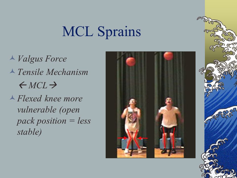
MCL Sprains Valgus Force Tensile Mechanism MCL Flexed knee more vulnerable (open pack position = less stable)
")
37
MCL Injuries Direct trauma in frontal plane injures MCL Combination of rotation can result in ACL and meniscus tears

38
MCL/LCL Injuries GRADE I: No instability Mild Effusion ROM full Mild tenderness w/ palpation

39
MCL/LCL Injuries GRADE II: Laxity w/ valgus or varus stress (more with 30 degrees of flexion) Decrease in ROM Increase medial (MCL) or lateral (LCL) pain GRADE III: Complete ligament rupture Complete loss of stability Immediate pain that transitions into dull ache
 Decrease in ROM Increase medial (MCL) or lateral (LCL) pain GRADE III: Complete ligament rupture Complete loss of stability Immediate pain that transitions into dull ache")
40
Treatment Based on severity of injury RICE Modify activity Crutches Exercises in sagittal plane Progress to functional exercise

Similar presentations


 articulating with 2 concave surfaces (tibia) Poor bony stability Stability increased.>")


>")


