Download presentation
Presentation is loading. Please wait.

1
Anatomy of the Reproductive System
Lab Activity 33 Anatomy of the Reproductive System Portland Community College BI 233

2
Scrotum Sac of skin and superficial fascia that hangs outside the abdominopelvic cavity at the root of the penis Contains paired testicles separated by a midline septum Its external positioning keeps the testes 3C lower than core body temperature

3
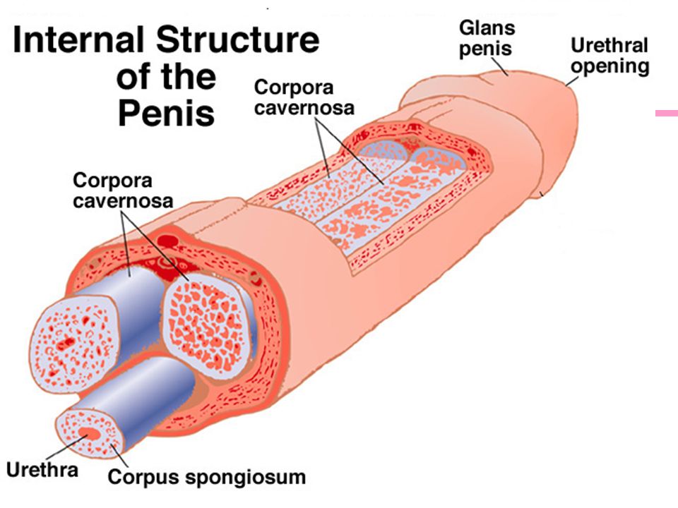
Penis Internal penis: The urethra and three cylindrical bodies of erectile tissue Corpus spongiosum: Surrounds the urethra and expands to form the glans and bulb of the penis Corpora cavernosa: Paired dorsal erectile bodies bound by fibrous tunica albuginea Crura: anchors the penis to the pubic arch

6
Penis Histology

7
Testicles Seminiferous tubules:
Produce the sperm Converge to form the tubulus rectus The straight tubulus rectus conveys sperm to the rete testis From the rete testis, the sperm: Leave the testis via efferent ductules Enter the epididymis Surrounding the seminiferous tubules are interstitial cells

8
Testicle

9
Seminiferous Tubules “Sperm Factory”
Interstitial cells (Leydig cells) In the soft connective tissue surrounding the seminiferous tubules. Produce Testosterone when stimulated by LH
 In the soft connective tissue surrounding the seminiferous tubules. Produce Testosterone when stimulated by LH.")
10
Seminiferous Tubules “Sperm Factory”
Sertoli Cells (sustentacular cells) Support and nourish the spermatogenic cells Completely surround the spermatic cells undergoing meiosis Creates the blood-testes barrier Stimulated by FSH Secretes: Androgen binding protein (ABP): Concentrates testosterone in the seminiferous tubules Inhibin: Released when sperm production is too high (slows it down). Inhibits the secretion of FSH and GnRH
 Support and nourish the spermatogenic cells. Completely surround the spermatic cells undergoing meiosis. Creates the blood-testes barrier. Stimulated by FSH. Secretes: Androgen binding protein (ABP): Concentrates testosterone in the seminiferous tubules. Inhibin: Released when sperm production is too high (slows it down). Inhibits the secretion of FSH and GnRH.")
12
Seminiferous Tubules

13
Seminiferous Tubules Histology

14
Epididymis Epididymis: Storage and maturation area for sperm
Its head joins the efferent ductules and caps the superior aspect of the testis The duct of the epididymis has stereocilia that: Absorb testicular fluid Pass nutrients to the sperm Nonmotile sperm enter, pass through its tubes and become motile (propelled by peristalsis) Upon ejaculation the epididymis contracts, expelling sperm into the ductus deferens
 Upon ejaculation the epididymis contracts, expelling sperm into the ductus deferens.")
15
Ductus Deferens and Ejaculatory Duct
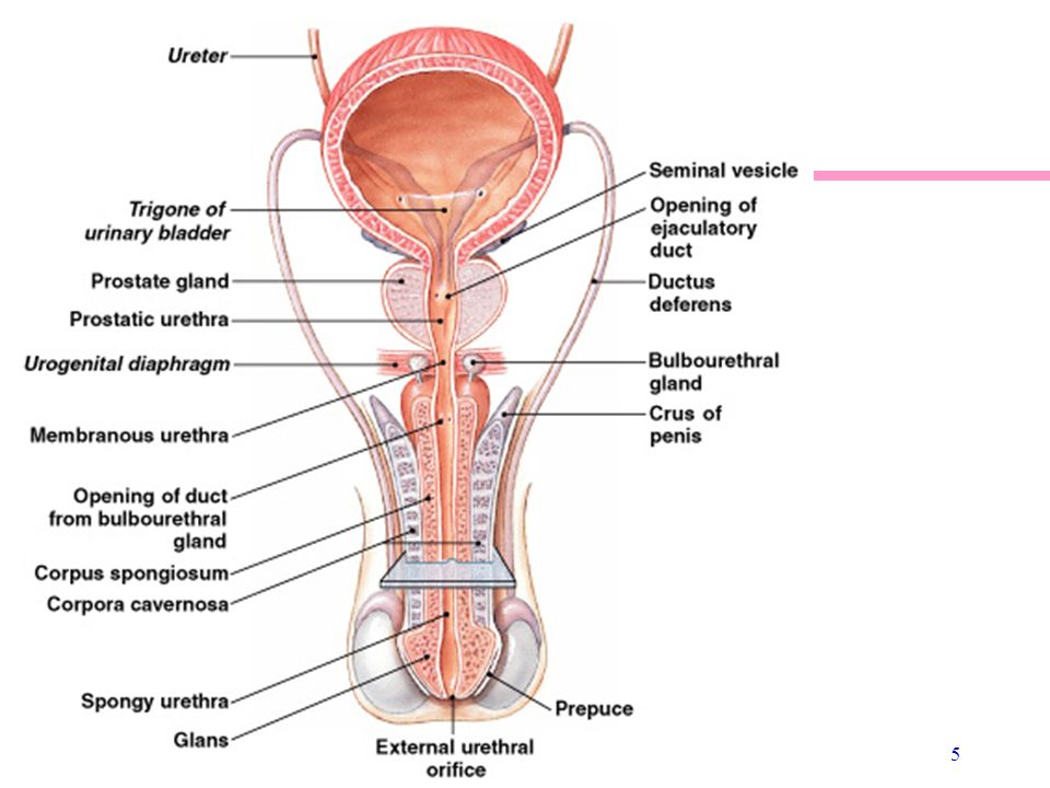
Ductus Deferens: Runs from the epididymis through the inguinal canal into the pelvic cavity Ampulla: The expanded terminal end on posterior bladder Ejaculatory Duct: Formed from the ampulla and the duct of the seminal vesicle Most of it is in the prostate gland Propels sperm from the epididymis to the urethra

16
Ampulla Seminal vesicle Ejaculatory Duct

17
Spermatic Cord Contains the structures running from the testicles to the pelvic cavity. Passes through the inguinal canal Contents: Vas Deferens Nerves Blood Vessels

18
Accessory Glands: Seminal Vesicles
Lie on the posterior wall of the bladder and secrete 60% of the volume of semen Seminal fluid: Fructose: provides energy for the sperm. Fibrinogen: helps turn semen into a bolus that can be readily propelled into the vagina. Prostaglandins: decrease cervical mucus viscosity and stimulate reverse peristalsis of the uterus. Join the ductus deferens to form the ejaculatory duct

19
Posterior Bladder

20
Accessory Glands: Prostate Gland
Doughnut-shaped gland that encircles part of the urethra inferior to the bladder Plays a role in the activation of sperm Enters the prostatic urethra during ejaculation Prostatic secretions include: Citrate: is a food source (TCA cycle) Proteolytic enzymes: acts to "decoagulate" the semen that was coagulated by seminal vesicle secretions, which helps the sperm begin their journey once inside the vagina
 Proteolytic enzymes: acts to decoagulate the semen that was coagulated by seminal vesicle secretions, which helps the sperm begin their journey once inside the vagina.")
21
Bulbourethral Glands (Cowper’s Glands)
Pea-sized glands inferior to the prostate Produce alkaline mucus prior to ejaculation that neutralizes traces of acidic urine in the urethra

22
Sperm Summary Produced: Seminiferous tubules Stored: Epididymis
Transported through epididymis by rhythmic peristaltic contractions as they mature Epididymis Vas Deferens Ejaculatory duct (ampulla of vas deferens fuses with duct of seminal vesicle “ejaculatory duct”) prostate prostatic urethra (then passes the bulbourethral gland) membranous urethra penile urethra
 prostate prostatic urethra (then passes the bulbourethral gland) membranous urethra penile urethra.")
23
Female External Genitalia
Mons pubis: fatty pad over the pubic symphysis Labia majora & minora: folds of skin encircling vestibule where find urethral and vaginal openings Clitoris: small mass of erectile tissue Bulb of vestibule: masses of erectile tissue just deep to the labia on either side of the vaginal orifice Perineum: Area between the vagina and anus

24
Female External Genitalia
Perineum

25
Vagina Thin-walled tube lying between the bladder and the rectum, extending from the cervix to the exterior of the body Wall consists of three coats: fibroelastic adventitia, smooth muscle muscularis, and a stratified squamous mucosa Mucosa near the vaginal orifice forms an incomplete partition called the hymen Vaginal fornix: upper end of the vagina surrounding the cervix

26
Bartholin’s Glands (aka: Vestibular Glands)
The Bartholin's glands are located on each side of the vaginal opening. They secrete fluid that helps lubricate the vagina. Sometimes the ducts of these glands become obstructed. Fluid backs up into the gland and causes swelling (Bartholin's cyst)
")
27
Female: Lateral View

28
Cervix Narrow lower neck of the uterus which projects into the vagina inferiorly Cervical canal – cavity of the cervix that communicates with: The vagina via the external os The uterine body via the internal os Cervical glands secrete mucus that covers the external os and blocks sperm entry except during midcycle

29
Endocervical canal Fornix

30
Uterine Tubes (Fallopian Tubes)
Receive the ovulated oocyte and provide a site for fertilization Empty into the superolateral region of the uterus via the isthmus Expand distally around the ovary forming the ampulla The ampulla ends in the funnel-shaped, ciliated infundibulum containing fingerlike projections called fimbriae

31
Uterine Tubes (Fallopian Tubes)
Function: events occurring in the uterine tube Fimbriae sweep oocyte into tube, cilia & peristalsis move it along, sperm reaches oocyte in ampulla, fertilization occurs within 24 hours after ovulation & zygote reaches uterus about 7 days after ovulation

32
Fallopian Tube Histology
Cilia sweep egg/zygote toward the uterus

33
Uterus Hollow, thick-walled organ located in the pelvis anterior to the rectum and posterosuperior to the bladder Body: Major portion of the uterus Fundus: Rounded region superior to the entrance of the uterine tubes Isthmus: Narrowed region between the body and cervix

34
Uterus

35
Uterine Histology Endometrium Simple columnar epithelium
Stroma of connective tissue and endometrial glands Stratum functionalis: Shed during menstruation Stratum basalis: Replaces stratum functionalis each month Myometrium 3 layers of smooth muscle Perimetrium Visceral peritoneum

36
Uterine Histology

37
Endometrium Simple columnar epithelium Endometrial glands

38
Ovaries Each follicle consists of an immature egg called an oocyte
Cells around the oocyte are called: Follicle cells (one cell layer thick) Stimulated to mature by FSH from the pituitary gland Granulosa cells (when more than one layer is present) Thecal cells: Cells in the ovarian stroma Thecal & granulosa cells work together to produce estrogen
 Stimulated to mature by FSH from the pituitary gland. Granulosa cells (when more than one layer is present) Thecal cells: Cells in the ovarian stroma. Thecal & granulosa cells work together to produce estrogen.")
39
Follicle Development Primordial follicle: one layer of squamous-like follicle cells surrounds the oocyte Primary follicle: two or more layers of cuboidal granulosa cells enclose the oocyte Secondary follicle: has a fluid-filled space between granulosa cells that coalesces to form a central antrum Graafian follicle: secondary follicle at its most mature stage that bulges from the surface of the ovary Corpus luteum : ruptured follicle after ovulation

40
Ovary Histology

41
Ovary Histology

42
Primary Follicle 1° Oocyte (arrested in prophase I) Nucleus
Primordial follicle Zona pellucida Thecal cells Granulosa cells

43
Secondary Follicle

44
Graafian Follicle Fluid filled antrum Oocyte 2° Granulosa cells Stalk
Corona radiata Zona pellucida

45
Ovulation LH from the pituitary gland will cause the Graafian follicle to rupture The oocyte will be released (ovulation) The follicle is now a corpus luteum Secretes estrogen and progesterone

46
Mammary Glands Modified sweat glands that produce milk (lactation)
Amount of adipose determines size of breast Milk-secreting glands open by lactiferous ducts at the nipple Areola is pigmented area around nipple Suspensory ligaments suspend breast from deep fascia of pectoral muscles (aging & Cooper’s droop)
")
47
Breast

48
Breast Prolactin from the pituitary gland stimulates the synthesis of milk Oxytocin from the posterior pituitary gland stimulates milk ejection

49
Lymphatic Drainage Lymph nodes draining the breast are located in the axilla.

50
Lab Activity 34 Gametogenesis

51
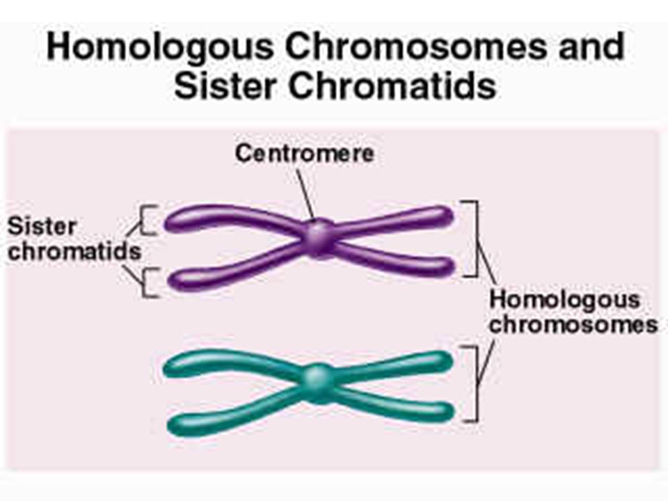
Gametogenesis Each cell has two sets of chromosomes (one maternal, one paternal) and is said to be diploid (2n chromosomal number) Humans have 23 pairs of homologous chromosomes Gametes only have 23 chromosomes and are said to be haploid (n chromosomal number) Gamete formation is by meiosis, in which the number of chromosomes is halved (from 2n to n)
 Gamete formation is by meiosis, in which the number of chromosomes is halved (from 2n to n)")
53
Review of Mitosis DNA replication happens during interphase and is not part of mitosis (or meiosis) Mitosis (and then cytokinesis) Results in the formation of two daughter cells identical to the original cells Prophase Metaphase Anaphase Telophase
 Results in the formation of two daughter cells identical to the original cells. Prophase. Metaphase. Anaphase. Telophase.")
54
Mitosis Original cell Cell with replicated DNA just prior to starting mitosis (X represents sister chromatids: 2 copies of the same chromosome) Daughter cells

55
Prophase First part of cell division Centromeres migrate to the poles

56
Metaphase Sister chromatids align in the center of the cell along the spindle that radiates from the centromeres

57
Anaphase Sister chromatids are pulled toward the poles so that each new cell will get only one copy of the chromosome The cell begins to elongate

58
Telophase Daughter nuclei begin forming
A cleavage furrow (for cell division) begins to form
 begins to form.")
59
Meiosis DNA replication happens during interphase
Meiosis consists of two nuclear and cellular divisions. The first division is known as meiosis I and results in two haploid cells. During meiosis II, those two haploid cells divide, yielding a total of four haploid cells.

60
Meiosis I Meiosis II Original cell
Cell with replicated DNA just prior to starting meiosis Cells at the end of meiosis I (sister chromatids stay together)
")
61
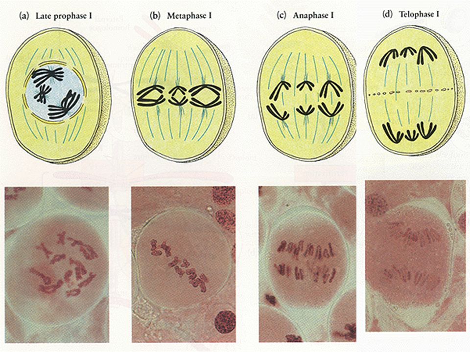
Prophase I: Synapsis Homologous pairs of chromosomes undergo synapsis and form tetrads with their homologous partners (4 chromosomes) Crossover: Genetic material is exchanged between the homologous chromosomes. Parts of maternal chromosomes may be exchanged with paternal ones Genetic recombination produces gametes unlike either parent

62
Exchange of Genetic Material
Genes are exchanged between homologous chromosomes

63
Meiosis I Metaphase I: Tetrads line up at the spindle equator
Anaphase I: Homologous chromosomes (still composed of joined sister chromatids) are distributed to opposite ends of the cell Telophase I: The nuclear membranes re-form around the chromosomal masses Cytokinesis: The cytoplasm is split in two.
 are distributed to opposite ends of the cell. Telophase I: The nuclear membranes re-form around the chromosomal masses. Cytokinesis: The cytoplasm is split in two.")
65
Results of Meiosis I At the end of meiosis I each daughter cell has:
Two copies of either a maternal or paternal chromosome (Compare to mitosis where you get one copy of maternal and one copy of paternal) A 2n amount of DNA and haploid number of chromosomes
 A 2n amount of DNA and haploid number of chromosomes.")
66
Meiosis II Mirrors mitosis except that chromosomes are not replicated before it begins Meiosis II accomplishes two tasks: It reduces the chromosome number by half (2n to n) Each of the daughter cells produced by meiosis I divides during meiosis II and the net result is 4 genetically unique haploid cells or gametes.
 Each of the daughter cells produced by meiosis I divides during meiosis II and the net result is 4 genetically unique haploid cells or gametes.")
68
Meiosis

69
Spermatogenesis Spermatogenic stem cells of the seminiferous tubules give rise to sperm in a series of events Mitosis of spermatogonia, forming spermatocytes Meiosis forms spermatids from spermatocytes Spermiogenesis: spermatids form sperm

70
Spermatogenesis

71
Spermiogenesis: Spermatids to Sperm

72
Sperm Sperm have three major regions
Head :contains DNA and has a helmet-like acrosome containing hydrolytic enzymes that allow the sperm to penetrate and enter the egg Midpiece: contains mitochondria spiraled around the tail filaments Tail :a typical flagellum produced by a centriole

73
Sperm

74
Oogenesis: Before birth
During fetal development, oogonia (stem cells) divide by mitosis to make primary oocytes Primary oocytes begin meiosis and stop in prophase I until puberty Primordial follicles: Support cells that surround the oocyte in the ovary 2 million present at birth 400,000 remain at puberty
 divide by mitosis to make primary oocytes. Primary oocytes begin meiosis and stop in prophase I until puberty. Primordial follicles: Support cells that surround the oocyte in the ovary. 2 million present at birth. 400,000 remain at puberty.")
75
Oogenesis: After Puberty
Each month, hormones cause several follicles to develop, which triggers the primary oocyte to resume meiosis I Polar bodies: When the cell divides, all the cytoplasm and organelles stay with one of the new cells, the other cell is just DNA, and is called a polar body and is discarded Secondary oocyte: The stage at which ovulation occurs.

76
Oogenesis: After Puberty
The secondary oocyte begins meiosis II, but stops in metaphase II The secondary oocyte is ovulated Meiosis II is completed only if it is fertilized.

77
Oogenesis

78
Life History of Oogonia
As a fetus, oogonia divide to produce millions by mitosis but most degenerate (atresia) Some develop into primary oocytes & stop in prophase stage of meiosis I 200,000 to 2 million present at birth 40,000 remain at puberty but only 400 mature during a woman’s life Each month, hormones cause meiosis I to resume in several follicles so that meiosis II is reached by ovulation Penetration by the sperm causes the final stages of meiosis to occur
 Some develop into primary oocytes & stop in prophase stage of meiosis I. 200,000 to 2 million present at birth. 40,000 remain at puberty but only 400 mature during a woman’s life. Each month, hormones cause meiosis I to resume in several follicles so that meiosis II is reached by ovulation. Penetration by the sperm causes the final stages of meiosis to occur.")
79
The End The End

Similar presentations


 Gametogenesis Process of gamete formation with the reduction by half.>")


