Download presentation
Presentation is loading. Please wait.

2
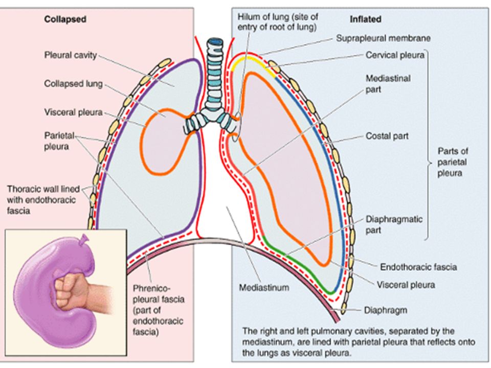
The pleura is divided into two major types, based on location: 1. Parietal pleura 2. Visceral pleura Each pleural cavity is the potential space enclosed between the visceral and parietal pleurae.

4
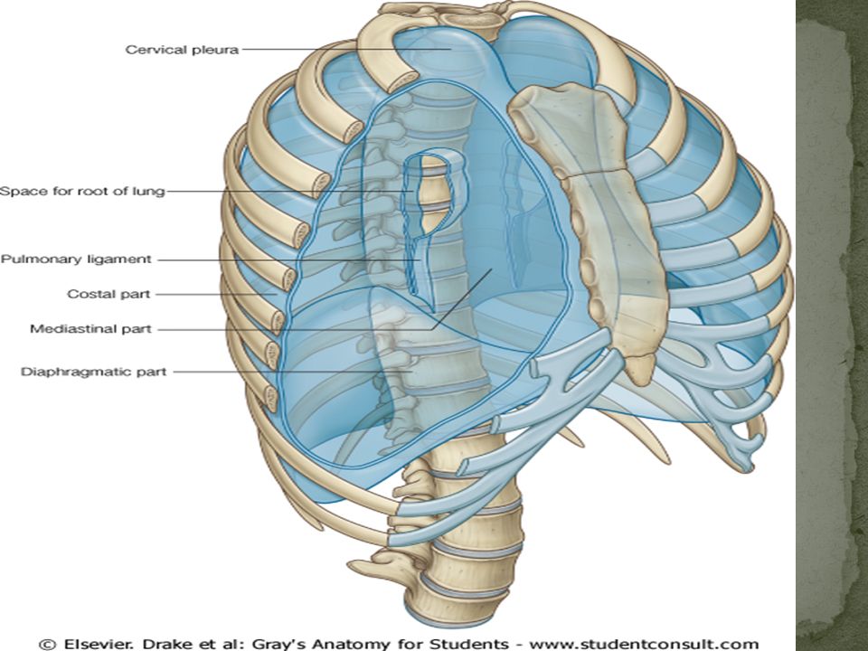
1. Costal part 2. Diaphragmatic part 3. Mediastinal part 4. Cervical part

5
Costal pleura- Lining internal surface of thoracic wall Mediastinal pleura- Covering sides of mediastinum Diaphragmatic pleura- Covering superior surface of dome of each hemidiaphragm Cervical pleura- A dome of pleura extending superiorly into superior thoracic aperture

7
Covers the lungs Cannot be dissected from lung Insensitive to pain

8
The parietal pleura is sensitive to pain, temperature, touch, and pressure and is supplied as follows: The costal pleura is segmentally supplied by the intercostal nerves. The mediastinal pleura is supplied by the phrenic nerve. The diaphragmatic pleura is supplied over the domes by the phrenic nerve and around the periphery intercostal nerves.

9
The visceral pleura covering the lungs is sensitive to stretch but is insensitive to common sensations such as pain and touch. It receives an autonomic nerve supply from the pulmonary plexus.

10
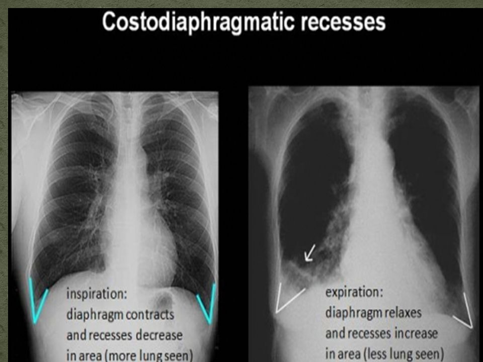
1. Costomediastinal recesses 2. Costodiaphragmatic recesses

11
Costomediastinal recesses Costodiaphragmatic recesses

13
MCL MAL Vertebral Lungs : 6 th rib 8 th rib 10 th vert Pleura : 8 th rib 10 th rib 12 th vert

14
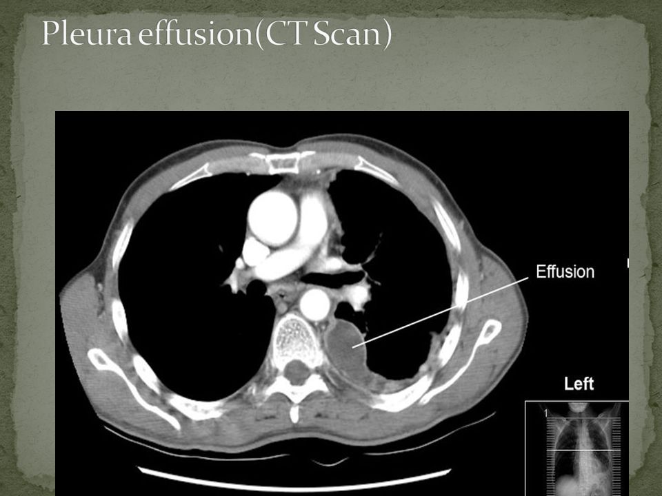
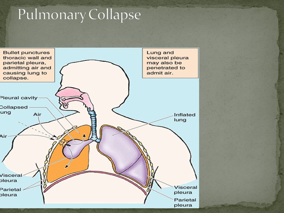
Excess fluid that accumulates in pleural cavity Can impair breathing by limiting the expansion of lungs during inhalation Types Serous fluid (hydrothorax) Blood (hemothorax) Chyle (chylothorax) Pus (pyothorax or empyema)
 Blood (hemothorax) Chyle (chylothorax) Pus (pyothorax or empyema)")
16
At least 300 ml of fluid must be present before upright chest films can pick up signs of pleural effusion

17
Lateral decubitus position more sensitive and can pick up as little as 50 ml of fluid

20
To obtain a sample of pleural fluid or to remove blood or pus or air To avoid damage to intercostal nerve and vessels, needle is inserted superior to rib, high enough to avoid collateral branches It is performed at Mid-Axillary Line, one or two intercostal spaces below the fluid level but not below the ninth intercostal space. The ideal site is eighth, or ninth intercostal space, and this site avoids possible accidental puncture of the lung, liver, spleen, and diaphragm.

24
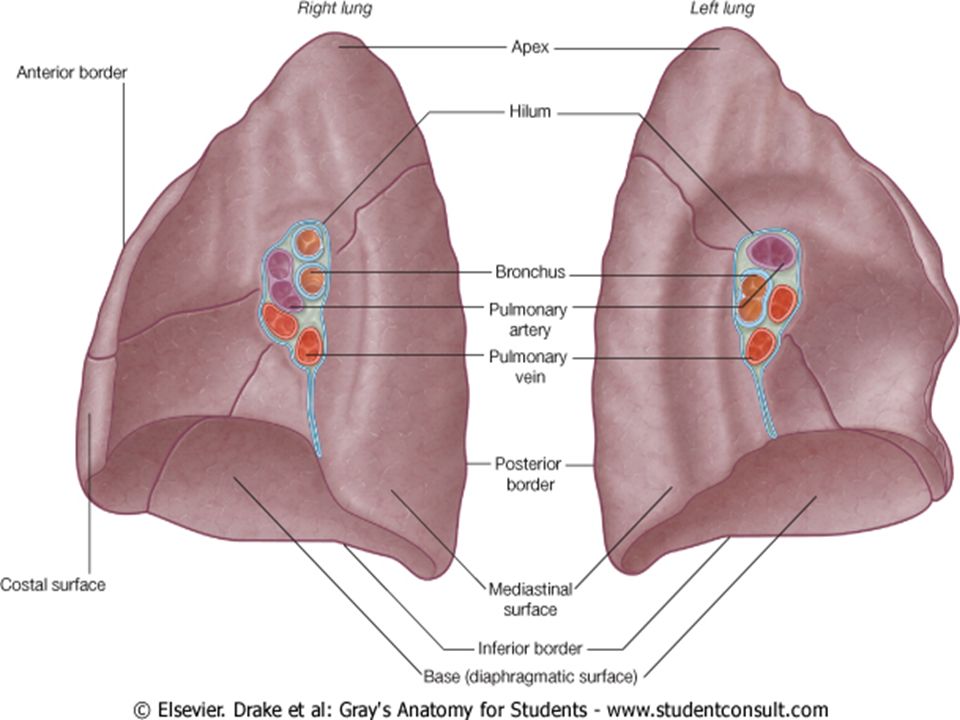
The right lung is normally a little larger than the left lung because the middle mediastinum, containing the heart, bulges more to the left than to the right. Each lung has a half-cone shape, with a base, apex, two surfaces and three borders. 1. Base 2. Apex 3. The two surfaces: Costal surface, mediastinal surface 4. Three borders: Inferior border, Anterior and Posterior borders

26
1.Pulmonary artery 2. Two pulmonary veins 3. Main bronchus 4. Bronchial vessels 5. Nerves and lymphatics.

27
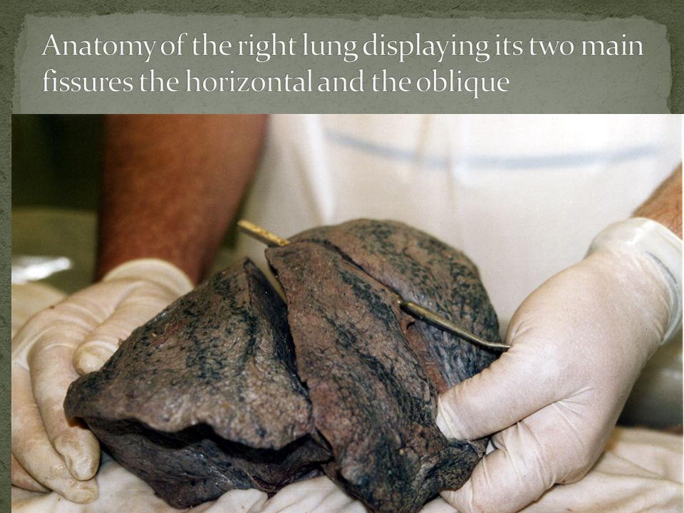
The right lung has three lobes and two fissures. Fissures 1. Oblique fissure 2. Horizontal fissure

29
1. Heart 2.Inferior vena cava 3.Superior vena cava 4.Azygos vein 5.Esophagus

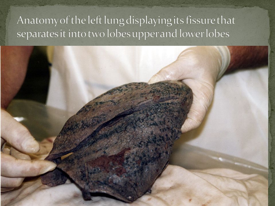
30
The left lung is smaller than the right lung and has two lobes separated by an oblique fissure. On the anterior surface of the lower part of the superior lobe a tongue-like extension (the lingula of left lung) projects over the heart bulge.
 projects over the heart bulge..")
32
1.Heart 2.Aortic arch 3.Thoracic aorta 4. Esophagus

34
Trachea Bronchi Right and left [primary] Lobar [secondary] [3 or 2] Segmental [Tertiary] [10] Bronchiole Terminal Respiratory Alveoli Alveolar duct Alveolar Sac Alveoli
![Trachea Bronchi Right and left [primary] Lobar [secondary] [3 or 2] Segmental [Tertiary] [10] Bronchiole Terminal Respiratory Alveoli Alveolar duct Alveolar Sac Alveoli](http://images.slideplayer.com/26/8294579/slides/slide_34.jpg "Trachea Bronchi Right and left [primary] Lobar [secondary] [3 or 2] Segmental [Tertiary] [10] Bronchiole Terminal Respiratory Alveoli Alveolar duct Alveolar Sac Alveoli")
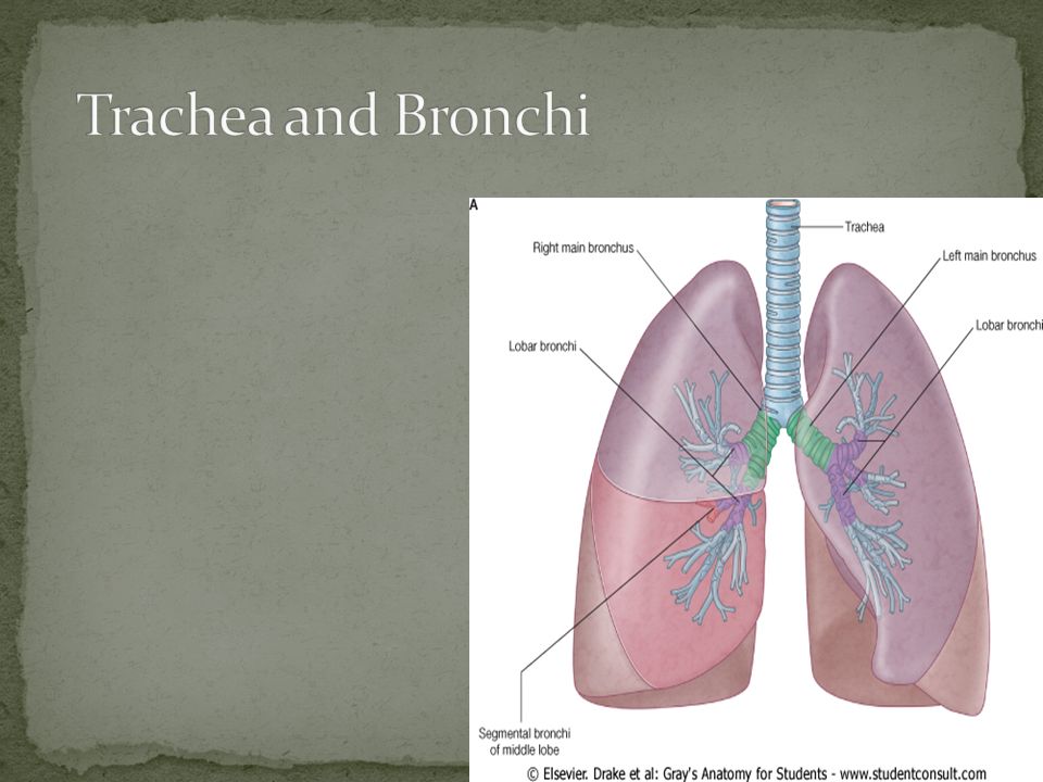
35
Trachea bifurcates → two main stem bronchi, right and left Carina- keel-like ridge between two openings of main stem bronchi Main stem bronchus divides into lobar bronchi 3 lobar bronchi on right: upper, middle, and lower 2 lobar bronchi on left: upper and lower Each lobar bronchus branches into segmental bronchi that supply a bronchopulmonary segment

37
If food, liquids, or foreign bodies are aspirated, they often will lodge in the right mainstem bronchus. Because right bronchus is wider and shorter and runs more vertically than left bronchus Encountered by dentists Aspiration of piece of tooth, filling material, or a small instrument. If the endotracheal tube used for intubation is inserted too far, it usually lodges in the right mainstem bronchus. This allows ventilation of the right lung, but leaves the left lung useless.endotracheal tubeintubation

38
A bronchopulmon ary segment is the area of lung supplied by a segmental bronchus and its accompanying pulmonary artery branch.

39
A bronchopulmonary segment Is a pyramidally shaped section of lung with its base covered by visceral pleura Is separated from adjacent segments by connective tissue septa Is aerated by segmental bronchus Has its own segmental bronchus and segmental branch of pulmonary artery and segmental branch of bronchial artery but not pulmonary vein Pulmonary veins are intersegmental

40
THERE ARE 10 BRONCHOPULMONARY SEGMENTS ON EACH SIDE

41
Has its own Bronchus Has its own Pulmonary artery (Blue) Drains to multiple pulmonary veins (Red) between segments So, each segment has its own bronchus and artery but not its own vein
 Drains to multiple pulmonary veins (Red) between segments So, each segment has its own bronchus and artery but not its own vein")
42
2 sets of Blood Supply 1.Pulmonary Vessels: for Gas Exchange 2. Bronchial Vessels: for blood supply to lung substance like any other organ

43
Pulmonary artery Carries unoxygenated blood from heart to lungs Each artery gives lobar and segmental arteries Pulmonary veins Intrasegmental veins drain to intersegmental veins in pulmonary septa, which drain to two pulmonary veins for each lung Carry oxygenated blood from lungs to heart

45
Bronchial arteries Basically supply lung substance From thoracic aorta Carry oxygenated blood to tissue of lungs, traveling along posterior surface of bronchi Left bronchial arteries- arise from thoracic aorta Right bronchial artery- arise from 3 rd posterior intercostal A.

46
Drain to azygos and accessory hemiazygos veins

47
Via pulmonary plexuses Located anterior and posterior to lung roots Plexus contains both sympathetic and parasympathetic fibers [2 types of autonomic fibers]
![Via pulmonary plexuses Located anterior and posterior to lung roots Plexus contains both sympathetic and parasympathetic fibers [2 types of autonomic fibers]](http://images.slideplayer.com/26/8294579/slides/slide_47.jpg "Via pulmonary plexuses Located anterior and posterior to lung roots Plexus contains both sympathetic and parasympathetic fibers [2 types of autonomic fibers]")
48
Innervate smooth muscle of bronchial tree, pulmonary vessels, and glands of bronchial tree Bronchodilators, vasoconstrictors, and inhibit glandular secretion Parasympathetic fibers Preganglionic parasympathetic fibers from vagus nerve Postganglionic parasympathetic nerves Innervate smooth muscle of bronchial tree, pulmonary vessels, and glands of bronchial tree Bronchoconstrictors, vasodilators, and secretomotor to glands

49
Lymph from lungs drains to Pulmonary lymph nodes (along lobar bronchi) Bronchopulmonary lymph nodes (along main stem bronchi) Superior and inferior tracheobronchial lymph nodes (superior and inferior to bifurcation of trachea) Right and left bronchomediastinal trunks Thoracic duct [left side] and Right lymphatic duct [right side]
![Lymph from lungs drains to Pulmonary lymph nodes (along lobar bronchi) Bronchopulmonary lymph nodes (along main stem bronchi) Superior and inferior tracheobronchial lymph nodes (superior and inferior to bifurcation of trachea) Right and left bronchomediastinal trunks Thoracic duct [left side] and Right lymphatic duct [right side]](http://images.slideplayer.com/26/8294579/slides/slide_49.jpg "Lymph from lungs drains to Pulmonary lymph nodes (along lobar bronchi) Bronchopulmonary lymph nodes (along main stem bronchi) Superior and inferior tracheobronchial lymph nodes (superior and inferior to bifurcation of trachea) Right and left bronchomediastinal trunks Thoracic duct [left side] and Right lymphatic duct [right side]")
Similar presentations


 Mrs. Benish Islam Coordinator Lecturer Surgical (IPMS) KMU.>")


 Pleura Parietal –Costal –Mediastinal –Diaphragmatic –Cupola.>")
