Download presentation
Presentation is loading. Please wait.

1
بسم الله الرحمن الرحيم

2
PITUITARY GLAND AND THYROID GLAND

3
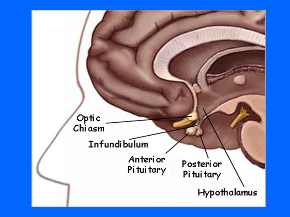
HYPOPHYSIS CEREBRI ( PITUITARY GLAND )
")
6
PITUITARY GLAND

8
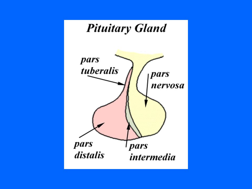
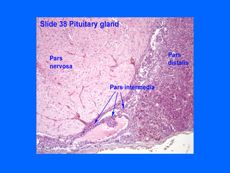
COMPONENTS (A)ADENOHYPOPHYSIS CEREBRI: 1- Pars Distalis (pars anterior) 2- Pars Tuberalis 3- Pars Intermedia (B) NEUROHYPOPHYSIS CEREBRI: 1- Median eminence 2- Infundibulum: Neural (Infundibular) Stalk 3- Pars Nervosa
ADENOHYPOPHYSIS CEREBRI: 1- Pars Distalis (pars anterior) 2- Pars Tuberalis 3- Pars Intermedia (B) NEUROHYPOPHYSIS CEREBRI: 1- Median eminence 2- Infundibulum: Neural (Infundibular) Stalk 3- Pars Nervosa")
9
DEVELOPMENT

10
(A)ADENOHYPOPHYSIS: Arises as outpocketing (evagination) of ectoderm from the roof of the primitive mouth of embryo → Rathke’s Pouch → Separates from oral cavity. (B) NEUROHYPOPHYSIS: Arise as a down growth from the floor of diencephalon, without detaching from the brain.
 NEUROHYPOPHYSIS: Arise as a down growth from the floor of diencephalon, without detaching from the brain..")
11
PITUITARY GLAND

13
NEUROHYPOPHYSIS xxxx XXXXX xxxxxxxx

15
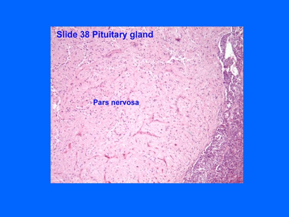
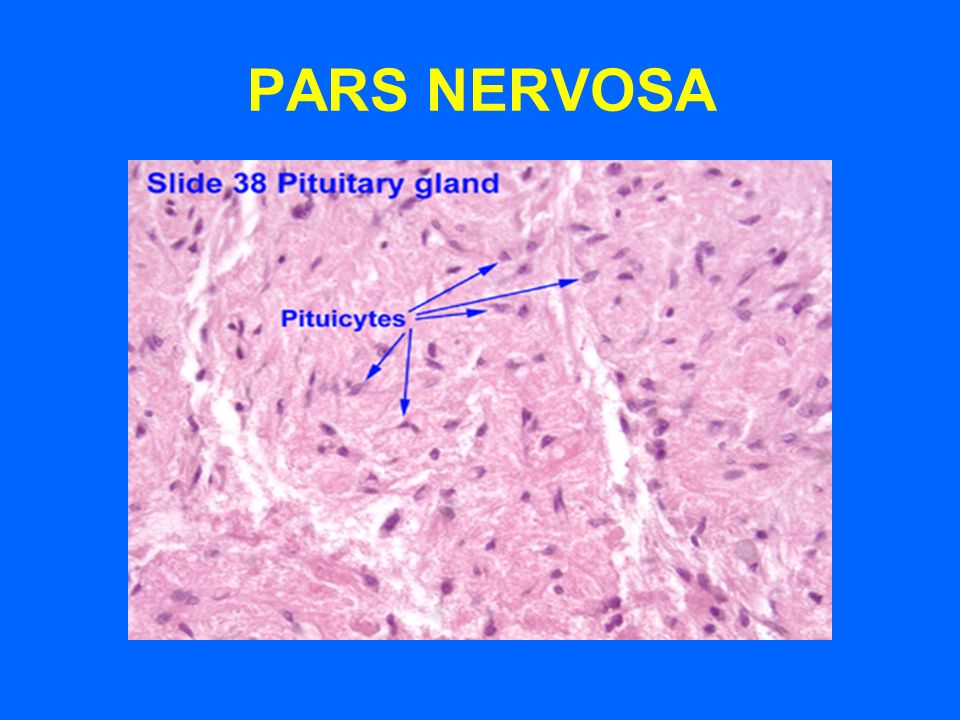
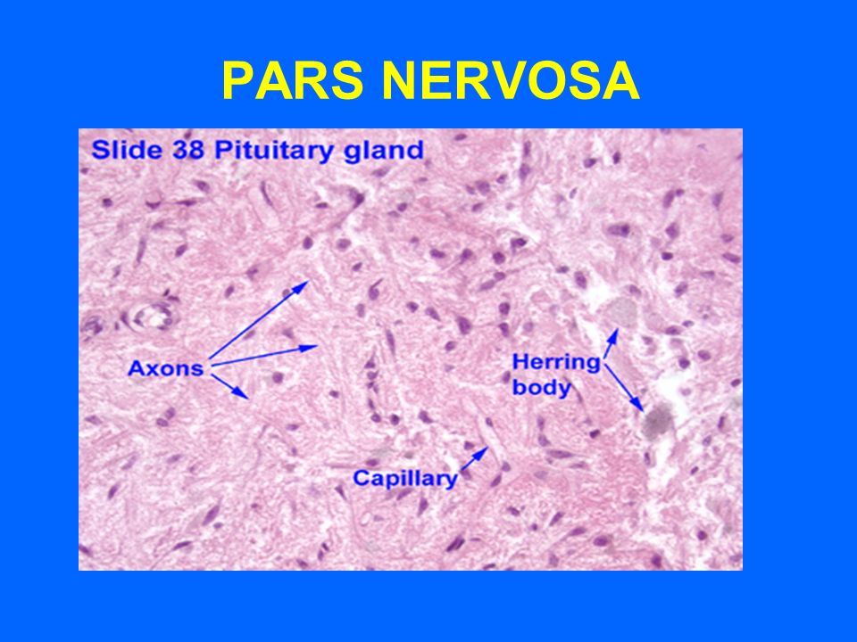
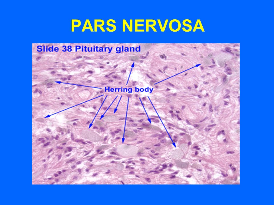
PARS NERVOSA

20
NEUROHYPOPHYSIS (A) PARS NERVOSA CONTENTS: 1- Unmyelinated axons of secretory neurons situated in supraoptic & paraventricular nuclei (i.e. Axons of hypothalamhypophyseal tract). 2- Herring’s Bodies: contain neurosecretory granules. 3- Fenestrated blood capillaries. 4- Pituicytes: branched glial-like cells. N.B. No secretory cells in pars nervosa.
. 2- Herring’s Bodies: contain neurosecretory granules. 3- Fenestrated blood capillaries. 4- Pituicytes: branched glial-like cells. N.B. No secretory cells in pars nervosa..")
21
HERRING BODIES - Are blue-black stained distentions of the axons in p. nervosa. - Representing accumulation of neurosecretory granules at axon termini and along the length of the axons in p. nervosa.

22
PITUICYTES Are glial-like cells in p. nervosa. Structure: Have numerous cytoplasmic processes with gap junctions. Functions: 1- Support the axons of the p. nervosa. 2- May have a trophic function.

23
Function of p. nervosa: Storage & release of: 1- Vasopressin (ADH) 2- Oxytocin
 2- Oxytocin")
24
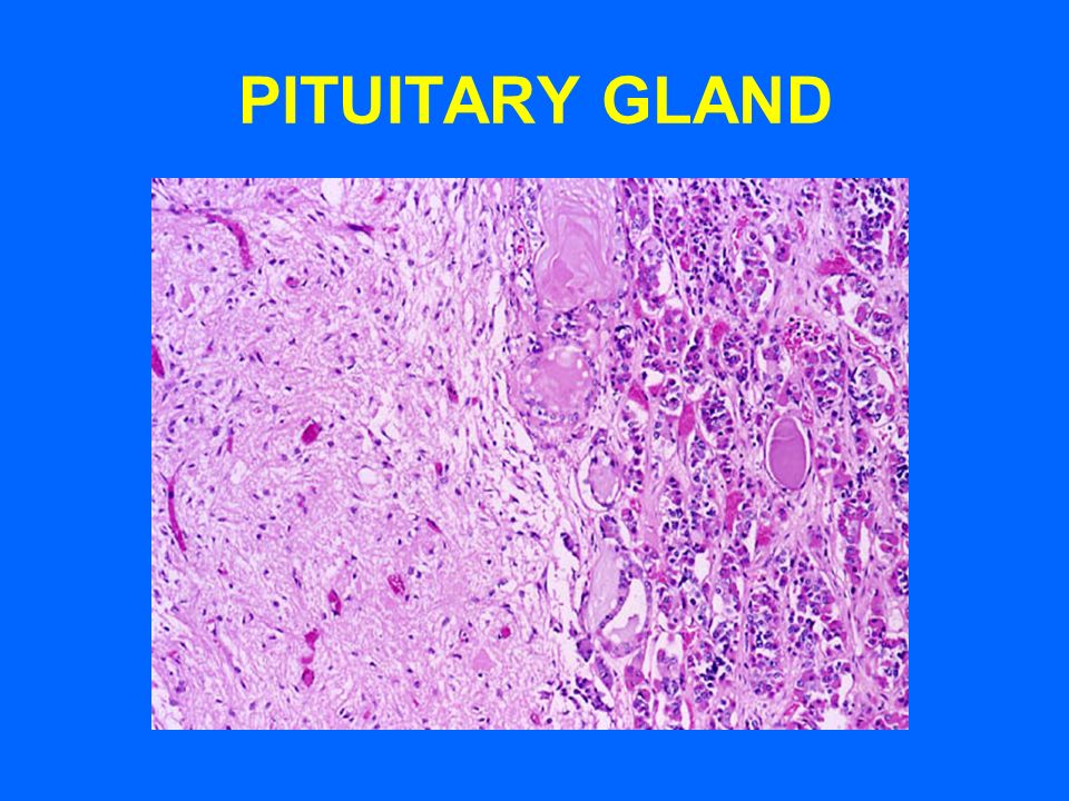
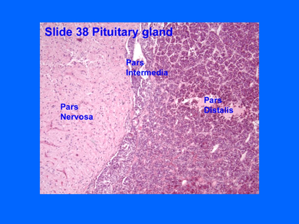
P. DISTALIS, P. INTERMEDIA & P. NERVOSA

31
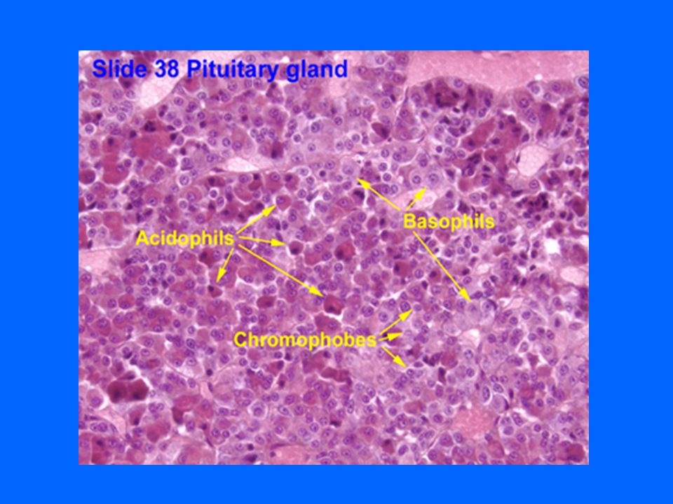
PARS DISTALIS Blue arrow: acidophils Red arrow: basophils Yellow arrow: chromophobes

32
Blue arrow: acidophils Red arrow: basophils Yellow arrow: chromophobes

33
PARS DISTALIS

35
GH CELLS

36
PROLACTIN CELLS

37
ACTH CELLS

39
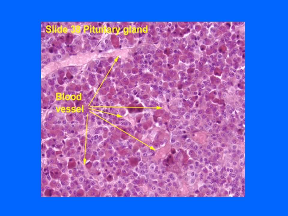
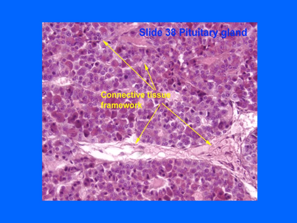
PARS DISTALIS Stroma: 1- Fibrous capsule. 2- Scant C.T. located mainly around aa &vv. (hypophyseal arteries & portal veins). 3- Reticular fibers: around the cords of cells & “ the sinusoidal capillaries.
. 3- Reticular fibers: around the cords of cells & the sinusoidal capillaries..")
40
PARS DISTALIS Parenchyma: 1- Cords of parenchymal cells. 2- Sinusoidal capillaries: With fenestrated endothelium.

41
PARS DISTALIS Types of parenchymal cells (1)Chromophils: a- Acidophils: 1- Somatotrophs (Somatotropic cells). 2- Mammotrophs (Mammotropic cells). b- Basophils: 1- Thyrotrophs (Thyrotropic cells)(TSH) 2- Gonadotrophs (Gonadotropic cells) (FSH, LH) 3- Corticotrophs (ACTH, Lipotropic H) 4- Melanotropes ?????????????
. b- Basophils: 1- Thyrotrophs (Thyrotropic cells)(TSH) 2- Gonadotrophs (Gonadotropic cells) (FSH, LH) 3- Corticotrophs (ACTH, Lipotropic H) 4- Melanotropes .")
42
Types of parenchymal cells (2) Chromophobes: may represent: 1- stem cells. 2- degranulated chromophils. 3- degenerated cells. (3) Folliculostellate cells: Structure: have long processes with gap j. Function: Not clear ( are non-secretory) ???(may be:1-supporting, 2-phagocytic).
 Folliculostellate cells: Structure: have long processes with gap j. Function: Not clear ( are non-secretory) (may be:1-supporting, 2-phagocytic)..")
43
PARS TUBERALIS Structure: Cuboidal basophilic cells. Function: Possibly contain FSH & LH.

44
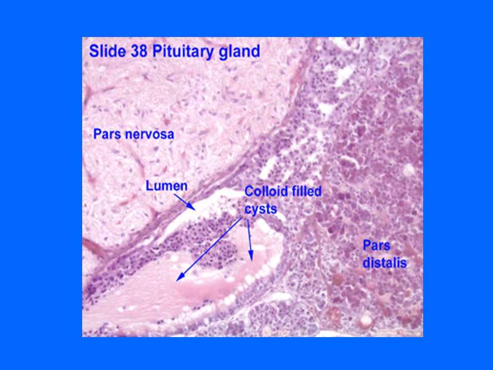
PARS INTERMEDIA (ZONA INTERMEDIA) L/M: 1-Numerous colloid-containing cysts (Rathke’s cysts): Are lined with cuboidal cells. Are remnants of Rathke’s pouch. 2-Cords of weakly basophilic cells. 3- Network of blood capillaries. Function: Secrete proopiomelanocortin→ Gives: α-MSH, Corticotropin, ß-lipotropin & ß-endorphin. N.B. α-MSH in human stimulates prolactin release.

45
BLOOD SUPPLY

46
------ Xxx

47
BLOOD SUPPLY (1)Sup. Hypoph. Arteries (Rt & Lt): To median eminence & Neural stalk → 1ry capillary plexus of fenestrated capillaries → Hypophyseal portal Veins (or venules) → 2ry capillary plexus of capillaries in adenohyp [ Hypophyseal Portal System ] [ “ “ Circulation ] It carries neurohormones from median eminence to adenohypophysis. (2) Inf. Hypoph. Arteries (Rt & Lt): Mainly to pars nervosa, They are Not participating in hypophyseal portal circulation. !!!!!!!!!!!!!!!!!!!!!!!!!!!!!!!!!!!!!
: To median eminence & Neural stalk → 1ry capillary plexus of fenestrated capillaries → Hypophyseal portal Veins (or venules) → 2ry capillary plexus of capillaries in adenohyp [ Hypophyseal Portal System ] [ Circulation ] It carries neurohormones from median eminence to adenohypophysis. (2) Inf. Hypoph. Arteries (Rt & Lt): Mainly to pars nervosa, They are Not participating in hypophyseal portal circulation. !!!!!!!!!!!!!!!!!!!!!!!!!!!!!!!!!!!!!.")
48
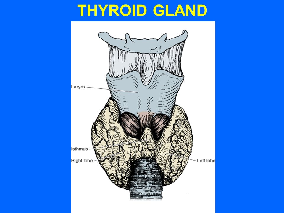
THYROID GLAND

59
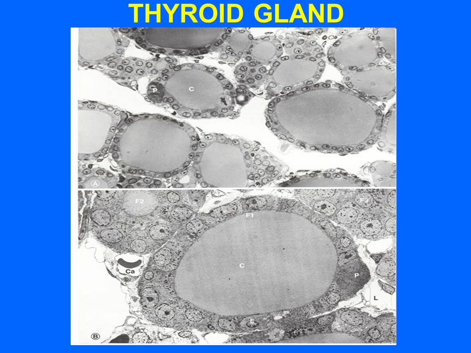
THYROID GLAND (E/M)
")
60
PARAFOLLICULAR CELL

61
STROMA OF THYROID GLAND 1- Capsule: dense irregular collagenous C.T. 2- Septa (Interlobular septa) : “ “ “ “. 3- Reticular fibers: Thin C.T., composed mostly of reticular fibers with rich capillary plexus surrounds each thyroid follicle.
 : . 3- Reticular fibers: Thin C.T., composed mostly of reticular fibers with rich capillary plexus surrounds each thyroid follicle..")
62
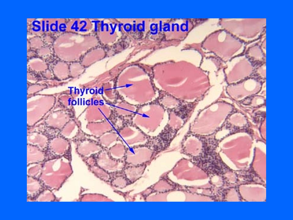
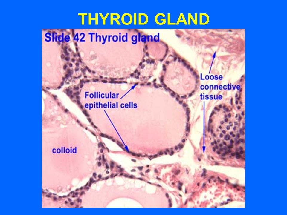
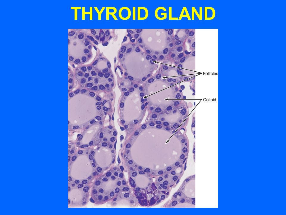
PARENCHYMA OF THYROID GLAND THYROID FOLLICLES: Are the structural and functional units of the thyroid gland.

63
THYROID FOLLICLES L/M: 1- Simple cuboidal epithelium: a- Follicular cells. b- Parafollicular cells. 2- Colloid: central colloid-filled lumen. N.B. Each follicle is surrounded by thin basal lamina.

64
FOLLICULAR (PRINCIPAL) CELLS L/M: Simple cuboidal cells May be squamous or low columnar. Round nucleus with prominent nucleoli. Basophilic cytoplasm.

65
FOLLICULAR (PRINCIPAL) CELLS E/M: - Mitochondria. - RER - Supranuclear Golgi Complex. - Numerous apically-located lysosomes. -Numerous dispersed small vesicles: contain newly formed thyroglobulin. - Numerous apical short microvilli.

66
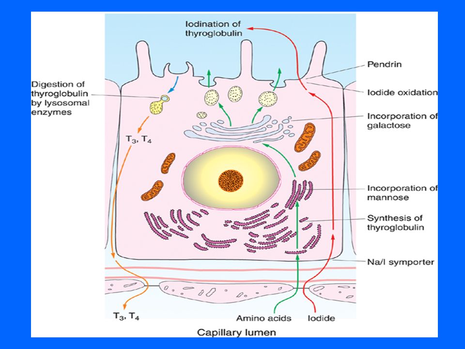
FOLLICULAR (PRINCIPAL) CELLS Function: Synthesis of thyroid hormones (T4 & T3). 1- Synthesis of thyroglobulin (glycoprotein). 2- Trapping of circulating iodide. 3- Oxidation of iodide into activated iodide (by lysosomal peroxidase). 4- Iodination of tyrosine of thyroglobulin in the colloid at the colloid-follicular cell interface. 5- Endocytosis of hormone-bound thyroglobulin→ Cleavage of T4 & T3 by lysosomal proteases.
. 2- Trapping of circulating iodide. 3- Oxidation of iodide into activated iodide (by lysosomal peroxidase). 4- Iodination of tyrosine of thyroglobulin in the colloid at the colloid-follicular cell interface. 5- Endocytosis of hormone-bound thyroglobulin→ Cleavage of T4 & T3 by lysosomal proteases..")
67
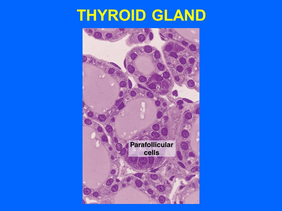
PARAFOLLICULAR CELLS (CLEAR CELLS) (C CELLS) L/M: -Pale stained cells. -Are found singly or in clusters in between the follicular cells. -Do not reach the lumen of the follicle. -Are larger than follicular cells (2-3 times). -Only 0.1% of the epithelial cells. -Have round nucleus
. -Only 0.1% of the epithelial cells. -Have round nucleus.")
68
PARAFOLLICULAR CELLS (CLEAR CELLS) (C CELLS) E/M: -Mitochondria. -RER (moderate). -Well-developed Golgi. -Small dense secretory granules located mainly in the basal cytoplasm. Function: Secrete calcitonin.
. -Well-developed Golgi. -Small dense secretory granules located mainly in the basal cytoplasm. Function: Secrete calcitonin..")
Similar presentations


 Bruce Durham Sam Michaels Tandy Sutton Jonathan Bylund.>")


 Rathke’s pouch –Roof of the embryonic mouth –Glandular tissue containing.>")
 glands are also named ductless glands, since they lack excretory ducts. Instead,>")


