Download presentation
Presentation is loading. Please wait.

1
The Blood

2
Blood Functions Transport oxygen, and nutrients to body tissues Transport oxygen, and nutrients to body tissues Remove CO2 and metabolic wastes from body tissues Remove CO2 and metabolic wastes from body tissues Regulates bodies Ph levels and ion concentrations Regulates bodies Ph levels and ion concentrations Maintenance of body temp (absorbs and distributes heat) Maintenance of body temp (absorbs and distributes heat) Restriction of fluid loss at injury sites (initiates the clotting process) Restriction of fluid loss at injury sites (initiates the clotting process) Defense against toxins and pathogens Defense against toxins and pathogens White Blood Cells (WBC) fight infections Antibodies fight specific pathogens or organisms
 Maintenance of body temp (absorbs and distributes heat) Restriction of fluid loss at injury sites (initiates the clotting process) Restriction of fluid loss at injury sites (initiates the clotting process) Defense against toxins and pathogens Defense against toxins and pathogens White Blood Cells (WBC) fight infections Antibodies fight specific pathogens or organisms")
3
Blood Composition Plasma: Liquid part of blood Plasma: Liquid part of blood Slightly thicker than water Slightly thicker than water Formed Elements: Formed Elements: Red Blood Cells Red Blood Cells White Blood Cells White Blood Cells Platelets Platelets

4
Formed Elements (RBC/Erythrocytes ) Biconcave disks (see pic.) Contain Hemoglobin Heme: Contains iron and binds O2 Globin: Binds CO2 Most abundant of the formed elements 4.8 million/mm3 of blood in females 5.4 million/mm3 of blood in males
 Biconcave disks (see pic.) Contain Hemoglobin Heme: Contains iron and binds O2 Globin: Binds CO2 Most abundant of the formed elements 4.8 million/mm3 of blood in females 5.4 million/mm3 of blood in males")
5
Formed Elements: RBC Live 120 days Live 120 days Wear and tear of transport Wear and tear of transport Erythropoeisis is the formation of RBC’s (within RED Bone Marrow) Erythropoeisis is the formation of RBC’s (within RED Bone Marrow) Broken down in spleen and liver Broken down in spleen and liver If destruction and creation aren’t equal = Anemia If destruction and creation aren’t equal = Anemia
 Erythropoeisis is the formation of RBC’s (within RED Bone Marrow) Broken down in spleen and liver Broken down in spleen and liver If destruction and creation aren’t equal = Anemia If destruction and creation aren’t equal = Anemia")
6
Iron Deficient Anemia Anemic BloodNormal Blood

7
Formed Elements: RBC Hemoglobin: Hemoglobin: Broken into heme and bilirubin (yellow pigment) Broken into heme and bilirubin (yellow pigment) Jaundice: Can’t metabolize and excrete bilirubin efficiently. Jaundice: Can’t metabolize and excrete bilirubin efficiently. Get a build up of yellow pigment within the bodies tissues (skin and eyes primarily) Get a build up of yellow pigment within the bodies tissues (skin and eyes primarily)
 Get a build up of yellow pigment within the bodies tissues (skin and eyes primarily).")
8
Jaundice Icterus : Jaundice of Sclera

9
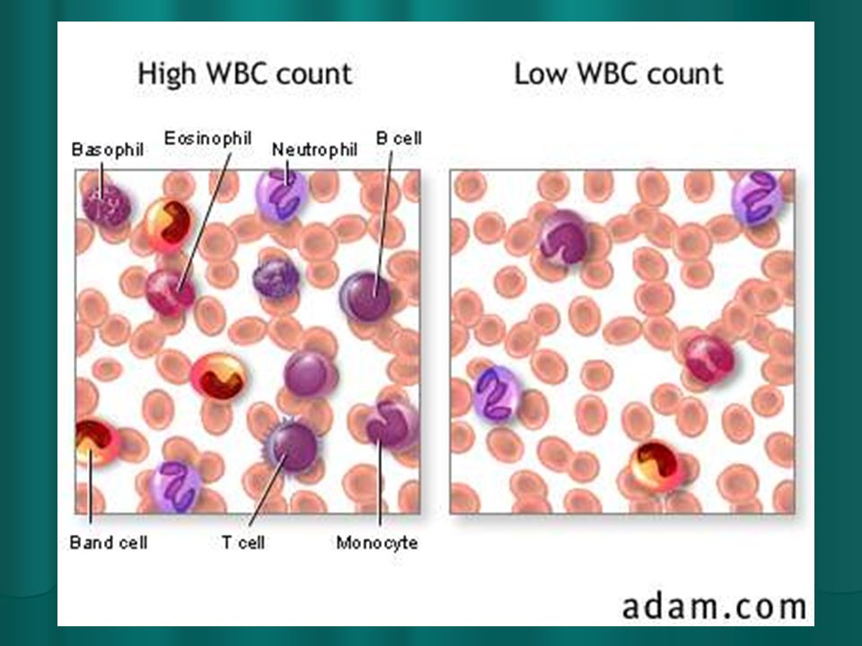
Formed Elements: White Blood Cells (Leukocytes) Six different types of leukocytes Six different types of leukocytes All function to protect the body against infection (immune system) All function to protect the body against infection (immune system) Produced in the White Bone Marrow Produced in the White Bone Marrow
 Six different types of leukocytes Six different types of leukocytes All function to protect the body against infection (immune system) All function to protect the body against infection (immune system) Produced in the White Bone Marrow Produced in the White Bone Marrow")
10
Formed Elements: WBC Leukocytes are destroyed and replaced daily Leukocytes are destroyed and replaced daily 5-10 thousand/mm3 of blood (normal) 5-10 thousand/mm3 of blood (normal) >10,000 = Infection >10,000 = Infection Leukemia is an excess in WBC Leukemia is an excess in WBC <5,000 = Leucopenia (can’t fight infections) <5,000 = Leucopenia (can’t fight infections)
 5-10 thousand/mm3 of blood (normal) >10,000 = Infection >10,000 = Infection Leukemia is an excess in WBC Leukemia is an excess in WBC <5,000 = Leucopenia (can’t fight infections) <5,000 = Leucopenia (can’t fight infections)")
12
Formed Elements: Platelets (thrombocytes) Membrane bound particles that house enzymes for clotting. Membrane bound particles that house enzymes for clotting.

13
Formed Elements: Platelets Number: 250-400,000/mm3 of blood Number: 250-400,000/mm3 of blood Production: Red bone marrow Production: Red bone marrow Structure: Disc shaped with no nucleus Structure: Disc shaped with no nucleus Function: Blood clotting Function: Blood clotting Lifespan: 5-9 days Lifespan: 5-9 days

14
Formed Elements

15
Hematocrit The percentage of a blood sample that is made up of formed elements The percentage of a blood sample that is made up of formed elements 46% in men 46% in men 42% in women 42% in women Upwards of 55% in athletes who are blood doping Upwards of 55% in athletes who are blood doping

16
Hemostasis/Blood Clotting 3 Phases 3 Phases Vascular Phase: Vascular spasms at the site of injury Vascular Phase: Vascular spasms at the site of injury Lasts for 30 minuets after injury Lasts for 30 minuets after injury Blood vessels constrict (restrict blood flow) Blood vessels constrict (restrict blood flow) Platelets begin releasing local hormones causing endothelial cells at injury site to become sticky Platelets begin releasing local hormones causing endothelial cells at injury site to become sticky Spasm is a reflex triggered by pain receptors at injury site Spasm is a reflex triggered by pain receptors at injury site
 Blood vessels constrict (restrict blood flow) Platelets begin releasing local hormones causing endothelial cells at injury site to become sticky Platelets begin releasing local hormones causing endothelial cells at injury site to become sticky Spasm is a reflex triggered by pain receptors at injury site Spasm is a reflex triggered by pain receptors at injury site")
17
Hemostasis Platelet Phase: Platelet Phase: Platelets begin to stick to endothelial and collagen fibers exposed by wound Platelets begin to stick to endothelial and collagen fibers exposed by wound More and more platelets arrive and stick together forming a “platelet plug” (not a clot yet) More and more platelets arrive and stick together forming a “platelet plug” (not a clot yet) Begins within 15 seconds of injury Begins within 15 seconds of injury
 More and more platelets arrive and stick together forming a platelet plug (not a clot yet) Begins within 15 seconds of injury Begins within 15 seconds of injury")
19
Hemostasis Coagulation Phase: Coagulation Phase: Begins 30 seconds after injury Begins 30 seconds after injury Fibrinogen within plasma is formed into insoluble fibrin. Fibrinogen within plasma is formed into insoluble fibrin. Fibrin forms into “mesh-like” network that catches passing blood cells and platelets. Fibrin forms into “mesh-like” network that catches passing blood cells and platelets. Blood Clot is formed Blood Clot is formed

20
Blood Clot Formation

22
Clotting Diseases Hemophilia: Hemophilia: Inherited disease Inherited disease Lack of clotting factors (chemicals) Lack of clotting factors (chemicals) Blood cannot clot properly Blood cannot clot properly
 Lack of clotting factors (chemicals) Blood cannot clot properly Blood cannot clot properly")
23
Clotting Diseases Sickle Cell Anemia: Sickle Cell Anemia: Production of abnormal hemoglobin, resulting in “sickle” shaped red blood cells Low oxygen carrying capacity Most predominant in African Americans RBC tangle together and form clots within the body

24
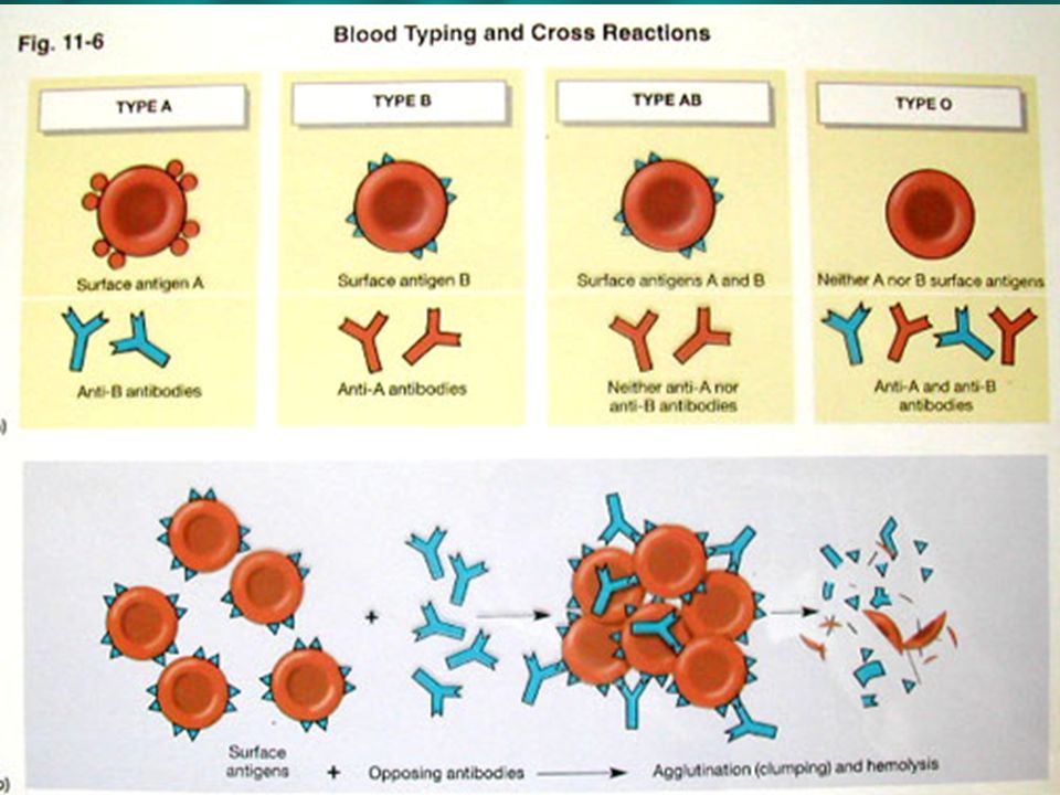
Blood Typing Blood type is determined by ANTIGENS on the surface of RBC’s. Blood type is determined by ANTIGENS on the surface of RBC’s. Blood type refers to antigens that are on your RBC’s Blood type refers to antigens that are on your RBC’s There are over 50 surface antigens There are over 50 surface antigens We predominantly use 3 We predominantly use 3 A B Rh factor Rh factor

25
Blood Typing Surface Antigens Surface Antigens If you have an antigen on your RBC’c you do not have the antibody. If you have an antigen on your RBC’c you do not have the antibody. Ex. Type A: Ex. Type A: Has A antigens Has A antigens Does not have B antigens Does not have B antigens Does not have A antibodies Does not have A antibodies Does have B Antibodies Does have B Antibodies Antigens are on RBC’s, antibodies are in the plasma Antigens are on RBC’s, antibodies are in the plasma

26
Blood Typing If both antibodies and antigens are present agglutination occurs. If both antibodies and antigens are present agglutination occurs. This is the “clumping” of blood when the antibodies and antigens combine. This is the “clumping” of blood when the antibodies and antigens combine. Ex. Type A blood has A antigens, if combined with type B blood or AB blood, agglutination occurs because type B blood has type B antigens. Ex. Type A blood has A antigens, if combined with type B blood or AB blood, agglutination occurs because type B blood has type B antigens.

28
Blood Typing Type A Blood: Type A Blood: Type B Blood: Type B Blood: Type AB Blood: Universal receiver (no antibodies to cause agglutination. Type AB Blood: Universal receiver (no antibodies to cause agglutination. Type O Blood: Universal donor (no antigens for receivers antibodies to agglutinate with) Type O Blood: Universal donor (no antigens for receivers antibodies to agglutinate with)
 Type O Blood: Universal donor (no antigens for receivers antibodies to agglutinate with).")
29
Blood Typing Rh factor: Rh factor: Rh– :no antibodies unless previously exposed to Rh factor through injection, pregnancy, etc… Rh– :no antibodies unless previously exposed to Rh factor through injection, pregnancy, etc… If mom has a second Rh+ baby, second baby’s blood will agglutinate as a result of mom’s Rh antibodies, which were formed when mom was exposed to her first child’s Rh+ blood. If mom has a second Rh+ baby, second baby’s blood will agglutinate as a result of mom’s Rh antibodies, which were formed when mom was exposed to her first child’s Rh+ blood. Cannot cross Rh- and Rh+ Cannot cross Rh- and Rh+

30
Blood Typing Type A: 40% Type A: 40% Type B: 10% Type B: 10% Type AB: 4% Type AB: 4% Type O: 46% Type O: 46% Rh+ : 85% Rh+ : 85% Rh- : 15% Rh- : 15% What are the overall most common and least common blood types??? What are the overall most common and least common blood types???

Similar presentations