Download presentation
Presentation is loading. Please wait.

1
Hook worms

2
1. Ancylostoma duodenale 2. Necator americanus A. duodenale N americanus

3
Morphology Ancylostoma duodenale: females: 10 to 13 mm adult males: 8 to 11 mm) Necator americanus. Female: 9 to 11 mm male: 7 to 9 mm

4
Buccal cavity Ancylostoma duodenale Necator americanus

5
Ancylostoma duodenale

6
Necator americanus

7
Hook worms

8
Hook worm

9
Hook worms Male’s posterior end is expanded to form a copulatory bursa.

10
Ancylostoma duodenale copulatory bursa and spines of male

11
N. americanus Copulatory bursa of

12
Ancylostoma duodenale

14
The Morphological Differences between Two species of Hookworms _____________________________________________________ A. duodenale N. americanus A. duodenale N. americanus______________________________________________________ Size larger smaller Size larger smaller______________________________________________________ Shape single curve, looks like C double curves, looks like S Shape single curve, looks like C double curves, looks like S______________________________________________________ Mouth 2 pairs of ventral teeth 1peir of ventral cutting plates Mouth 2 pairs of ventral teeth 1peir of ventral cutting plates____________________________________________________________ Copulatory circle in shape oval in shape Copulatory circle in shape oval in shape Bursa (a top view) (a top view) Bursa (a top view) (a top view)____________________________________________________________ Copulatory 1pair with separate 1pair of which unite to form Copulatory 1pair with separate 1pair of which unite to form spicule endings a terminal hooklet spicule endings a terminal hooklet______________________________________________________________________________________________________________
 (a top view) Bursa (a top view) (a top view)____________________________________________________________ Copulatory 1pair with separate 1pair of which unite to form Copulatory 1pair with separate 1pair of which unite to form spicule endings a terminal hooklet spicule endings a terminal hooklet______________________________________________________________________________________________________________.")
15
Epidemiology The second most common human helminthic infection (after ascariasis). Worldwide distribution, mostly in areas with moist, warm climate. A. duodenale: in south of Iran (Khozestan) N. americanus (North) Hook worm محلسالمحقق 9/8لاهيجان-روستاها1371رضائيان 6/4تنكابن- شهر و روستا1375رضائيان 19/فريدون كنار-مازندران- شهر و روستا 1378رضويون 2شهريار1379شهابي 25/2ساري-روستاها1380روحاني
 N. americanus (North) Hook worm محلسالمحقق 9/8لاهيجان-روستاها1371رضائيان 6/4تنكابن- شهر و روستا1375رضائيان 19/فريدون كنار-مازندران- شهر و روستا 1378رضويون 2شهريار1379شهابي 25/2ساري-روستاها1380روحاني.")
16
Life cycle Eggs are passed in the stool larvae (rhabditiform) hatch in 1 to 2 days. After 5 to 10 days (and two molts) filariform (third-stage) larvae (infective. After 5 to 10 days (and two molts) filariform (third-stage) larvae (infective. These infective larvae can survive 3 to 4 weeks in favorable environmental conditions. These infective larvae can survive 3 to 4 weeks in favorable environmental conditions. On contact with the human host, the larvae penetrate the skin and are carried through the veins to the heart and then to the lungs. On contact with the human host, the larvae penetrate the skin and are carried through the veins to the heart and then to the lungs. The larvae reach the small intestine, where they reside and mature into adults. The larvae reach the small intestine, where they reside and mature into adults. Adult worms live in the lumen of the small intestine, where they attach to the intestinal wall with resultant blood loss by the host. Most adult worms are eliminated in 1 to 2 years, but longevity records can reach several years. In addition, infection by A. duodenale may probably also occur by the oral and transmammary route. In addition, infection by A. duodenale may probably also occur by the oral and transmammary route.
 filariform (third-stage) larvae (infective. After 5 to 10 days (and two molts) filariform (third-stage) larvae (infective. These infective larvae can survive 3 to 4 weeks in favorable environmental conditions. These infective larvae can survive 3 to 4 weeks in favorable environmental conditions. On contact with the human host, the larvae penetrate the skin and are carried through the veins to the heart and then to the lungs. On contact with the human host, the larvae penetrate the skin and are carried through the veins to the heart and then to the lungs. The larvae reach the small intestine, where they reside and mature into adults. The larvae reach the small intestine, where they reside and mature into adults. Adult worms live in the lumen of the small intestine, where they attach to the intestinal wall with resultant blood loss by the host. Most adult worms are eliminated in 1 to 2 years, but longevity records can reach several years. In addition, infection by A. duodenale may probably also occur by the oral and transmammary route. In addition, infection by A. duodenale may probably also occur by the oral and transmammary route..")
17
Life cycle 1-2 days 7 days 2 weeks

18
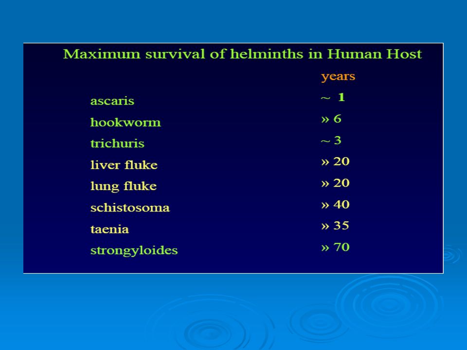
Life Cycle Final host: man Infective stage: Larva 3 or filariform Infection route: by skin Food: blood and tissue fluid Site of inhabitation: small intestine Long lasting infection (up to 18 years)
")
19
Life cycle

20
Clinical Manifestations 1. Larval migration: Dermatitis (ground itch). allergic reaction, petechiae or papule with itching and burning sensation. Scratching leads to secondary infection respiratory symptoms can be observed during pulmonary migration of the larvae (not as sever as Ascaris)
.")
21
Epigastric pain as that of a duodenal ulcer. * Iron deficiency anemia ( microcytic hypochromic anemia) Allotriophagy (or pica) is due to the lack of trace element iron trace element iron Gastrointestinal bleeding 2. Adults worm in small intestine * More sever in A. duodenale
 Allotriophagy (or pica) is due to the lack of trace element iron trace element iron Gastrointestinal bleeding 2. Adults worm in small intestine * More sever in A. duodenale.")
22
Treatment Albendazole: 400 mg/kg (single dose, drug of choice) Mebendazole: 100 mg/kg 2 times/day/ for 3 days Pyrantel pamoate: 10 mg/kg (single dose)
 Mebendazole: 100 mg/kg 2 times/day/ for 3 days Pyrantel pamoate: 10 mg/kg (single dose)")
23
Prevention and control

24
Diagnosis

25
Eggs Morphologically it is not possible to differentiate between A. duodenale and N. americanus.

26
Strongyloides stercoralis

29
Morphology Rhabditiform larvae (non-infective form) It’s a nematode, so it has two larval forms: Filariform larvae (pathogenic form)
 It’s a nematode, so it has two larval forms: Filariform larvae (pathogenic form)")
30
Morphology Size: about 2 mm Size: about 2 mm Has parasitic and free living Has parasitic and free living Adult worm in small intestine Adult worm in small intestine Adult worm can live for up to 30 years in intestine Adult worm can live for up to 30 years in intestine

31
Epidemiology Found worldwide An estimated 50 to 100 million cases An estimated 50 to 100 million cases Favors warmer tropical and subtropical climates Endemic in sub-Saharan Africa, Latin America, southeast Asia, and United States

32
Epidemiology More frequently found in rural areas, institutional settings, and lower socioeconomic groups.

33
Epidemiology Worms can be free-living in the soil or live in a host. Worms can be free-living in the soil or live in a host. The definitive host is humans, but may also affect other primates, cat and dogs The definitive host is humans, but may also affect other primates, cat and dogs

34
Epidemiology in Iran Strongyloides stercoralis محلسالمحقق 5/8لاهيجان-روستاها1371رضائيان 3/10تنكابن- شهر و روستا1375رضائيان 74/فريدون كنار- مازندران- شهر و روستا 1378رضويون 06/31تهران-كودكان عقب مانده ذهني 1379يوسفي 1ساري-روستاها1380روحاني 3/اسلامشهر1381عسگري

35
Life cycle

36
The females live threaded in the epithelium of the small intestine and by parthenogenesis produce eggs, which yield rhabditiform larvae. The rhabditiform larvae can either be passed in the stool (see "Free-living cycle" above), or can cause autoinfection.
, or can cause autoinfection..")
37
Symptoms Commonly asymptomatic Commonly asymptomatic But symptoms may include: But symptoms may include: Gastrointestinal (diarrhea, abdominal pain, malabsorption) Respiratory (coughing, wheezing) Dermatologic (ground itch, rash) Anemia People with weaker immune systems such as elderly people and children are more susceptible.
 Respiratory (coughing, wheezing) Dermatologic (ground itch, rash) Anemia People with weaker immune systems such as elderly people and children are more susceptible.")
38
Strongyloidiasis Strongyloides stercoralis has a direct parasitic life cycle, meaning it can complete its entire life cycle in the definitive host. Strongyloides stercoralis has a direct parasitic life cycle, meaning it can complete its entire life cycle in the definitive host. This causes an autoinfection in the human, because the worm keeps infecting them without ever leaving. This causes an autoinfection in the human, because the worm keeps infecting them without ever leaving. Disseminated strongyloidiasis, or hyperinfection, occurs in immunocomprised victims when the worms spread throughout the body, leading to sepsis and secondary bacterial infections. Disseminated strongyloidiasis, or hyperinfection, occurs in immunocomprised victims when the worms spread throughout the body, leading to sepsis and secondary bacterial infections. Hyperinfection has an 85% fatality rate. Hyperinfection has an 85% fatality rate.

42
Treatment Albendazole 400 mg 2 times per day for 2 days In hyperinfection: 400 mg/day for 15 days Ivermectin 200 mg/kg for 2 days

43
Prevention Properly dispose of human wastes. Wear Shoes.

44
Trichinella

45
Trichinella Trichinosis is caused by infection with: T. spiralis (found worldwide in many carnivorous and omnivorous animals), T. pseudospiralis (mammals and birds worldwide) larva does not encyst cases of human infection from Thailand, T. nativa (Arctic bears), T. nelsoni (African predators and scavengers), T. britovi (carnivores of Europe and western Asia). Infection of all hosts occurs through larvae encysted in muscle tissue (carnivorism)
, T. pseudospiralis (mammals and birds worldwide) larva does not encyst cases of human infection from Thailand, T. nativa (Arctic bears), T. nelsoni (African predators and scavengers), T. britovi (carnivores of Europe and western Asia). Infection of all hosts occurs through larvae encysted in muscle tissue (carnivorism).")
46
Trichinella In Europe (italy, France, Spain,..), US 10,000 cases annually fron China
, US 10,000 cases annually fron China")
47
Trichinella Female: 3-5 mm Male: 1.4-1.6 mm

48
Life cycle of Trichinella Larva in muscle of pigs, mice, Jackal, Bear, and human Adult in intestine of human, and carnivores Female is larviparous

49
Life cycle of Trichinella Larva will be calcified after 6-12 months

50
Trichinella The larvae enters a muscle cell and lives as an intracellular parasite developing in the cytoplasm of the host cell The larvae manipulates the host cell to its needs (probably by secretion of suitable effector proteins, the molecular mechanism is not well understood) The end product is a nurse cell A fine net of blood vessels forms around the nurse cell
 The end product is a nurse cell A fine net of blood vessels forms around the nurse cell")
51
Trichinella The host cell looses its myofilament and several additional subcellular changes occur Both host cell and worm are enclosed by a collagen capsule (collagen mRNA has been detected in nurse cell, but some authors suggest the capsule is secreted by surrounding fibroblasts Cize are 1 mm

52
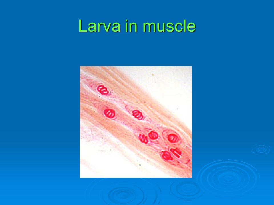
Larva in muscle

54
Clinical Features: Light infections may be asymptomatic. Intestinal invasion can be accompanied by gastrointestinal symptoms (diarrhea, abdominal pain, vomiting). Larval migration into muscle tissues (one week after infection) can cause periorbital and facial edema, conjunctivitis, fever, myalgias, splinter hemorrhages, rashes, and blood eosinophilia. Occasional life-threatening manifestations include myocarditis, central nervous system involvement, and pneumonitis. Occasional life-threatening manifestations include myocarditis, central nervous system involvement, and pneumonitis. Larval encystment in the muscles causes myalgia and weakness, Photophobia Diplopia
. Larval migration into muscle tissues (one week after infection) can cause periorbital and facial edema, conjunctivitis, fever, myalgias, splinter hemorrhages, rashes, and blood eosinophilia. Occasional life-threatening manifestations include myocarditis, central nervous system involvement, and pneumonitis. Occasional life-threatening manifestations include myocarditis, central nervous system involvement, and pneumonitis. Larval encystment in the muscles causes myalgia and weakness, Photophobia Diplopia.")
55
Trichinellosis periorbital and facial edema splinter hemorrhages

56
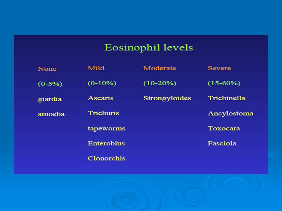
Diagnosis of trichinellosis (trichinosis) clinical symptoms and eosinophilia specific diagnostic tests, including antibody detection, muscle biopsy, and microscopy.
 clinical symptoms and eosinophilia specific diagnostic tests, including antibody detection, muscle biopsy, and microscopy.")
57
Trichinella Muscle samples (highly active muscle like columns of the diaphragm, tongue) were squeezed between two glass plates and then microscoped at low power Modern surveillance pools samples from multiple animals and uses digestion and filtration to detect Trichinella
 were squeezed between two glass plates and then microscoped at low power Modern surveillance pools samples from multiple animals and uses digestion and filtration to detect Trichinella")
59
Treatment Corticosteroid (prednisone) and Mebendazole (drug of choice)
 and Mebendazole (drug of choice)")
60
Prevention Freezing of meat can kill larva (-18 C for 24 hours) T. nativa is resistant to freezing and can cause sever disease Proper cooking of meat (58.3 C can inactivate larva

Similar presentations


>")


 N. americanus (New World) Greyish white or pinkish in color.>")


