Download presentation
Presentation is loading. Please wait.

1
RTEC 111 Bushong Ch 2, 3 &15 Technique Math

2
Fundamentals of Radiologic Science RTEC 111 Bushong Ch. 2

4
Units of Measurement This allows scientists to describe quantities. The fundamental units of measurement are mass, length and time. Two widely used systems of measurement

5
UNITS OF RADIATION MEASUREMENT TO QUANTIFY THE AMOUNT OF RADIATION A PATIENT OR WORKER RECEIVES.

6
Conventional (British)Units vs. SI Units Conventional (British) Units Used Since The 1920’s (foot, pound, second) also called the US customary system 1948 - A System Of Units Based On Metric Measurements Was Developed By The International Committee For Weights And Measures. SI Units
 Units Used Since The 1920’s (foot, pound, second) also called the US customary system A System Of Units Based On Metric Measurements Was Developed By The International Committee For Weights And Measures. SI Units.")
8
Commonly Used SI Prefixes

9
Important Radiology Units Roentgen (R) is coulomb per kilogram (C/kg) Radiation absorbed dose (rad) is gray (Gy) Radiation equivalent man (rem) is seivert (Sv)
 is coulomb per kilogram (C/kg) Radiation absorbed dose (rad) is gray (Gy) Radiation equivalent man (rem) is seivert (Sv)")
10
Conv. Units SI Units RADS REMS R - ROENTGEN GRAYS SIEVERT C/KG

11
ROENTGEN (R) SI = coulomb per kilogram (C/kg) or air kerma Gy a The quantity of X-ray radiation Only exposure in air Output of the x-ray tube Does not indicate actual patient exposure or absorption
 SI = coulomb per kilogram (C/kg) or air kerma Gy a The quantity of X-ray radiation Only exposure in air Output of the x-ray tube Does not indicate actual patient exposure or absorption")
12
RADIATION ABSORBED DOSE (RAD) SI = GRAY (Gy) Measures the amount of energy absorbed in any medium (the patient) 1 Gy = 100 rads 1/100 or 0.01 Gy = 1 rad 1 centigray = 1 rad
 SI = GRAY (Gy) Measures the amount of energy absorbed in any medium (the patient) 1 Gy = 100 rads 1/100 or 0.01 Gy = 1 rad 1 centigray = 1 rad")
13
REM / SIEVERT 1 Sv = 100 rem 1/100 Sv = 1 rem 1 centisievert = 1 rem Used for occupational exposure EMPLOYEE EXPOSURE

14
RADIATION EQUIVALENT MAN (rem) OR Effective dose SI UNITS = SIEVERT Not all types of radiation produce the same responses in living tissue The unit of dose equivlaence, as expressed as the product of absorbed dose in RAD and the quality factor
 OR Effective dose SI UNITS = SIEVERT Not all types of radiation produce the same responses in living tissue The unit of dose equivlaence, as expressed as the product of absorbed dose in RAD and the quality factor")
15
Rem OR Sievert SI UNITS = SIEVERT 1 Sv = 100 rem THE PRODUCT OF THE GRAY AND THE QUALITY FACTOR.

16
Rad VS. Rem RAD’S X QUALITY FACTOR = REM GRAY’S X QUALITY FACTOR = SIEVERT QUALITY FACTOR FOR X-RAYS = 1 So…… Rads = Rems

17
Rems & Rads Sieverts & Grays PAtient = r A ds & gr A ys Employee (technologists) = r E ms & si E v E rts
 = r E ms & si E v E rts")
18
U.S. to S.I. Conversion a trick to remember U.S. = rads & rems Pennies S.I. = grays & sieverts Dollars

19
Metric System milli m 10 -3 5 mrem = 0.005 rem 5000 mrem = 5 rems 500 mrem = 0.5 rems 50 mrem = 0.05 rems 5 mrem = 0.005 rems

20
Radiology – units of measurement What units correlate with: Exposure Dose Effective dose

21
The Structure of Matter RTEC 111

22
Radiology Mechanics Velocity The motion of an object can be described by the use of two terms velocity and acceleration. Velocity = speed What is the speed of x-rays in a vacuum?

23
X-Ray Properties Travel in straight lines. Travel at the speed of light, 3 X 10 8 meters per second in a vacuum or 299,792,458 m/s or 29,979,245,800 cm/s Can ionize matter.

24
Kinetic energy (KE) The energy associated with the motion of an object Kinetic energy depends on the mass of the object and the square of its velocity
 The energy associated with the motion of an object Kinetic energy depends on the mass of the object and the square of its velocity")
25
Potential energy (PE) The stored energy of position or configuration
 The stored energy of position or configuration")
26
Heat Is the kinetic energy of the random motion of molecules

27
Atoms Elements: 112 substances have been identified 92 are naturally occurring and 20 more have been artificially produced The atom is the smallest particle of matter that has the properties of an element

29
Atomic Structure What does Z # mean? Atomic mass?

30
Combining atoms Atom + Atom = molecule Molecule + Molecule = Compound The smallest particle of an element is an atom; the smallest particle of a compound is a molecule

31
Elements Chemical elements – determined by the # of protons Isotopes – neutrons, atomic mass Shells – electron orbits Ion or Ionization?

32
e- shell configuration is dependent on the size of the atom

33
Ionization of carbon Ion pair 34 eV of energy is required

34
Electron Arrangement The maximum number of e- that can exist in each shell increase with distance from the nucleus See table No outer shell can contain more than eight e-

35
Periodic table of the elements The table is organized by the number of e- in the outer or valence shell of an atom # of e- in the outermost shell = the period of that atom on the table The valence shell is important because it determines how that element will react and interact with other elements

37
Electrons Can exist only in certain shells Each shell has different electron binding energies or energy levels

38
Electron binding energy The closer the e- is to the nucleus the more tightly it is bound and the higher the binding energy Also the larger the Z# of the atom the higher the binding energy for any given shell….therefore more difficult of ionize larger atoms

39
Ionization potential The energy required to ionize tissue atoms How much energy is required to ionize tungsten’s K shell? Pg 46

40
Types of Ionizing Radiation All ionizing radiation can be classified into two categories Particulate or electromagnetic radiation What type of radiation is used for diagnostic ultrasound or magnetic resonance imaging? Ionizing or Nonionizing?

41
Particulate Radiation The emission of particles and energy from the nucleus in order to become stable Radioactive elements are called radionuclides or radioisotopes

42
Radioisotopes Occur when atoms have too many or too few neutrons Can occur naturally or can be man made

43
Radioisotope decay Decay from the nucleus to become stable Beta emission and Alpha emission Alpha & Beta particles can cause ionization because of high kinetic energy What form of energy does x-rays use to ionize

44
Radioactive Half-Life The time required for a quantity of radioactivity to be reduced to one-half its original value For radiology: Half-value layer To reduce the strength of the x-ray beam by 1/2

45
Photons vs Particles Particles cause ionization through kinetic energy Photons have no mass, no charge, travel at the speed of light and are considered energy disturbances in space. A form of EM energy. Photons travel at the speed of light or not at all.

46
X-rays vs Gamma rays Forms of EM energy Only difference between x-rays and gamma rays is their origin Only difference between alpha and beta particles is their origin

47
Origins X-rays = outside the nucleus in the e- shells Alpha & Beta particles = from the nucleus Gamma rays = from the nucleus As part of radioactive decay

48
X-rays have low ionization rates and a very long range in tissue

49
Technique Calculations

50
What happens to primary? When x-rays pass through a patient's body, three things can happen: (1) the x-ray photon is transmitted, passing through the body, interacting with the film, and producing a dark area on the film; (2) the x- ray photon is absorbed in an area of greater tissue density, producing lighter areas on the film; and (3) the x-ray photon is scattered and reaches the film causing an overall gray fog.
 the x-ray photon is transmitted, passing through the body, interacting with the film, and producing a dark area on the film; (2) the x- ray photon is absorbed in an area of greater tissue density, producing lighter areas on the film; and (3) the x-ray photon is scattered and reaches the film causing an overall gray fog..")
51
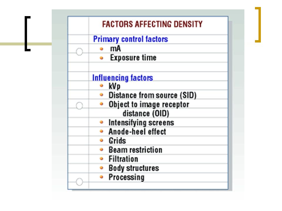
Density The degree of overall blackening from the black metallic silver deposited in the emulsion. Optical Density: range of human visibility

53
Densitometer

54
Density.25 TO -2.5 The straight line of the H&D curve (Hurter & Driffield)
")
55
Optical Density Controlling factor: mAs mAs determines the quantity of x- rays What is the formula to determine quantity of x-rays?

57
mAs mA X time (sec) = mAs The quantity of x-rays produced
 = mAs The quantity of x-rays produced")
58
To see changes in optical density In order to see changes in optical density on a radiograph you must increase you mAs by at least 20 – 30% To double the density on a radiograph you must double your mAs

59
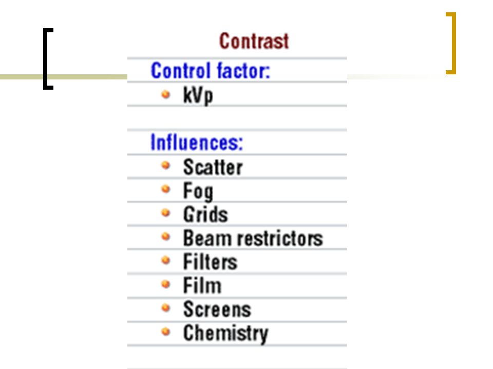
kVp Kilovolts peak kVp Controls the Contrast (for analog/film imaging)
")
60
Low vs High contrast As a radiologic technologist, you will learn to distinguish between low- and high-contrast radiographs. A radiograph with large differences in density is a high-contrast, or short- scale, radiograph. If the density differences aren't as great, you have a low-contrast, or long-scale, radiograph.

68
kVp: Effects density & contrast a. 15% rule: 15% kVp = doubling of exposure to the film 15% kVp = halving of exposure to the film b. 15% rule will always change the contrast of the image because kV is the primary method of changing image contrast.

69
Its math time……

Similar presentations


 Covered Keywords Roentgen, gray, exposure rates, absorbed dose, dose equivalent, quality factors, linear energy transfer, relative biological.>")
