Download presentation
Presentation is loading. Please wait.

1
Histology of endocrine glands
Dr Abubakr H Mossa MBBS, 8&11 /3/12

2
Outline Endocrine tissues/glands in the body
Basic structure of endocrine glands Pituitary gland Adenohypophysis Neurohypophysis Thyroid gland Parathyroid glands Adrenal glands Cortex Medulla Islets of Langerhans (pancreas)
")
4
Endocrine tissues/glands in the body
Endocrine gland produces/secretes hormones to control or alter the function of other cells/tissue/organs Endocrine function can be carried out by: Glands like: pituitary, thyroid, parathyroid, adrenal and pineal glands Clusters of cells in larger organs: ovarian follicles cells, islets of pancreas, Leydig cells of testes… Scattered Single cells: enteroendocrine cells (GIT)..
..")
5
Basic structure of endocrine glands
Endocrine glands have: Parenchyma (cells): polyhedral, arranged in cords or plates, contain secretory vesicles Blood vessels and capillaries: plenty, to convey the hormones and meet the high metabolic demand, in close relation to the cells Little CT
: polyhedral, arranged in cords or plates, contain secretory vesicles. Blood vessels and capillaries: plenty, to convey the hormones and meet the high metabolic demand, in close relation to the cells. Little CT.")
6
Pituitary gland Found in the ……….. of sphenoid bone at the base of brain (hypothalamus) Master gland Has two parts with different embryological origins Adenohypophysis (anterior pituitary) Neurohypophysis (posterior pituitary)
 Neurohypophysis (posterior pituitary)")
7
Pituitary gland: parts

8
Pituitary gland: cells
Adenohypophysis Chromophils Acidophils Somatotrophs (GH) Mammotrophs (prolactin) Basophils Thyrotrophes (TSH) Gonadotrophs (FSH, LH) Corticotrophs (ACTH) Chromophobes Neurohypophysis Axon terminals with Herring’s bodies (ADH, oxytocin) Pituicytes
 Mammotrophs. (prolactin) Basophils. Thyrotrophes. (TSH) Gonadotrophs. (FSH, LH) Corticotrophs. (ACTH) Chromophobes. Neurohypophysis. Axon terminals with Herring’s bodies. (ADH, oxytocin) Pituicytes.")
9
Pituitary stalk

10
Adenohypophysis In pars distalis:
Cells which take the stain are called chromophils: These are divided into acidophils (red) (A) & basophils (blue) (B) Cells which do not stain are called chromophobes (C)
 (A) & basophils (blue) (B) Cells which do not stain are called chromophobes (C)")
11
Adenohypophysis Pars intermedia: Pars tuberalis:
Contains mainly basophils Function in humans not clearly known Produces melanocytes stimulating hormones Pars tuberalis: Wraps partially the infundibulum Highly vascular No specific hormones secreted here in humans

12
Acromegaly? Gigantism ?

13
Neurohypophysis No production of hormones takes place here, only release of hormones form the nerve endings The cell bodies of these neurons are in the hypothalamus (supraoptic & paraventricular nuclei) The nerve endings contain secretory vesicles which are known as Hering’s (or neurosecretory) bodies (NB) Contain ADH & oxytocin The other type of cells in posterior pituitary is called pituicytes (P)which are glial cells sheathing the unmyelinated axons
 The nerve endings contain secretory vesicles which are known as Hering’s (or neurosecretory) bodies (NB) Contain ADH & oxytocin. The other type of cells in posterior pituitary is called pituicytes (P)which are glial cells sheathing the unmyelinated axons.")
14
Control of pituitary gland: vascular & neural connection to hypothalamus

15
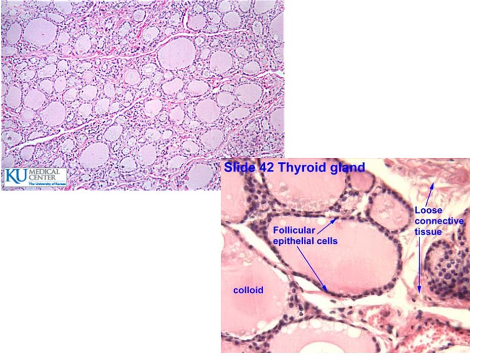
Thyroid gland In the anterior aspect of the neck Has Isthmus & 2 lobes
Covered by pretracheal fascia Divided into lobules by the CT septa Each lobule contains follicles, parafollicular cells & capillaries Follicles; sac like structure Each follicle is surrounded by a basal lamina and reticular fibers The follicle is lined by a simple cuboidal epithelium The follicular cells secrete thyroxine T4 & T3 The height of the cells depends on the activity of the cells Low or squamous: inactive or resting High or columnar: active or secretory The cavity of the follicle is full of colloid (acidophilic gel-like substance) for the storage of hormone-bound thyroglobulin Parafollicular cells :lie between follicles, pale cells with round nucleus, secrete calcitonin
 for the storage of hormone-bound thyroglobulin. Parafollicular cells :lie between follicles, pale cells with round nucleus, secrete calcitonin.")
18
Parathyroid glands 4 in number, 2 superior & 2 inferior
Lie posterior to the thyroid gland Have two types of cells: Chief (principal cells): most numerous, acidophilic cytoplasm with round nucleus, secretes parathyroid hormone (PTH) which increase blood calcium Oxyphil cells: less in number and have deeply acidophilic cytoplasmic granules
: most numerous, acidophilic cytoplasm with round nucleus, secretes parathyroid hormone (PTH) which increase blood calcium. Oxyphil cells: less in number and. have deeply acidophilic cytoplasmic granules.")
20
Adrenal gland Lies on the top of the kidney Highly vascularized
Has two regions: cortex (larger) & medulla
 & medulla.")
22
Adrenal cortex; Has three layers (zones) which are not well demarcated
Zona glomerulosa: (mineralocorticoids; aldosterone) Thin layer Cells are arranged in circles or arches Dark nucleus & acidophilic cytoplasm with few lipid droplets Sinusoids present between cells Zona Fasciculata: (glucocorticoids: cortisol) Widest zone Cells arranged in columns or cords Large cells with pale staining nuclei and acidophilic cytoplasm containing plenty of lipid droplets Zona reticularis: (androgens, estrogen) Thin zone near the medulla Cells are deeply staining with network like arrangement
 Thin layer. Cells are arranged in circles or arches. Dark nucleus & acidophilic cytoplasm with few lipid droplets. Sinusoids present between cells. Zona Fasciculata: (glucocorticoids: cortisol) Widest zone. Cells arranged in columns or cords. Large cells with pale staining nuclei and acidophilic cytoplasm containing plenty of lipid droplets. Zona reticularis: (androgens, estrogen) Thin zone near the medulla. Cells are deeply staining with network like arrangement.")
24
Adrenal medulla Cells here are large & arranged in groups or short cords with sinusoids in between The cytoplasm of medullary cells is clear or light basophilic in H & E stain These cells are called also chromaffin cells as they stain with dicromate salt in a reaction called chromaffin reaction (catecholamine containing cells take this stain). So these cells secrete adrenalin and nor adrenalin. They are derived from the neural crest cells and respond directly to sympathetic nerve endings. So they are considered as postganglionic cells. Other cells in medulla are the few but large parasympathetic ganglionic cells.
. So these cells secrete adrenalin and nor adrenalin. They are derived from the neural crest cells and respond directly to sympathetic nerve endings. So they are considered as postganglionic cells. Other cells in medulla are the few but large parasympathetic ganglionic cells.")
25
Medullary cells Adrenal medulla

26
Vascular anatomy of adrenal glands

27
Islets of Langerhans This is the endocrine part of pancreas
Oval or round cluster of endocrine cells with plenty of capillaries in between Has many types of cells 1 million islet and most are in the tail region

29
Cell types in islets of Langerhans

30
Thanks

Similar presentations


 glands are also named ductless glands, since they lack excretory ducts. Instead,>")


 in the body similar to the nervous.>")

 are released in the blood or tissue fluid); they have influence on organs and tissues.>")


