Download presentation
Presentation is loading. Please wait.

1
Angiography Arteriography Aortograms and Venography
SPRING 2011 FINAL

2
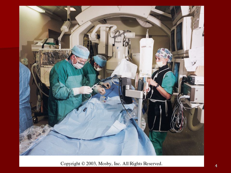
Angiography Is the general term that describes the radiologic examination of vascular structures within the body after the introduction of an iodinated contrast medium or gas

3
Types of Angiographic Procedures

5
Angiography Team Radiologist CIT (Radiologic Technologist)
Sometimes more than one Other specialists (if needed) Nurse Anesthesiologist (if needed)
 Nurse. Anesthesiologist (if needed)")
6
Indications Verify the presence of tumors Internal bleeding Stenosis
Blood supply to tumors Internal bleeding Possible anemia Stenosis Can be caused form atherosclerosis Occlusions Clots Thrombus Embolus Aneurysms Heart disease

7
Contraindications Previous severe reaction to contrast
Impaired renal function Impaired blood clotting factors Inability to undergo surgical procedure

8
Contrast Media Iodinated contrast media is used
Can produce nausea & an uncomfortable burning sensation Allergic reactions Severe: anaphylactic shock Shock, rapid shallow breathing, high pulse rate & ALOC Mild: Hives or slight difficulty breathing

9
What is this?

11
Angiographic Trays and Sterile Supplies

12
Other Supplies for Angiography

13
Needles Vascular access needles
Size based on external diameter of needle Allows for appropriate Guidewires matching So internal diameter must also be known

14
Guidewires Used as a platform over which a catheter is to be advanced
Once positioned guidewire is fixed and catheter is advanced until it meets the tip of the guidwire Mostly constructed on stainless steel & coated with Teflon

15
Introducer Sheaths Short catheters used when multiple catheters will be used Placed in lieu of a catheter

16
Catheters Angiographic Catheters, PTCA Balloon Catheters, Drug Delivery Catheters, Guide Catheters, Guide Wires, Intravascular Ultrasound Catheters, Stents, Atherectomy Devices, Valvuloplasty. Inflation Devices, Introducers Sheath, Y-Adapters, Manifolds, Stopcocks, Control Syringes, Pressure Monitoring Products, Disposable Transducers, Disposable Domes, Fluid Delivery System, Contrast Injection Lines.

18
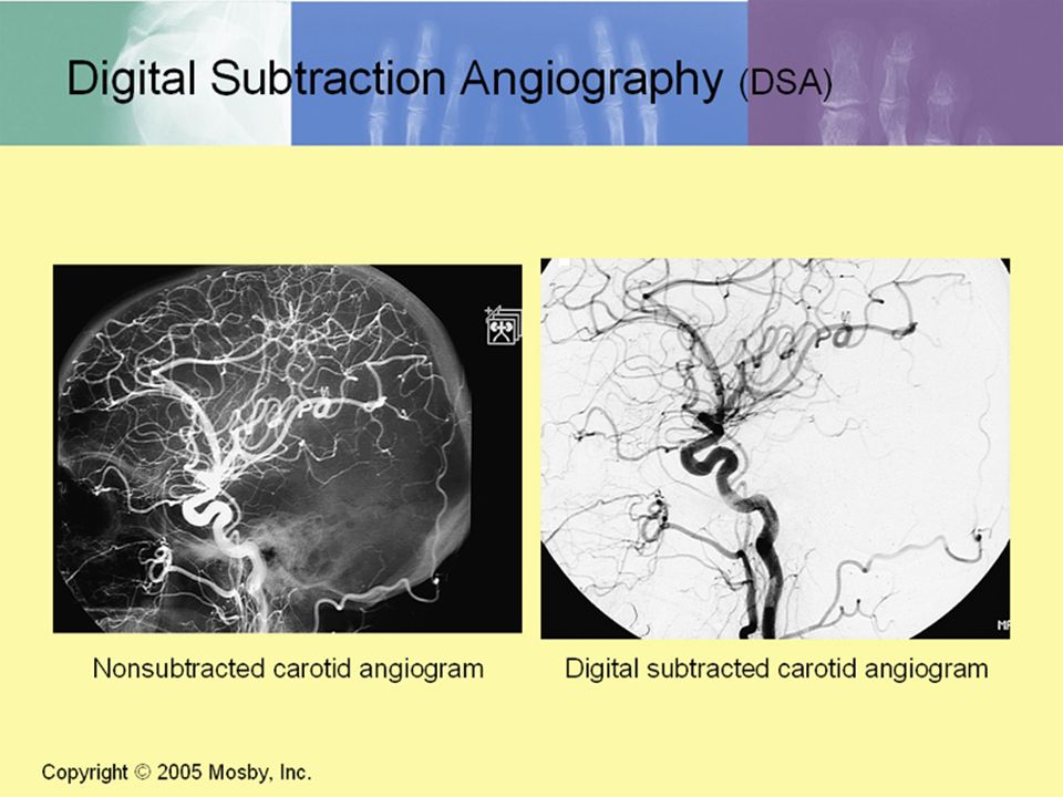
DSA A subtraction mask is taken before contrast injected
Each of digitized image is from the mask Images acquired form 1 image every 2-3 sec Up to 30 images per sec

19
Three Dimensional (3-D) Intraarterial Angiography
 Intraarterial Angiography")
20
What Method is this?

21
Catherization: Selinger Technique

22
Selinger Technique Catheters and Guidewires

23
Pre-Procedure PT’s are usually limited to a liquid diet and routine medications Adequate hydration An IV line placed Sedative may be given History taken and vitals taken Informed consent

24
Preparing the Patient Room
Must be extensively cleaned Equipment checked Room thoroughly stocked Extra supplies as needed

25
Radiation Protection PT is protected by no less than 2.5 mm of Aluminum Beam restriction Avoidance of repeat exposure Cardinal rules Time Distance Shielding

26
Post Procedure PTs usually can resume normal activity after 24 hours
Most often can go home after 24 hours Because internal bleeding can be life threatening Vitals are monitored Puncture site is monitored for bleeding

27
Stent Placement

28
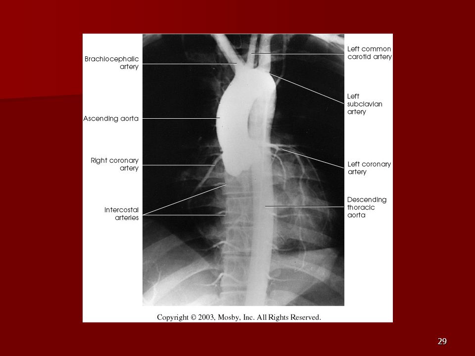
Aortogram

30
AORTOGRAM

31
Abdominal Aortoraphy

32
Abdominal Angiography

33
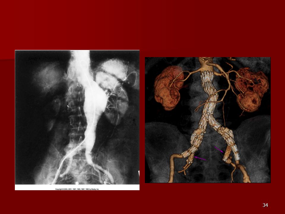
AAA Pre and Post Stent Placement

35
Abdominal Stent

36
AAA Open repair of abdominal aortic aneurysms is a major operation, and you would need to be in hospital for approx. 7 to 10 days. Under a general anesthetic, an incision (cut) is made in the abdomen. Then the abdominal aorta above and below the aneurysm is controlled and clamped to stop blood flow, which enables the aneurysm to be opened up and a new artificial artery sewn in to replace the dilated segment. The clamps are then removed to restore the blood flow, and the abdomen is sewn up. Back to top Risks and complications of an 'open' operation with a graft The major complications of the open operation are the risk of either bleeding or problems with the heart, which is put under some strain when the clamps are placed on the abdominal aorta. There can be problems resulting from a reduction in blood flow to the legs and kidneys during or after the operation. Overall, the incidence of serious complications is 5 to 7%. What alternatives are there to having an open operation? A relatively new technique that is being tested in hospitals is to use a 'stent' to help repair abdominal aortic aneurysms (see the diagram below). The aim of this technique is to perform a less major operation, which can be tolerated better by more patients. Depending on the shape and size of the arteries around the aneurysm, the abdominal aortic aneurysm can be repaired from the groin. This operation usually involves making small cuts on each side of the groin under general anesthetic. A graft is then inserted into the abdominal aortic aneurysm through an artery in the groin. Special stents are used to hold the graft in place.
 is made in the abdomen. Then the abdominal aorta above and below the aneurysm is controlled and clamped to stop blood flow, which enables the aneurysm to be opened up and a new artificial artery sewn in to replace the dilated segment. The clamps are then removed to restore the blood flow, and the abdomen is sewn up. Back to top. Risks and complications of an open operation with a graft. The major complications of the open operation are the risk of either bleeding or problems with the heart, which is put under some strain when the clamps are placed on the abdominal aorta. There can be problems resulting from a reduction in blood flow to the legs and kidneys during or after the operation. Overall, the incidence of serious complications is 5 to 7%. What alternatives are there to having an open operation A relatively new technique that is being tested in hospitals is to use a stent to help repair abdominal aortic aneurysms (see the diagram below). The aim of this technique is to perform a less major operation, which can be tolerated better by more patients. Depending on the shape and size of the arteries around the aneurysm, the abdominal aortic aneurysm can be repaired from the groin. This operation usually involves making small cuts on each side of the groin under general anesthetic. A graft is then inserted into the abdominal aortic aneurysm through an artery in the groin. Special stents are used to hold the graft in place.")
37
Pulmonary Circulation

38
Pulmonary Arteriogram

39
Celiac Ateriogram

40
Hepatic Arteriogram

41
Splenic Arteriorgram

42
Renal Arteriogram A) Large eccentric noncalcified stenosis of the proximal renal artery (arrowhead). (B) Angiography shows the absence of flow in the renal artery and in distal runoff arteries after initial predilation with a 2.5-mm balloon catheter. (C) Obstruction of the main renal artery after stent implantation; the delivery catheter is still in place (arrowhead). (D) The angiogram shows the guiding catheter deep inside the artery (arrowhead). (E) Angiogram after thrombolysis shows no modification in the clot. (F) Angiography after successful recanalization: the renal parenchyma is well contrasted apart from a small area of the lower pole (arrowhead)
 Large eccentric noncalcified stenosis of the proximal renal artery (arrowhead). (B) Angiography shows the absence of flow in the renal artery and in distal runoff arteries after initial predilation with a 2.5-mm balloon catheter. (C) Obstruction of the main renal artery after stent implantation; the delivery catheter is still in place (arrowhead). (D) The angiogram shows the guiding catheter deep inside the artery (arrowhead). (E) Angiogram after thrombolysis shows no modification in the clot. (F) Angiography after successful recanalization: the renal parenchyma is well contrasted apart from a small area of the lower pole (arrowhead)")
43
renal

47
Lower Limb Arteries

49
Leg Atherosclerosis

50
Atherosclerosis Left Leg

51
Upper Limb Arteries

52
Upper Extremity Anatomy

53
Brachial and Axillary Arteriogram

54
Axillary Arteriogram

55
Hand Arteriogram

56
Hand Arteriogram with Occlusion
Right upper extremity angiogram shows abrupt occlusion of the superficial palmar arch (arrow). The deep palmar arch remains patent (arrowheads). There is no visible filling of the proper palmar digital arteries of the fourth and fifth digits.
. The deep palmar arch remains patent (arrowheads). There is no visible filling of the proper palmar digital arteries of the fourth and fifth digits.")
57
A, Guidewire advanced through stenosis
A, Guidewire advanced through stenosis. B, Small catheter advanced through stenosis. C, Large catheter advanced through stenosis. D, Postangioplasty stenotic area. View JPG View PDF

58
Balloon Angioplasty

60
Balloon Angioplasty Procedure

61
Femoral Artery Angioplasty

62
Placing a Stent after Angioplasty with Balloon

63
Intravascular Stents

64
Let’s Review

65
B C A: RT subclavian B: Lt internal carotid C. Lt subclavina
D: brachicephalic Aortic bulb Ascending aorta Aortic Arch Descending Aorta

66
What is the name of this Procedure? What is it done for?

67
What is the name of this pathology?

68
What part of the body is being imaged
What part of the body is being imaged? What is the pathology is this image?

69
A Right common carotid B. RT external carotid C. RT internal carotid

70
What is this method callled?

71
A C B Arch of aorta Descending aorta Asending aorta

72
Venography

73
Venous Circulation

74
What is Venography? Vein study using x-ray and contrast media
Fluoroscopy and still images One of the most accurate tests for deep vein thrombosis (DVT) Most commonly done in legs for DVT
 Most commonly done in legs for DVT.")
75
Thrombosis and Embolism
Intravascular clot Commonly in veins more than arteries 3 factors Where blood is slow Change in the wall of vessels Change in the blood itself Thrombus that becomes detached from the vessel wall Can easily flow to heart causing PE Severity depends on location of embolism

76
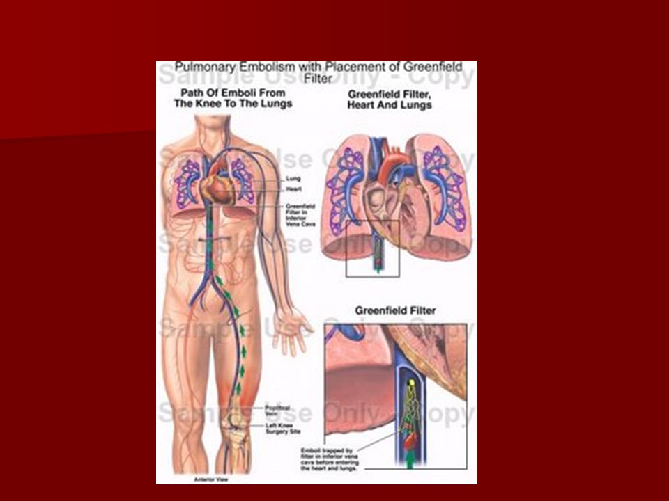
Pulmonary Embolism Occurs when a clot forms or becomes lodged in the pulmonary artery Most commonly thrombus originates in the lower limbs and migrates Can lead to resp distress, heart failure or cardiogenic shock Symptoms are acute: Sudden coughing SOB Chest pain

77
Pulmonary Emboli (PE)
")
78
Indications Diagnose deep vein thrombosis
Prevent pulmonary embolism Distinguish blood clots from obstructions in the veins Evaluate congenital vein problems Assess the functioning of deep leg vein valves Identify a vein for arterial bypass grafting Diagnose deep vein thrombosis , a blood clot deep within the leg that may lead to an obstruction of a blood vessel in the lungs ( pulmonary embolism ) Distinguish blood clots from obstructions in the veins Evaluate congenital vein problems Assess the functioning of deep leg vein valves Identify a vein for arterial bypass grafting
 Distinguish blood clots from obstructions in the veins. Evaluate congenital vein problems. Assess the functioning of deep leg vein valves. Identify a vein for arterial bypass grafting.")
79
Risk Factors and Complications
Previous thrombosis Dilution of the contrast dye in the lower limb Difficulty accessing the veins due to: Obesity Severe swelling (edema) Inflammation in the cells ( cellulitis )
 Inflammation in the cells ( cellulitis )")
80
Contraindications Bleeding disorders Allergy to iodine CHF
Severe pulmonary hypertension

81
Prior to Procedure Fast or drink only clear fluids for four hours before the test Thorough PT history obtained Informed consent If you are nervous about the test, your doctor may give you a sedative. You may be asked to fast or drink only clear fluids for four hours before the test. Tell your doctor if you have a history of allergies, hay fever, or bad reactions to an injected contrast dye. If you are nervous about the test, your doctor may give you a sedative

82
During Procedure PT will lie on a tilting x-ray table
Area of interest will be shaved and cleaned Local anesthetic Catheter will be inserted. A small incision may be made in that area as well You will lie on a tilting x-ray table. If necessary, you will be shaved in the area where the catheter (small tube used to inject the dye) will be inserted. A small incision may be made in that area as well. Anesthesia You may be given a local anesthetic to numb the area where the catheter will be inserted.
 will be inserted. A small incision may be made in that area as well. Anesthesia You may be given a local anesthetic to numb the area where the catheter will be inserted.")
83
Explanation of Procedure: Legs
The catheter is inserted into PT vein (usually a vein in the foot) Contrast is slowly injected. A tight band may be tied around your ankle and upper thigh or your lower body may be tilted Fluoro and/or x-ray images taken The procedure takes about minutes The catheter is inserted into your vein (usually a vein in the foot) and a special dye is slowly injected. A tight band may be tied around your ankle, or your lower body may be tilted, which helps to fill the deep venous system with dye. You will be asked to remain still as a clinician uses a fluoroscope to view the movement of the dye through your veins. A series of x-rays will be taken during this time. How Long Will It Take? The procedure takes about 30 minutes for an uncomplicated venography. This time may increase, depending on the specifics of the procedure. Furthermore, you will need to keep your leg straight for six hours after the procedure has been completed.
 Contrast is slowly injected. A tight band may be tied around your ankle and upper thigh. or your lower body may be tilted. Fluoro and/or x-ray images taken. The procedure takes about minutes. The catheter is inserted into your vein (usually a vein in the foot) and a special dye is slowly injected. A tight band may be tied around your ankle, or your lower body may be tilted, which helps to fill the deep venous system with dye. You will be asked to remain still as a clinician uses a fluoroscope to view the movement of the dye through your veins. A series of x-rays will be taken during this time. How Long Will It Take The procedure takes about 30 minutes for an uncomplicated venography. This time may increase, depending on the specifics of the procedure. Furthermore, you will need to keep your leg straight for six hours after the procedure has been completed.")
84
Post Procedure Rest and avoid strenuous activity Increase fluid intake
Stop bleeding with pressure Call DR if it won’t stop bleeding Observe for signs of infection PT will be sore for a few days Resume normal activity 24 hours after procedure When you get home from the test, take it easy for the rest of the day and try to avoid going up and down flights of stairs, or any strenuous activity. Drink large amounts of fluid for the next 24 hours, to help flush the remaining dye from your body. If any bleeding or swelling occurs at the injection or puncture site, put pressure on the site for at least 10 minutes. If this fails to stop the bleeding, go to the emergency room of a local hospital or call your physician for advice. You may remove the bandage the day after your test. Observe the injection site for any swelling, heat, redness, pain, or drainage. The injection area will be sore for a few days. Most people are able to resume normal activities the day after the procedure.

85
Possible Post Procedure Complications
Infection at the injection site Tissue damage Phlebitis (inflammation of a vein) Allergic reactions to the contrast dye Congestive heart failure Acute renal insufficiency Venous thrombosis in a healthy leg Dislodging a clot, perhaps resulting in pulmonary embolus or other complications People with kidney problems or diabetes , especially those taking metformin (Glucophage), may have a higher risk for complications resulting from venography.
 Allergic reactions to the contrast dye. Congestive heart failure. Acute renal insufficiency. Venous thrombosis in a healthy leg. Dislodging a clot, perhaps resulting in pulmonary embolus or other complications. People with kidney problems or diabetes , especially those taking metformin (Glucophage), may have a higher risk for complications resulting from venography.")
86
Lower Limb Veins

87
Lower Limb Venograms To rule out thrombosis of the deep veins of the leg Deep vein thrombosis (DVT) Contrast media injected in superficial veins of the foot with a needle

88
Lower Limb Venograms

89
DVT

90
Inferior Venacavagram
Primarily to rule out thrombus or occlusion Catheter inserted into femoral vein and positioned inside the common iliac vein or inferior aspect of inferior vena cava Contrast injected at 20 ml/sec for total of 40ml

91
Upper Limb Veins

92
Upper Limb Venograms Most often for thrombosis or occlusion
Contrast injected in a superficial vein in the elbow or wrist Using a catheter or needle 40-80ml at a rate of 1-4ml/sec

93
Superior Venacavagram
Primarily done to rule out thrombus or occlusion Needle or catheter is introduced into antecubital fossa Catheter is positioned in the axillary or subclavian vein and contrast is injected 30-50ml at 10-15ml/sec X-rays should include: Brachicephalic vein Subclavian vein Superior vena cava RT Atrium

94
Superior Venacavagram

95
Stenosis on a Superior Venacavogram
72-year-old man with non—small cell lung carcinoma and progressive superior vena cava syndrome due to severe stenosis. Superior venacavagram before endovascular treatment shows severe stenosis of vena cava and confluence of right innominate vein. Note collateral venous network from thoracic, neck, and azygos veins.

96
Inferior Venacavagram

97
Inferior Venacavagram

98
Inferior Vena Cava Filters

99
Inferior Vena Cava Filter Placement
Designed to trap thrombus before causing an embolization When anticoagulants are contraindicated this can be used

100
Inferior Vena Cava Filter Placement

102
Hepatic Venogram Performed to rule out stenosis or thrombus of the hepatic veins Obtain pressure measurements of the veins inside the liver Usually catheter enters jugular vein or upper limb veins

103
Hepatic Venogram

104
Portal Venogram

105
Portal System

106
Transjugular Intrahepatic Portosystemic Shunt
Intervention for creating an artificial low-pressure pathway Between portal & hepatic veins Hepatic venogram usually preformed b before placement US also useful

107
Transjugular Intrahepatic Portosystemic Shunt

108
Renal Venogram Rule out thrombosis of renal vein
Renal vein catheterized to take blood Measure the production of renin Catheter insertion site: femoral vein Contrast injected 8ml/sec for 16ml total 2 images per second for 4 seconds

109
Renal Venogram

Similar presentations


, awareness of heartbeat.>")


>")


 and Pulmonary Embolism (PE)>")
Hypertension 2)Coronary Artery Disease - arteriosclerosis - atherosclerosis - angina - myocardial infarction.>")


