Download presentation
Presentation is loading. Please wait.

1
Rehabilitation after ankle sprain Dr. Ali Abd El-Monsif Thabet

2
Introduction The LCL is composed of three separate bands that are commonly referred to as separate ligaments. These are the anterior and posterior talofibular ligaments and the calcaneofibular ligament. The LCL is composed of three separate bands that are commonly referred to as separate ligaments. These are the anterior and posterior talofibular ligaments and the calcaneofibular ligament.

4
Introduction The MCL is most commonly called the deltoid ligament. As its name implies, the deltoid ligament is a fan-shaped. The MCL is most commonly called the deltoid ligament. As its name implies, the deltoid ligament is a fan-shaped.

6
Kinematics The LCL helps control varus stresses that result in lateral distraction of the joint and helps check extremes of joint ROM, particularly calcaneal inversion. The LCL helps control varus stresses that result in lateral distraction of the joint and helps check extremes of joint ROM, particularly calcaneal inversion.

7
Kinematics This ligament helps control medial distraction stresses on the ankle joint and also helps check motion at the extremes of joint range, particularly with calcaneal eversion This ligament helps control medial distraction stresses on the ankle joint and also helps check motion at the extremes of joint range, particularly with calcaneal eversion

8
Pathomechanics Acute ankle trauma is responsible for 10 to 40 percent of sports-related injuries and sprains account for 75- 85 percent of ankle injuries. Acute ankle trauma is responsible for 10 to 40 percent of sports-related injuries and sprains account for 75- 85 percent of ankle injuries.

9
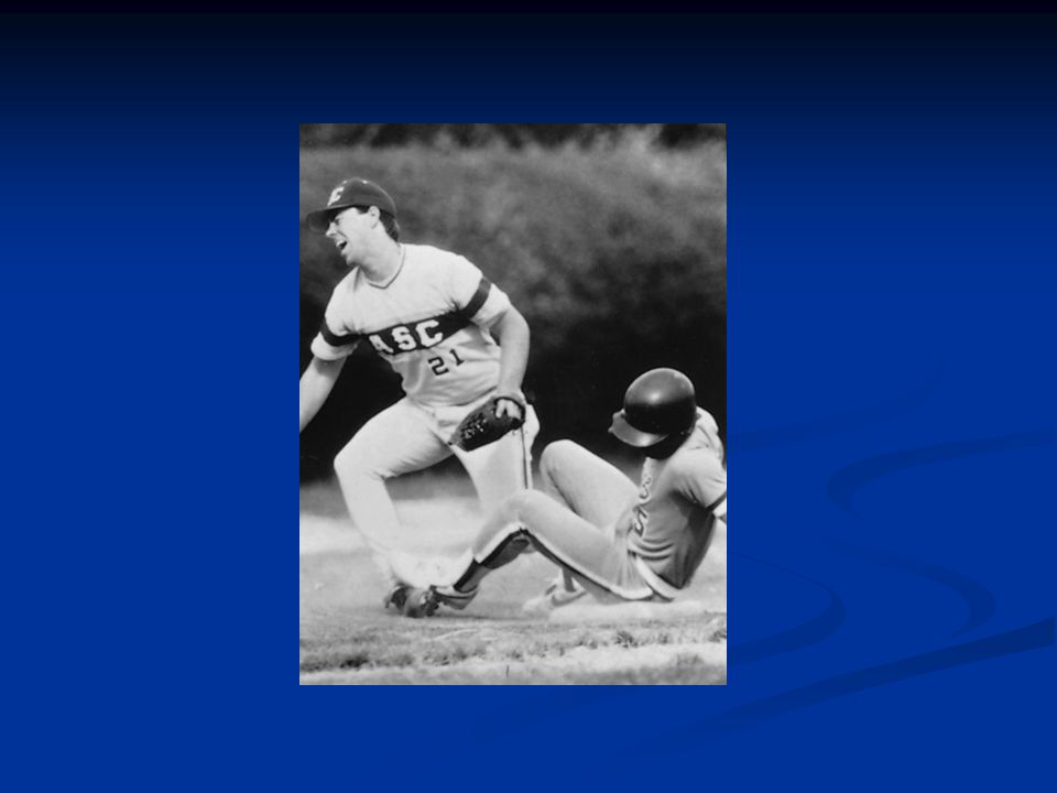
Injury mechanism The most common mechanism of injury in ankle sprains is a combination of plantar flexion and inversion The most common mechanism of injury in ankle sprains is a combination of plantar flexion and inversion

11
A twisting injury or going over on the ankle usually results in an inversion of the foot and ankle A twisting injury or going over on the ankle usually results in an inversion of the foot and ankle

13
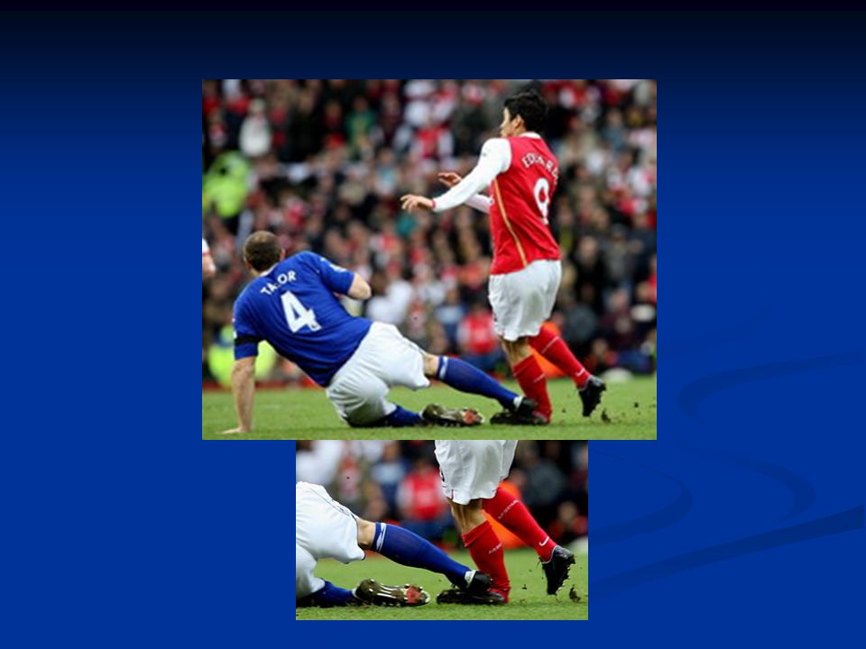
Tackling to the medial aspect of the foot (varus stress) Tackling to the medial aspect of the foot (varus stress)
 Tackling to the medial aspect of the foot (varus stress)")
15
A medial ligament sprain is rare but can occur particularly with a fracture. This happens when the ankle rolls with the sole of the foot faces outwards, damaging the ligaments on the inside of the ankle. A medial ligament sprain is rare but can occur particularly with a fracture. This happens when the ankle rolls with the sole of the foot faces outwards, damaging the ligaments on the inside of the ankle.

17
Diagnosis Tenderness along the joint, pain in weight bearing Tenderness along the joint, pain in weight bearing Severe swelling Severe swelling Diagnosing Ankle Sprains tests Anterior drawer test Anterior drawer test Talor tilt test Talor tilt test

18
Anterior drawer test Perform the anterior drawer test with the ankle at 90° to the leg. Grasp the heel and pull forward while, with the other hand, placing posterior force on the tibia. If the test is positive, the so-called suction sign occurs. Dimpling is observed at the anterolateral aspect of the ankle, indicating compromise of the anterior talofibular ligament. A firm endpoint will be absent Perform the anterior drawer test with the ankle at 90° to the leg. Grasp the heel and pull forward while, with the other hand, placing posterior force on the tibia. If the test is positive, the so-called suction sign occurs. Dimpling is observed at the anterolateral aspect of the ankle, indicating compromise of the anterior talofibular ligament. A firm endpoint will be absent

19
Talar tilt test The talar tilt test is used to examine the integrity of the calcaneofibular or the deltoid ligament. The patient is seated comfortably on the end of an exam table. Possible alternate positions can be side lying or supine. The examiner grasps the foot with the ankle at 90° to the leg, while stabilizing the tibia and fibula. To test the calcaneofibular ligament the examiner will adduct and invert the calcaneous into a varus position. The deltoid ligament is examined by abducting and everting the calcaneous into a valgus position. A positive test will result in laxity and/or pain The talar tilt test is used to examine the integrity of the calcaneofibular or the deltoid ligament. The patient is seated comfortably on the end of an exam table. Possible alternate positions can be side lying or supine. The examiner grasps the foot with the ankle at 90° to the leg, while stabilizing the tibia and fibula. To test the calcaneofibular ligament the examiner will adduct and invert the calcaneous into a varus position. The deltoid ligament is examined by abducting and everting the calcaneous into a valgus position. A positive test will result in laxity and/or pain

20
FIGURE. Inversion stress test to assess the integrity of the calcaneofibular ligament.

21
Squeeze test To perform the squeeze test, place the thumb on the tibia and the fingers on the fibula at the midpoint of the lower leg; then squeeze the tibia and fibula together. Consider pain along the length of the fibula, which indicates a positive test result To perform the squeeze test, place the thumb on the tibia and the fingers on the fibula at the midpoint of the lower leg; then squeeze the tibia and fibula together. Consider pain along the length of the fibula, which indicates a positive test result

22
The common causes of chronic ankle pain * Poor rehabilitation * Poor rehabilitation * A fracture that was not initially diagnosed * A fracture that was not initially diagnosed * Post traumatic arthritis * Post traumatic arthritis * Osteochondritis dissecans (loose bit of bone in the joint) * Osteochondritis dissecans (loose bit of bone in the joint) * Syndesmotic ligament injury * Syndesmotic ligament injury * Functional instability (a feeling of 'giving way') * Functional instability (a feeling of 'giving way') * Ankle impingement * Ankle impingement
 * Osteochondritis dissecans (loose bit of bone in the joint) * Syndesmotic ligament injury * Syndesmotic ligament injury * Functional instability (a feeling of giving way ) * Functional instability (a feeling of giving way ) * Ankle impingement * Ankle impingement")
23
Ankle sprain types The sprained ankle is often classified as to how severe it is: First degree ankle sprain: First degree ankle sprain: * Some stretching or mild tearing of the ligament. * Little or no functional loss - the joint can still function and bear some weight * Some stretching or mild tearing of the ligament. * Little or no functional loss - the joint can still function and bear some weight * Mild pain * Some swelling * Some joint stiffness * Mild pain * Some swelling * Some joint stiffness

24
Second degree ankle sprain: Second degree ankle sprain: *Moderate tearing of the ligament fibers *Moderate instability of the joint * Moderate to severe pain – weight bearing is very painful (difficulty walking ) * Swelling and stiffness *Moderate tearing of the ligament fibers *Moderate instability of the joint * Moderate to severe pain – weight bearing is very painful (difficulty walking ) * Swelling and stiffness A more effective means of immobilizing the ankle (splints) may be needed A more effective means of immobilizing the ankle (splints) may be needed
 * Swelling and stiffness *Moderate tearing of the ligament fibers *Moderate instability of the joint * Moderate to severe pain – weight bearing is very painful (difficulty walking ) * Swelling and stiffness A more effective means of immobilizing the ankle (splints) may be needed A more effective means of immobilizing the ankle (splints) may be needed")
25
Third degree ankle sprain: Third degree ankle sprain: * Total rupture of a ligament - there is a loss of motion * Gross instability of the joint - joint function is lost * Severe pain initially followed by no pain * Severe swelling Cast immobilization is needed for at least 2-3 weeks. * Total rupture of a ligament - there is a loss of motion * Gross instability of the joint - joint function is lost * Severe pain initially followed by no pain * Severe swelling Cast immobilization is needed for at least 2-3 weeks.

26
Rehabilitation (sprained ankle) All ankle sprains recover through three phases:All ankle sprains recover through three phases: Phase 1 includes resting, protecting the ankle and reducing the swelling (one week).Phase 1 includes resting, protecting the ankle and reducing the swelling (one week). Phase 2 includes restoring range of motion, strength and flexibility (one week to two weeks).Phase 2 includes restoring range of motion, strength and flexibility (one week to two weeks). Phase 3 includes gradually returning to activities that do not require turning or twisting the ankle and doing maintenance exercises. This will be followed later by being able to do activities that require sharp, sudden turns (cutting activities) such as tennis, basketball or football (weeks to months).Phase 3 includes gradually returning to activities that do not require turning or twisting the ankle and doing maintenance exercises. This will be followed later by being able to do activities that require sharp, sudden turns (cutting activities) such as tennis, basketball or football (weeks to months).
.Phase 2 includes restoring range of motion, strength and flexibility (one week to two weeks). Phase 3 includes gradually returning to activities that do not require turning or twisting the ankle and doing maintenance exercises. This will be followed later by being able to do activities that require sharp, sudden turns (cutting activities) such as tennis, basketball or football (weeks to months).Phase 3 includes gradually returning to activities that do not require turning or twisting the ankle and doing maintenance exercises. This will be followed later by being able to do activities that require sharp, sudden turns (cutting activities) such as tennis, basketball or football (weeks to months)..")
27
Image 2 - Aircast gel ankle brace Ankle tape

28
Achilles tendon stretching using a towel.

29
Use of elastic tubing in strengthening exercises for eversion. Single-leg toe raises done on a step

30
high-top shoes

31
Thank you

Similar presentations


>")


