Download presentation
Presentation is loading. Please wait.

1
Pathology Lap Pictures and Explanation POD II BLOCK Abdullah Al Garni

2
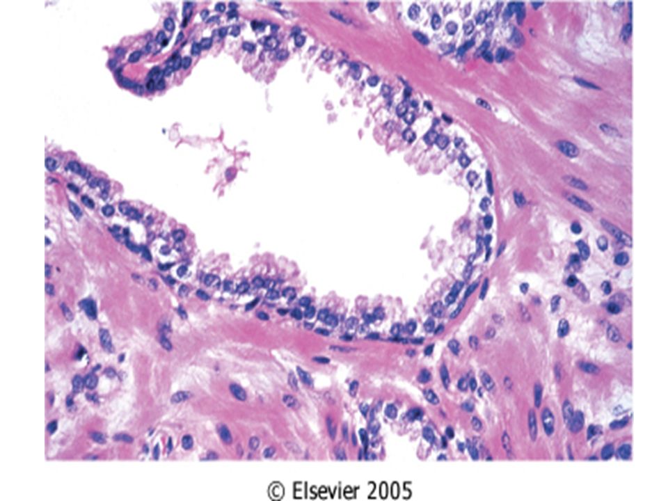
Prostate gland, nodular hyperplasia تــــضخــم غـــدة الـــبروســـتاتا ( الموثة )
")
3
well-circumscribed nodules

4
hyperplastic glands With two layers well-circumscribed nodules margin the stroma

6
Breast, Fibroadenoma ســــرطــــان الثــــــدي

8
fibroadenomanormal breast tissuewell-demarcated border

9
Large irregularly shaped glandular spacesfibrous stroma

10
columnar epithelium Delicate stroma

11
Breast, Invasive ductal carcinoma ســـــرطان الـــــثدي

12
duct-like spacesfibrous stroma

13
duct-like spacesfibrous stroma

14
Non-neoplastic ductsInvading tumor cells Fat Stroma

15
Tumor cells

16
Invasive malignant glands

18
Invasive squamous cell carcinoma EXAMPLE: Bronchous ســـرطـــان القصبات الهوائية

19
central areas of keratinization

20
intercellular bridges the large hyperchromatic nuclei

21
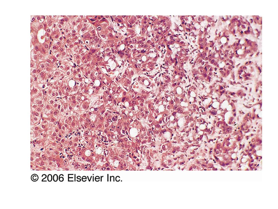
Lung, Metastatic Carcinoma ســـــرطان الـرئة

22
The lung tissue has been replaced by large sheets of malignant cells The sheets is formed of pleomorphic cells with hyperchromatic nuclei There is also extensive necrosis The characteristic architecture of the residual normal lung can be seen.

24
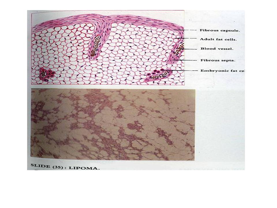
Lipoma

26
mature white fat cellsThe nucleus

27
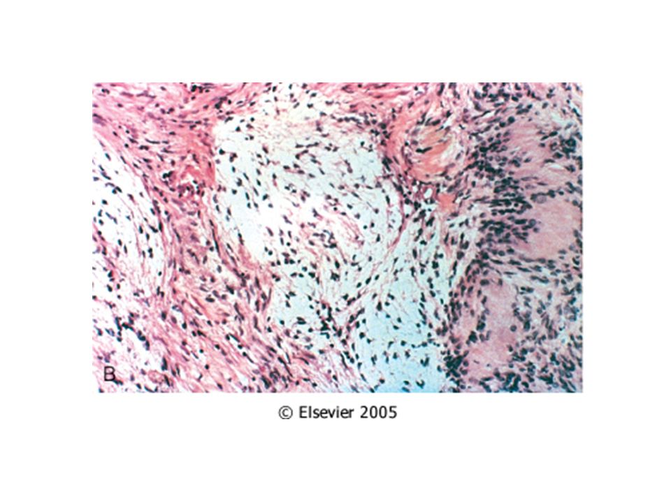
Schwannoma

28
The tumor shows a mixture of two growth patterns: -In the Antoni A pattern of growth, elongated cells with cytoplasmic processes are arranged in fascicles in areas of moderate to high cellularity with little stromal matrix. The nuclear palisading can be seen. - In the Antoni B pattern of growth, the tumor is less densely cellular with a loose meshwork of cells along with microcysts and myxoid changes. In both areas, the cytology of the individual cells is similar, with elongated cell shape and regular oval nuclei A A B B

30
Black arrow: Antoni A ; blue arrow: Antoni B

31
Bone, osteosarcoma ســـــرطان العـــظـــم

32
Spicules of tumor bone malignant osteoblasts that have produced spicules of bone surrounded by osteoid matrix (( characteristic of osteosarcomas)) External information
) External information")
33
Mitosis

34
eosinophilic osteoid

35
Uterus, leiomyosarcoma ســـــرطان الــــرحم

36
high cellularity

37
High (N/C ratio) enlarged nuclei
 enlarged nuclei")
38
Heart, acute myocardial infarction احتشاء عضلة القلب

39
coagulative necrosis (SCAR)
")
40
neutrophils

41
Coagulative necrosis Granulation tissue

42
Myocardial infarct, old احتشاء عضلة القلب, كبار السن

43
(SCAR) fibroblasts
 fibroblasts")
44
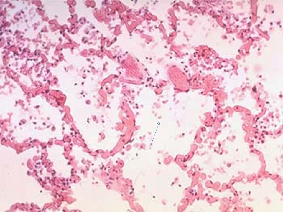
Pulmonary edema تورم الرئة

45
Congested alveolar septa Intra-alveolar transudate

Similar presentations


 {AJCC} from Cummings.please see handouts as well for updated AJCC Tx Minimum requirements.>")


>")


