Download presentation
Presentation is loading. Please wait.

1
Joints, Palpations, & ROM
Foot and Ankle Joints, Palpations, & ROM

2
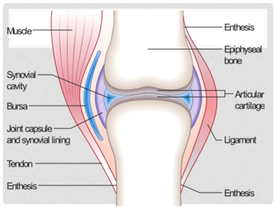
Joints: A synovial joint, also known as a diarthrosis , is the most common and most movable type of joint in the body Other types: Fibruous and Cartlaginous Main structural differences between synovial and fibrous joints are capsules surrounding the articulating surfaces of a synovial joint and the presence of lubricating synovial fluid within those capsules (synovial cavities).
.")
4
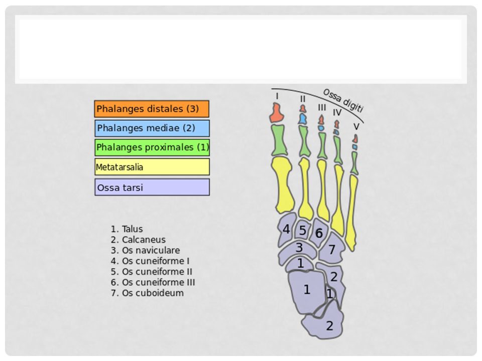
Joints Toes and Metatarsals:
Interphalangeal joints: These joints connect the phalanges. They’re synovial joints strengthened by collateral and plantar ligaments, and they let you flex and extend your toes. Metatarsophalangeal joints: They allow you to flex and extend your toes as well as move them apart and closer together. Intermetatarsal joints Tarsometatarsal joints Cuboideonavicular joints

6
joints Foot: These two joints allow you to invert and evert the foot Subtalar joint: This joint is the posterior joint formed between the talus and the calcaneus. It’s a synovial joint, and it’s stabilized by medial, lateral, and interosseous talocalcaneal ligaments. Transverse tarsal joint: The transverse tarsal joint is actually a combination of the following two joints: Talocalcaneonavicular joint Calcaneocuboid

7
Subtalar Joint

8
Transverse Tarsal Joint

9
Joints Ankle: The ankle joint is a synovial hinge joint, so you can plantarflex and dorsiflex The ankle joint is made up of distal ends of the tibia and fibula

10
ROM Toes Ankle Flexion/extension Abduction/Adduction
Dorsiflexion/plantarflexion Inversion/eversion Circumduction

11
ROM

12
What to palpate – Bony Landmarks
From distal to proximal: Distal Phalangeals Heads of the Metatarsals Styloid process of fifth metatarsal Sinus Tarsi - soft tissue depression just anterior to the lateral malleolus. (Sinus Tarsi is filled with EDB & fat pad) Medial and Lateral Malleoli Head of the Talus Calcaneous Shaft of the Tibia and Fibula Head of the fibula Tibial Tuberosity
 Medial and Lateral Malleoli. Head of the Talus. Calcaneous. Shaft of the Tibia and Fibula. Head of the fibula. Tibial Tuberosity.")
13
What to Palpate – Muscles and Tendons
Gastrocnemius Soleus Achilles Tendon Tibialis Anterior Extensor Digitorum Longus Flexor Digitorum Longus Peroneus Longus

14
Palpations - Foot

15
How to palpate “Palpate with a purpose”
Head of Talus - felt just behind the navicular, by everting & inverting the midfoot. Sustentaculum Tali - one fingerbreadth below medial malleolus. (serves as an attachment for the spring ligament & supports the talus); can be painful when palpated
; can be painful when palpated.")
16
Palpations - Ankle

17
In class… Pair off and palpate 3 different people’s foot and ankle
Identify bones and ligaments Continue to work on foot diagram if needed

18
What you need to know for the exam
Where to Palpate specific bones/ligaments ALL ROM

19
Homework Color pages 4 and 5 in the packet

Similar presentations