Download presentation
Presentation is loading. Please wait.

1
Essentials of Human Anatomy & Physiology Copyright © 2003 Pearson Education, Inc. publishing as Benjamin Cummings Slides 16.21 – 16.37 Seventh Edition Elaine N. Marieb Chapter 16 The Reproductive System Lecture Slides in PowerPoint by Jerry L. Cook

2
FEMALE REPRODUCTIVE SYSTEM Much more complex than the male. Needs to be able to produce gametes, AND nurture and protect the developing fetus during 9 months of pregnancy.

3
FEMALE REPRODUCTIVE SYSTEM INTERESTING FACTS The largest cell in the female body is the egg. A woman never runs out of eggs. At birth she has between 1 and 2 million potential eggs (follicles) and by puberty has 300,000 to 400,000 viable eggs (follicles) that can be fertilized. The average lifespan of an egg once it is released from the ovary is 12-24 hours after which it either disintegrates or is flushed out of the body with the menstrual flow. The average life span of the sperm is 2-3 days.
 and by puberty has 300,000 to 400,000 viable eggs (follicles) that can be fertilized. The average lifespan of an egg once it is released from the ovary is hours after which it either disintegrates or is flushed out of the body with the menstrual flow. The average life span of the sperm is 2-3 days..")
4
CONTINUED... In the uterus, prior to birth, the baby’s body is covered by a thin layer of hair. As soon as the baby is born that hair soon disappears. The hair is called lanugo (lan-oo-go). When a baby is born, both male and female, the mammary glands are sometimes so stimulated by the mothers hormones that they give tiny drops of milk for about a week.
. When a baby is born, both male and female, the mammary glands are sometimes so stimulated by the mothers hormones that they give tiny drops of milk for about a week..")
5
CONTINUED... Female babies can actually bleed vaginally for several days after birth in response to the removal of the mothers hormones during pregnancy. The number of times a woman's ovaries are stimulated to produce a mature ovum may be related to her lifetime risk for developing breast and/or ovarian cancer. Women who experience "ovarian rest" through taking the birth control pill, through pregnancies, through breast feeding, are at a lower lifetime risk for development of these diseases.

6
CONTINUED... The female human body is capable of giving birth to 35 children in one lifetime!!

7
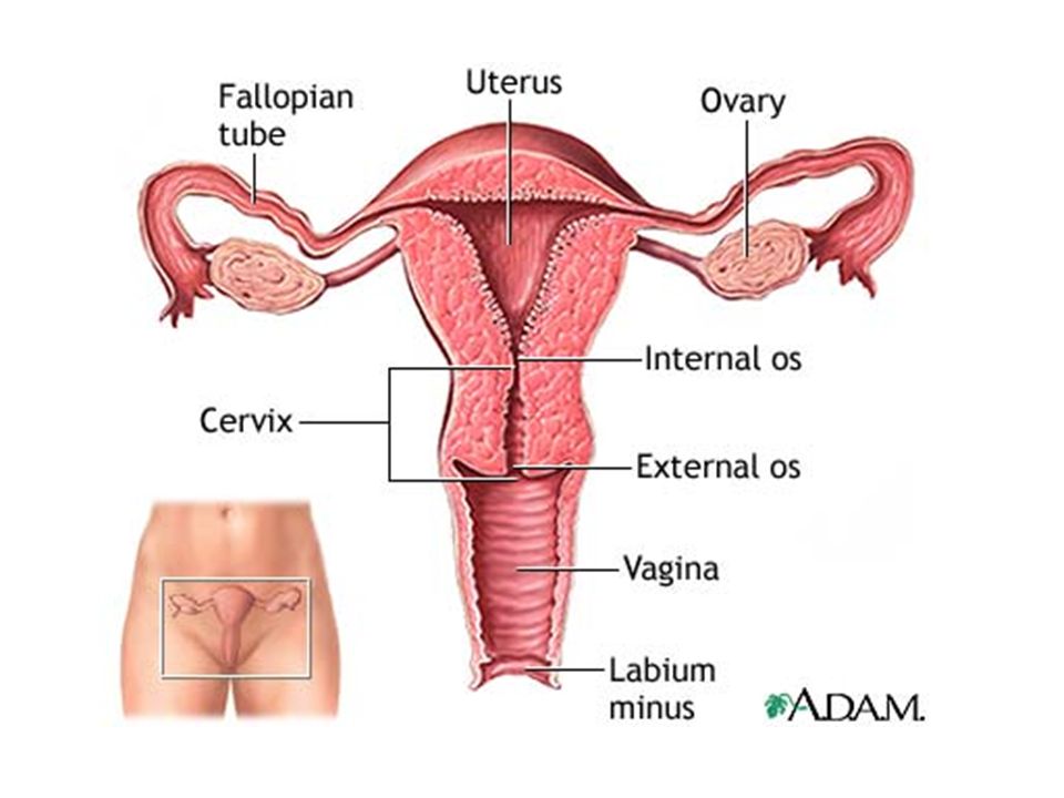
Female Reproductive System Slide 16.21a Copyright © 2003 Pearson Education, Inc. publishing as Benjamin Cummings Ovaries = primary sex organ that produce ova & endocrine sex organs Duct System: Uterine tubes (fallopian tubes) Uterus Vagina External genitalia
 Uterus Vagina External genitalia.")
8
Ovaries Internal Structure Each ovary is subdivided into a: 1. Medulla = contains blood, lymph vessels, & nerves = for nourishment & support 2. Cortex = ovarian follicles covered by germinal epithelial cells

9
Ovaries Slide 16.22 Copyright © 2003 Pearson Education, Inc. publishing as Benjamin Cummings Composed of ovarian follicles (sac-like structures) Structure of an ovarian follicle: Oocyte (immature egg) Follicular cells Figure 16.7
 Structure of an ovarian follicle: Oocyte (immature egg) Follicular cells Figure")
10
FOLLICLE MATURATION FIG. 16.7 During child bearing years, each month FSH stimulates one primordial follicle to mature: the following events occur over a 14 day period (approximately):
:.")
11
Ovarian Follicle Stages Slide 16.23 Copyright © 2003 Pearson Education, Inc. publishing as Benjamin Cummings 1. Primary follicle – contains an immature oocyte and grows (contains fluid filled cavity called antrum) 2. Graafian (vesicular) follicle – growing follicle with a maturing oocyte that is ready to be ejected 3. Ovulation – when the egg is mature the follicle ruptures to release egg Occurs about every 28 days The ruptured follicle is transformed into a 4. corpus luteum & eventually degenerates
 2. Graafian (vesicular) follicle – growing follicle with a maturing oocyte that is ready to be ejected 3. Ovulation – when the egg is mature the follicle ruptures to release egg Occurs about every 28 days The ruptured follicle is transformed into a 4. corpus luteum & eventually degenerates.")
12
Support for Ovaries Slide 16.24a Copyright © 2003 Pearson Education, Inc. publishing as Benjamin Cummings Suspensory ligaments – secure ovary to lateral walls of the pelvis Ovarian ligaments – attach to uterus Broad ligament – a fold of the peritoneum, encloses suspensory ligament

13
Support for Ovaries Slide 16.24b Copyright © 2003 Pearson Education, Inc. publishing as Benjamin Cummings Figure 16.8b

14
Uterine (Fallopian) Tubes Slide 16.25 Copyright © 2003 Pearson Education, Inc. publishing as Benjamin Cummings Receive the ovulated oocyte Provide a site for fertilization Attaches to the uterus (endpoint) Does not physically attach to the ovary: distal ends are expanded over ovary and form extensions called fimbriae Supported by the broad ligament
 Does not physically attach to the ovary: distal ends are expanded over ovary and form extensions called fimbriae Supported by the broad ligament.")
15
Uterine Tube Function Slide 16.26 Copyright © 2003 Pearson Education, Inc. publishing as Benjamin Cummings Fimbriae – finger-like projections at the distal end that receive the oocyte Cilia inside the uterine tube slowly move the oocyte towards the uterus (takes 3–4 days) Fertilization typically occurs inside the uterine tube (fallopian tube)
 Fertilization typically occurs inside the uterine tube (fallopian tube).")
16
http://www.youtube.com/watch?v=dwtFYOLFeN w&feature=topicshttp://www.youtube.com/watch?v=dwtFYOLFeN w&feature=topics

17
Uterus Slide 16.27 Copyright © 2003 Pearson Education, Inc. publishing as Benjamin Cummings Musclular organ, Located between the urinary bladder and rectum Hollow organ Functions of the uterus: Receives a fertilized egg from fallopian tube Retains the fertilized egg Nourishes the fertilized egg

18
Female Reproductive System Slide 16.21b Copyright © 2003 Pearson Education, Inc. publishing as Benjamin Cummings Figure 16.8a

20
Support for the Uterus Slide 16.28a Copyright © 2003 Pearson Education, Inc. publishing as Benjamin Cummings Broad ligament – attached to the pelvis Round ligament – anchored interiorly Uterosacral ligaments – anchored posteriorly

21
Support for the Uterus Slide 16.28b Copyright © 2003 Pearson Education, Inc. publishing as Benjamin Cummings Figure 16.8b

22
Regions of the Uterus Slide 16.29 Copyright © 2003 Pearson Education, Inc. publishing as Benjamin Cummings Body – main portion Fundus – area where uterine tube enters Cervix – narrow outlet that protrudes into the vagina: pap smears are taken from cervical tissue

23
Walls of the Uterus Slide 16.30 Copyright © 2003 Pearson Education, Inc. publishing as Benjamin Cummings Endometrium Inner layer Allows for implantation of a fertilized egg Sloughs off if no pregnancy occurs (menses) Myometrium – middle layer of smooth muscle: makes bulk of uterus Serous layer – outer visceral peritoneum / covering
 Myometrium – middle layer of smooth muscle: makes bulk of uterus Serous layer – outer visceral peritoneum / covering.")
25
Vagina Slide 16.31 Copyright © 2003 Pearson Education, Inc. publishing as Benjamin Cummings Extends from cervix to exterior of body Behind bladder and in front of rectum Serves as the birth canal Receives the penis during sexual intercourse Hymen – partially closes the vagina until it is ruptured

26
External Genitalia (Vulva) Slide 16.32a Copyright © 2003 Pearson Education, Inc. publishing as Benjamin Cummings Mons pubis Fatty area overlying the pubic symphysis (cartilage) Covered with pubic hair after puberty Figure 16.9
 Covered with pubic hair after puberty Figure")
27
External Genitalia (Vulva) Slide 16.32b Copyright © 2003 Pearson Education, Inc. publishing as Benjamin Cummings Labia – skin folds Labia majora Labia minora Figure 16.9

28
External Genitalia Slide 16.33 Copyright © 2003 Pearson Education, Inc. publishing as Benjamin Cummings Vestibule Enclosed by labia majora Contains opening of the urethra and the greater vestibular glands (produce mucus) Clitoris Contains erectile tissue, external excitatory organ Corresponds to the male penis: becomes engorged with blood and swells during sexual stimulation
 Clitoris Contains erectile tissue, external excitatory organ Corresponds to the male penis: becomes engorged with blood and swells during sexual stimulation.")
29
Oogenesis Slide 16.34 Copyright © 2003 Pearson Education, Inc. publishing as Benjamin Cummings The total supply of eggs are present at birth Ability to release eggs begins at puberty Reproductive ability ends at menopause Oocytes are matured in developing ovarian follicles

30
Oogenesis Slide 16.35 Copyright © 2003 Pearson Education, Inc. publishing as Benjamin Cummings Oogonia – female stem cells found in a developing fetus. **This occurs with the primordial germ cells within the embryo. Step 1 (*@ embryo): Oogonia (46 chromosomes) undergo mitosis to produce primary oocytes (** Note that human females are born with all potential ova as primary oocytes) Primary oocytes are surrounded by cells that form primary follicles in the ovary Oogonia no longer exist by the time of birth
: Oogonia (46 chromosomes) undergo mitosis to produce primary oocytes (** Note that human females are born with all potential ova as primary oocytes) Primary oocytes are surrounded by cells that form primary follicles in the ovary Oogonia no longer exist by the time of birth.")
32
Oogenesis Slide 16.36 Copyright © 2003 Pearson Education, Inc. publishing as Benjamin Cummings Primary oocytes are inactive until puberty @ puberty, once each month, Follicle stimulating hormone (FSH) causes some primary follicles to mature & grow: 1. Oocyte (92) undergoes Meiosis inside maturing follicle 2. Produces a secondary oocyte (46 chromosomes) and the first polar body(46 chromosomes)
 causes some primary follicles to mature & grow: 1. Oocyte (92) undergoes Meiosis inside maturing follicle 2. Produces a secondary oocyte (46 chromosomes) and the first polar body(46 chromosomes).")
33
FUNCTION OF POLAR BODIES: Although the polar bodies do not become anything they do serve a purpose in reproduction/oogenesis. The polar bodies are the byproducts of the primary and secondary oocyte at each point of meiotic division in oogenesis. The polar body allows for the oocyte to get rid of chromosomes while at the same time taking the least amount of resources (cytoplasm) from the oocyte. Each meiotic division serves as a means of moving the oocyte toward its need haploid number of chromosomes for fertilization. So you could say that the polar bodies function as a means of cellular structure conservation. They help ensure that the oocyte remains nutrient/resource rich while at the same time helping the oocyte reach its haploid number.
 from the oocyte. Each meiotic division serves as a means of moving the oocyte toward its need haploid number of chromosomes for fertilization. So you could say that the polar bodies function as a means of cellular structure conservation. They help ensure that the oocyte remains nutrient/resource rich while at the same time helping the oocyte reach its haploid number..")
34
Meiosis II is completed after ovulation (*Triggered by LH) only if sperm penetrates When & if Meiosis II is complete: A second polar body is separated from the large ovum *& the haploid nuclei of the sperm and the now- matured ovum fuse
 only if sperm penetrates When & if Meiosis II is complete: A second polar body is separated from the large ovum *& the haploid nuclei of the sperm and the now- matured ovum fuse")
35
Oogenesis Slide 16.37 Copyright © 2003 Pearson Education, Inc. publishing as Benjamin Cummings Figure 16.10

36
http://www.youtube.com/watch?v=_dYxH9MxRp whttp://www.youtube.com/watch?v=_dYxH9MxRp w http://www.youtube.com/watch?v=tvikQMfKPx M&feature=relatedhttp://www.youtube.com/watch?v=tvikQMfKPx M&feature=related

37
TWINS http://www.pennmedicine.org/encyclopedia/em_ DisplayAnimation.aspx?gcid=000058&ptid=17http://www.pennmedicine.org/encyclopedia/em_ DisplayAnimation.aspx?gcid=000058&ptid=17

38
Oogenesis vs. Spermatogenesis

Similar presentations


.>")


