Download presentation
Presentation is loading. Please wait.

1
ANTERIOR, POSTERIOR COMPARTMENTS & CUBITAL FOSSA Dr. Kumar K. V. Associate Professor 26.10.2009

2
Biceps brachii Origin: long head, supraglenoid tubercle; short head, coracoid process Insertion: radial tuberosity Action: supinator of forearm, flexor of elbow joint, weak flexor of should joint Pronator teres Origin: medical epicondyle of humerus and deep fascia of forearm Insertion: middle of lateral surface of radius Action: pronation of forearm and flexion of elbow

3
Muscles of arm Antererior group –Biceps brachii Coracobrachia lis –Brachialis Posterior group – triceps brachii

4
Musculocutaneous n. Median n. Medial brachial cutaneous n. Medial antebrachial cutaneous n. Superior ulnar coleteral a.Deep brachial a. Ulnar n. Biceps Median n.

5
Musculocutaneous n. Median n. Axillary a. Brachial a. Medial antebrachial cutaneous n. Medial brachial cutaneous n. Deep brachial a. Superior ulnar coleteral a. & Ulnar n. Inferior ulnar coleteral a.

6
Musculocutaneous Distribution: Biceps brachii, brachalis and coracobrachialis ‘BBC nerve’; skin on anterior aspect of forearm

7
Brachial artery Continuation of axillary artery Divides into radial and ulnar arteries at level of neck of radius Branches –Deep brachial a. - accompanies with radial nerve –Superior ulnar collaeral a. – - accompanies with ulnar nerve –Inferior ulnar collateral a.

8
Radial artery and branches Radial recurrent a. Superfical palmar branch Principal artery of thumb Ulnar artery and branches Ulnar recurrent a. Common interosseous artery –Anterior interossous a. –Posterior interosseous a. Deep palmar branch

9
Supraspinatus Axillary nerve in the quadrangular space Triceps muscles Tendon of Triceps Ulnar n. Teres Major Deltoid Unconeus POSTERIOR COMPARTMENT OF ARM

10
Triceps brachii Origin: long head, infraglenoid tubercle; lateral head, above groove for radial n., medial head, below groove for radial n. Insertion: olecranon of ulna Action: extends elbow joint), long head can extend and adduct shoulder joint
, long head can extend and adduct shoulder joint.")
11
Radial Nerve in the Arm & Forearm –Distribution: Extensor muscles of arm and forearm, brachioradialis; skin on back of arm, forearm, and radial side of dorsum of hand and radial two and one-half fingers –Injury: Wristdrop

12
Joints of the Upper Extremity Elbow Joint –Synovial – hinge –Diarthrosis Articulations –Humerus & Ulna –Humerus & Radius Many Ligaments

13
Cubital fossa Boundaries Base - line drawn through epicondyles of humerus Apex - brachioradialis laterally and pronator teres medially Roof - skin, superficial fascia, deep fascia and aponeurosis of biceps Floor - brachialis, supinator and capsule of elbow joint

14
Contents from lateral to medial Biceps brachii tendon Brachial a.divides into radial and ulnar a.usually at apex of fossa Median n. Lateral to the biceps brachii tendon,radial n. and lateral antebrachial cutaneous n.

15
Basilic vein –Arises from the medial side of the dorsal venous arch of hand –Ascends on the ulnar side of forearm to the elbow and then in the medial bicepital brachii furrow to middle of the arm where it pierces the deep fascia and joins the brachial vein or axillary vein Median cubital vein links cephalic vein and basilic vein in the cubital fossa. It is a frequent site for venipuncture to remove a sample of blood or add fluid to the blood

16
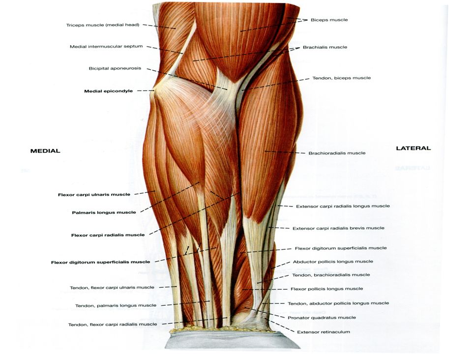
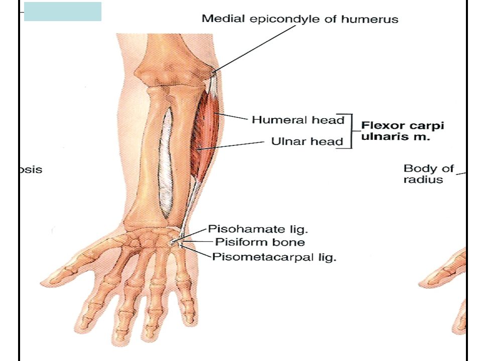
Anterior Compartment Forearm Flexor Carpi Radialis Flexor Retinaculum Medial Epicondyle Flexor Digitorum Superficialis is deep to other flexors Flexor Carpi Ulnaris Brachioradialis Pronator Teres Anterior View

17
Muscles of forearm Superficial layer –Brachioradialis –Pronator teres –Flexor carpi radialis –Palmaris longus –Flexor carpi ulnaris

19
Muscolocutaneous n. Brachial a. Radial n. Radial a. Median n. Common interosseous a. Ulnar a., v. & n. FOREARM VESSELS & NERVES

20
Radial recurrent a. Radial a. Radial n. Median n. Ulnar n. Ulnar a. Ulnar recurrent a. Ulnar n. Brachial a. CUBITAL NERVES & ARTERIES

21
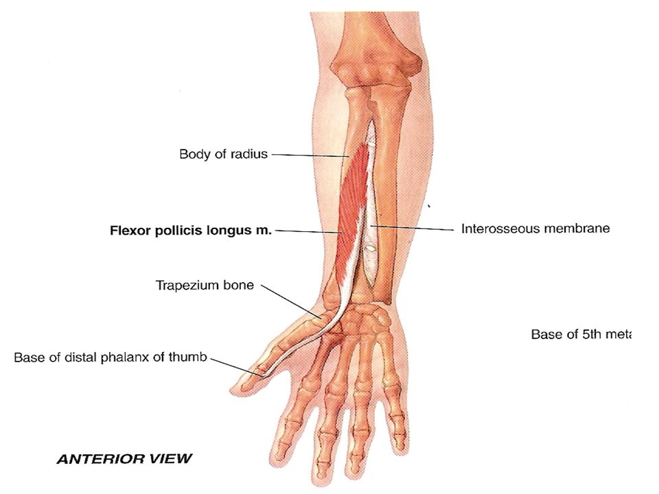
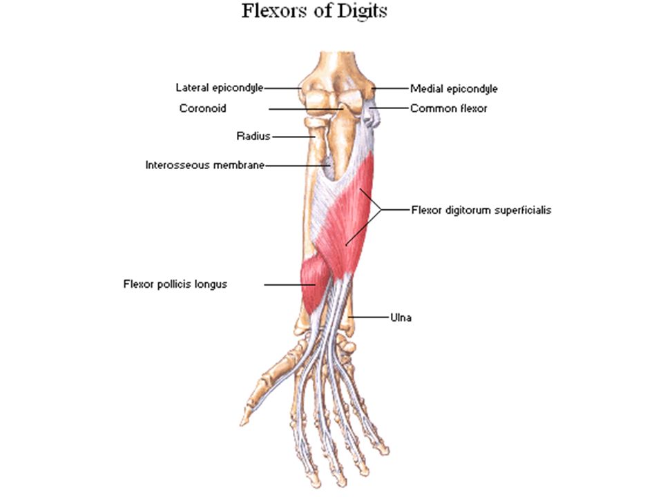
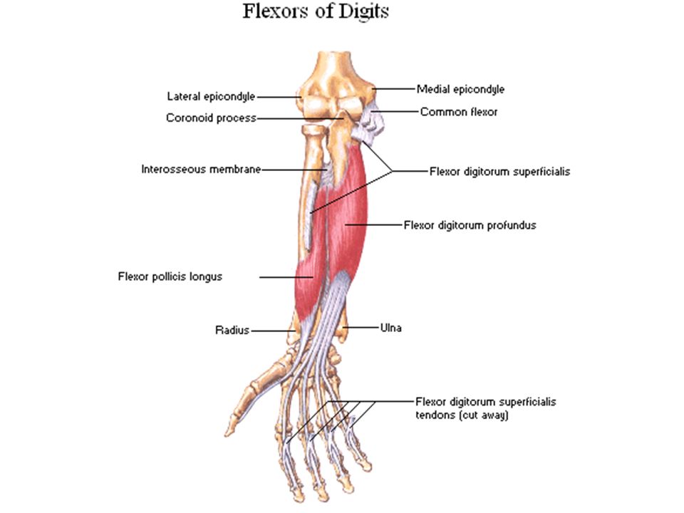
Superficial Layer: (1) Pronator teres, (2) flexor carpi radialis, (3) palmaris longus, (4) flexor digitorum superficialis and (5) flexor carpi ulnaris. Deep Layer: (1) Flexor pollicis longus, (2) flexor digitorum profundus and (3) pronator quadratus. Muscles of the Anterior Compartment of the forearm
 Flexor pollicis longus, (2) flexor digitorum profundus and (3) pronator quadratus. Muscles of the Anterior Compartment of the forearm.")
22
Pronators of the forearm

23
Pronator teresOrigin: Humeral head: from the medial epicondyle of the humerus (common flexor origin). Ulnar head: from the medial border of the coronoid process of the ulna. Insertion: Into the pronator tuberosity on the lateral surface of the middle part of the shaft of the radius. Nerve Supply: From the median nerve. Action: Pronation of the forearm at the radio-ulnar joints. Flexion of the forearm at the elbow joint.

24
Flexor carpi radialis

26
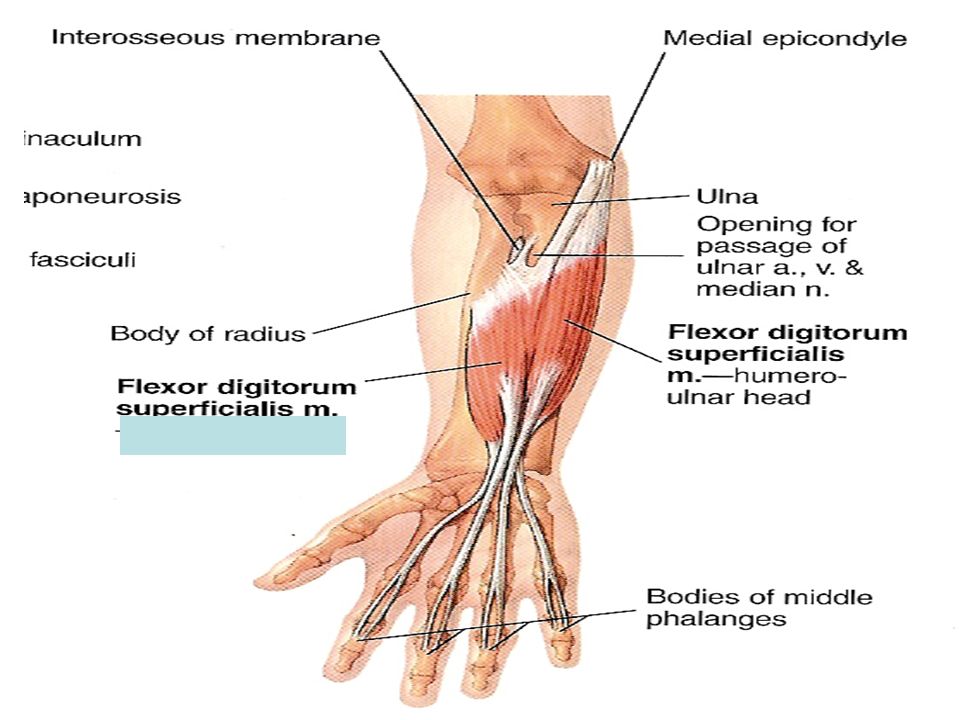
Flexor digitorum superficialis Origin: 1.Humero-ulnar head: from the medial epicondyle of the humerus (common flexor origin) and from the medial border of the coronoid process of the ulna. 2.Radial head: from the oblique line on the anterior surface of the shaft of the radius. Insertion: Its tendon divides into 4 tendons which are inserted into the sides of the middle phalanges of the medial 4 fingers.Its tendon divides into 4 tendons which are inserted into the sides of the middle phalanges of the medial 4 fingers. Nerve Supply: From the median nerve.From the median nerve. Action: 1.Flexion of the proximal interphalageal joints and metacarpophalangeal joints of the medial 4 fingers. 2.Helps of flexion of the hand at the wrist joint.

30
Flexor digitorum profundus Origin: From the upper ¾ of the anterior surface of the shaft of the ulna and adjoining part of the interosseus membrane.From the upper ¾ of the anterior surface of the shaft of the ulna and adjoining part of the interosseus membrane. Insertion: The muscle divides into 4 tendons which pierce the tendons of the flexor digitorum superficialis and are inserted into the bases of the distal phalanges of the medial 4 fingers.The muscle divides into 4 tendons which pierce the tendons of the flexor digitorum superficialis and are inserted into the bases of the distal phalanges of the medial 4 fingers. Nerve Supply: Its lateral half: from the anterior interosseus nerve (branch of the median nerve).Its lateral half: from the anterior interosseus nerve (branch of the median nerve). It medial half: from the ulnar nerve.It medial half: from the ulnar nerve. Action: 1.Flexion of the distal interphalangeal joints of the medial 4 fingers. 2.Helps in flexion of the proximal interphalangeal joints and metacarpophalangeal joints of the medial 4 fingers. 3.Helps of flexion of the hand at the wrist joint.
.Its lateral half: from the anterior interosseus nerve (branch of the median nerve). It medial half: from the ulnar nerve.It medial half: from the ulnar nerve. Action: 1.Flexion of the distal interphalangeal joints of the medial 4 fingers. 2.Helps in flexion of the proximal interphalangeal joints and metacarpophalangeal joints of the medial 4 fingers. 3.Helps of flexion of the hand at the wrist joint..")
33
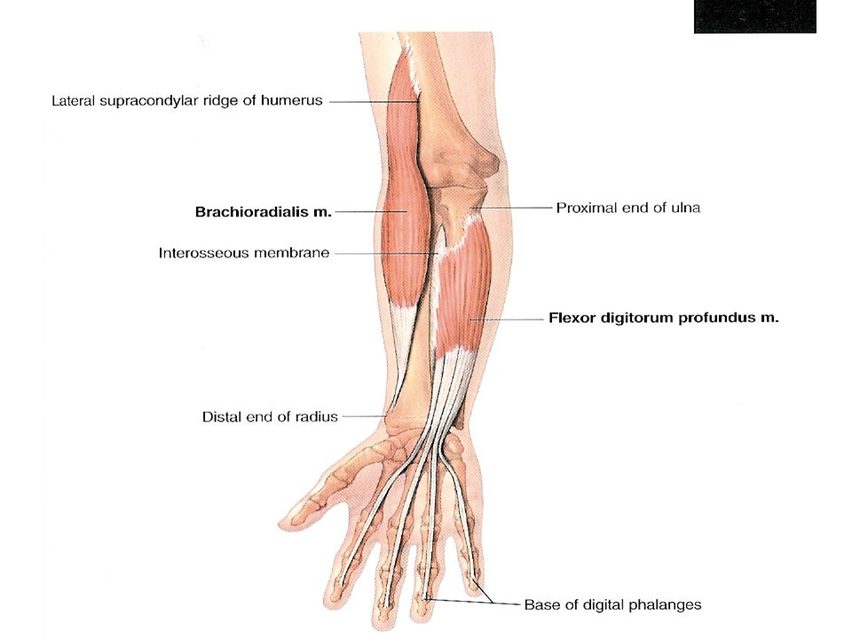
Brachioradialis Origin: from the upper 2/3 of the lateral supracondylar ridge of the humerus. Insertion: Into the base of the styloid process of the radius. Nerve Supply: From the radial nerve. Action: Flexion of the forearm (specially in midprone position). Restoration of the forearm into the midprone position.
. Restoration of the forearm into the midprone position..")
34
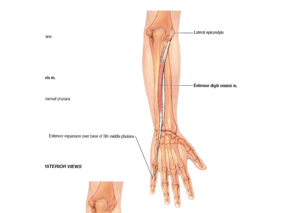
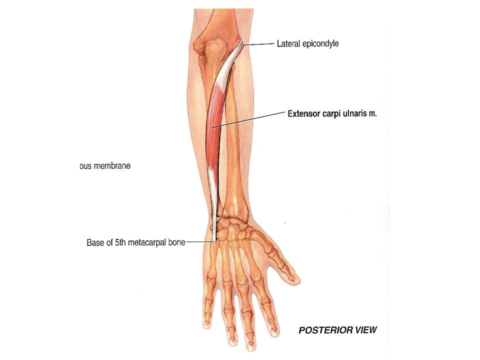
ExtensorCompartments Of the Forearm & The Hand

37
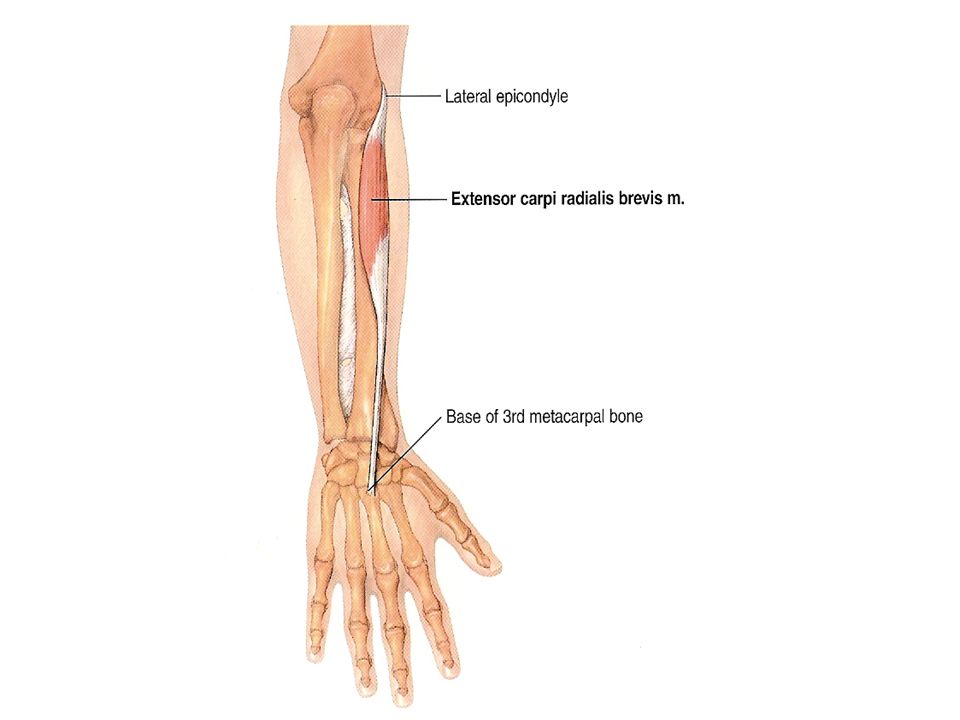
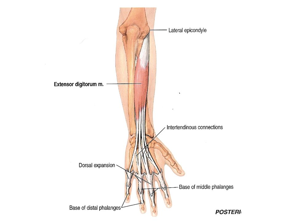
Extensor digitorum Origin: from the lateral epicondyle of the humerus (common extensor origin). Insertion: Into the extensor expansion of the medial 4 fingers. Nerve Supply: From the deep branch of radial nerve. Action: Extension of metacarpophalangeal and interphalangeal joints of the medial 4 fingers. Extension of the hand at wrist joint.

42
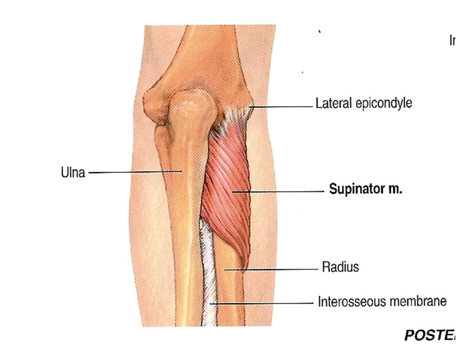
SupinatorOrigin: from (1) the lateral epicondyle of the humerus, (2) lateral collateral ligament of the elbow joint, (3) annular ligament of the superior radio-ulnar joint, and (4) supinator fossa and crest of the ulna. Insertion: Into the upper 1/3 of the lateral surface of the shaft of the radius. Nerve Supply: From the deep branch of the radial nerve. Action: It helps the biceps in supination of the forearm at the radio-ulnar joints.

44
Deep palmar fascia Superficial layer Thenar fascia Hypothenar fascia Palmar aponeurosis thick triangular membrane Deep layer palmar interosseous fascia

45
Palmar aponeurosis

46
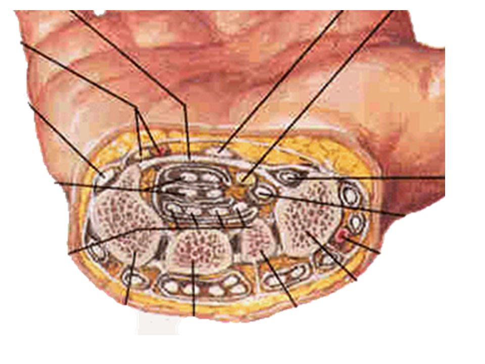
Carpal Tunnel Flexor retinaculum Thickening of deep fascia in the hand Attached laterally to scaphoid and trapeziun and medially to pisiform and hamate Carpal tunnel Formed by flexor retinaculum and carpal groove Transmits –Median n. –Flexor digitorum superficialis and flexor digitorum profundus enclosed by common flexor sheath –Flexor pollicus longus enclosed by tendinous sheath of flexor pollicus longus

48
Flexar Retinaculum & related structures Common flexor sheath Tendinous sheath of flexor pollicis longus

49
Muscles of hand Lateral group - thenar (4) –Abductor pollicis brevis –Flexor pollicis brevis –Opponens pollicis –Adductor pollicis Action: flex, abduct, adduct and oppose thumb Medial group - hypothenar (3) –Abductor digiti minimi –Flexor digiti minimi brevisOpponens digiti minimi –Action: flex, abduct, and oppose little finger
 –Abductor pollicis brevis –Flexor pollicis brevis –Opponens pollicis –Adductor pollicis Action: flex, abduct, adduct and oppose thumb Medial group - hypothenar (3) –Abductor digiti minimi –Flexor digiti minimi brevisOpponens digiti minimi –Action: flex, abduct, and oppose little finger")
50
Superficial palmar arch Formed by ulnar artery and superficial palmar branch of radial artery Curve of arch lies across the palm, level with the distal border of fully extended thumb Gives rise to three common palmar digital arteries each then divides into two proper palmar digital arteries

51
Superficial palmar a. recurrent n. Ulnar a. Superficial Palmar Arterial Arch

52
Nerves, Vessels & Hand Muscles

53
Deep palmar arch Formed by radial artery and deep palmar branch of ulnar artery Curve of arch lies across upper part of palmar at level with proximal border of extended thumb Gives rise to three palmar metacarpal arteries

54
Intermedial group Lumbricales (4) - flex fingers at MP joints; extend fingers at IP joints Palmar interossei (3) - adduct fingers towards middle finger at MP joints Dorsal interossei (3) - abduct fingers away from middle finger at MP joints
 - flex fingers at MP joints; extend fingers at IP joints Palmar interossei (3) - adduct fingers towards middle finger at MP joints Dorsal interossei (3) - abduct fingers away from middle finger at MP joints")
55
Intrinsic Muscles of Hand ABduction Palmar Interossei Lumbricals ADDuction 1st 2nd 3rd 4th Dorsal Interossei Interossei help the lumbricals to extend IP joints and flex MC-P joints

56
Lumbricals & Nerves

57
Anatomical snuff box When the thumb is abducted and extended, a triangular hollow appears between the tendon of the extensor pollicis longs medially and the tendons of the extensor pollicis brevis and abductor pollicis longus laterally. The floor of the snuff box is the scaphoid and trapezium bones and crossed by the radial a..

58
Extensor Retinaculum & Dorsum of Hand

59
Dorsum of hand Extensor retinaculum Thickening of deep faxcia of forearm a wrist Attached laterally to radius and medially to styloid process of ulna and triquetrum Forms six fibrous compartments for extensor tendons passing from forearm into hand:

60
tendons of abductor pollicis longus and extensor pollicis brevis and their synovial sheaths; tendons of extensor carpi radialis longus and brevis and their synovial sheaths; tendon of pollicis longus and its synovial sheath; tendons of extensor digitorum, extensor indicis and their synovial sheaths; tendon of extensor digiti minimi and its synovial sheaths; tendon of extensor carpi ulnaris and its synovial sheaths

61
Fascia of the dorsal hand The superficial fascia Deep fascia - the dorsal fascia of hand –Superficial layer (dorsal aponeurosis) –Deep layer (dorsal interosseous fascia)
 –Deep layer (dorsal interosseous fascia)")
62
Two spaces The dorsal subcutaneous space The dorsal subaponeurotic space Superficial fascia Dorsal aponeurosis Dorsal interosseous fascia Dorsal subcutaneous space Dorsal subaponeurotic space

63
Nerves of hand Median n. - thenar except adductor pollicis, first two lumbricals; skin of thenar, central part of palm, palmar aspect of radial three and one-half fingers, including middle and distal fingers on dorsum Ulnar n. - hypothenar muscles, interossei, 3rd and 4th lumbricals and adductor pollicis; skin of hypothenar, palmar surface of ulnar one and one-half fingers

64
Radial n. skin of radial side of dorsum of hand and radial two and one-half fingers

65
Intrinsic Muscles of Hand Little finger –All digiti minimiUlnar (Flexor, Abductor, Opponens) Thumb –Abductor pollicis brevisMedian –Flexor pollicis brevisMedian –Opponens pollicisMedian –Adductor pollicisUlnar Other Intrinsic Muscles –Palmar + Dorsal InterosseiUlnar –LumbricalsMedian, Ulnar MuscleNerve
 Thumb –Abductor pollicis brevisMedian –Flexor pollicis brevisMedian –Opponens pollicisMedian –Adductor pollicisUlnar Other Intrinsic Muscles –Palmar + Dorsal InterosseiUlnar –LumbricalsMedian, Ulnar MuscleNerve")
Similar presentations


>")


 Joint and Hand>")

