Download presentation
Presentation is loading. Please wait.

1
NEPHROLOGY: THE MAKING OF URINE
DR. NAEEM

3
Outline Introduction Function Cross Section of the kidney Blood supply
Nephron Nephron Physiology

4
INTRODUCTION The kidneys are a pair of bean shaped organs found along the posterior wall of the abdominal cavity. The left kidney is located slightly more superior than the right kidney due to the larger size of the liver on the right side of the body.

5
Function of the kidneys
Homeostatic functions Regulation of electrolytes Maintenance of acid-base Regulation of blood pressure Filtration of blood Formation of URINE Production of hormones - calcitriol, erythropoietin and the enzyme renin.

6
The kidneys are bean-shaped with the convex side of each organ located laterally and the concave side medial. The indentation on the concave side of the kidney, known as the renal hilus, provides a space for the renal artery, renal vein, and ureter to enter the kidney. Every day, a person's kidneys filter about 120 to 150 quarts of blood to produce about 1 to 2 quarts of waste products and extra fluid. The wastes and extra fluid become urine, which flows to the bladder through tubes called ureters.

7
Blood Supply The renal arteries branch directly from the abdominal aorta and enter the kidneys. Once in the kidney, the renal arteries diverge into smaller arterioles. Afferent arterioles carry blood into the renal cortex, where it separated into a bundle of capillaries known as a GLOMERULUS. From the glomerulus, the blood recollects into smaller efferent arterioles that descend into the renal medulla. The efferent arterioles separate into the peritubular capillaries that surround the renal tubules. Next, the peritubular capillaries merge to form veins that merge again to form the large renal vein. From the renal vein, the blood joins the IVC which carries blood back to the heart.

8
Blood Supply cont…

9
Nephrons The actual removal of wastes occurs in tiny units inside the kidneys called nephrons. Each kidney has about a million nephrons. In the nephron, a glomerulus—which is a tiny blood vessel, or capillary—intertwines with a tiny urine-collecting tube called a tubule. The glomerulus acts as a filtering unit, or sieve, and keeps normal proteins and cells in the bloodstream, allowing extra fluid and wastes to pass through.

11
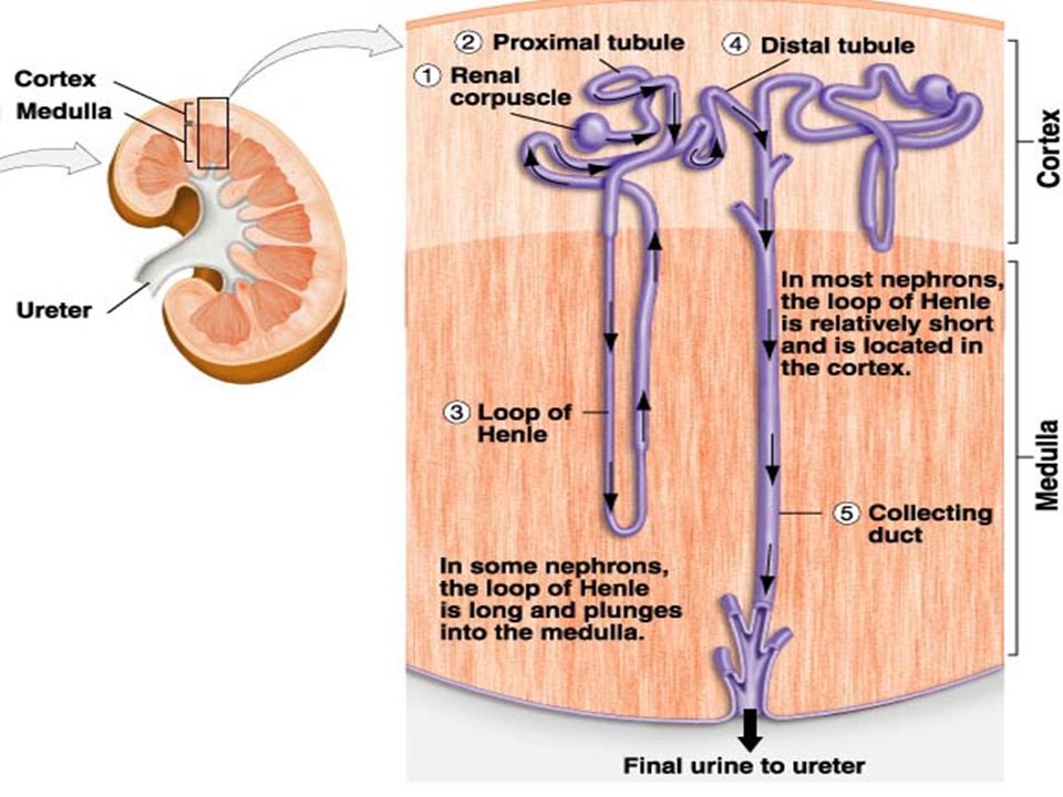
Renal Corpuscle The renal corpuscle is the initial blood- filtering component of a nephron. It consists of 2 structures: the glomerulus and a Bowmans capsule. The glomerulus is a small tuft of capillaries Fluid from blood in the glomerulus is collected in the Bowman's capsule to form "glomerular filtrate", which is then further processed along the nephron to form urine.

12
Renal Corpuscle Continued…
The structures of the layers of the Glomerulus determines the permeability. Factors that influence the selectivity are the negative charges on the Basement Membrane , the epithelium and the pore size of the glomeruli. As a result, small ions like Na & K will pass freely while large molecules like proteins, albumin and hemoglobin have no permeability.

13
Renal Corpuscle Continued
Basic Filtration unit of the kidney. GFR: Glomerular Filtration Unit Rate at which blood is filtered through all of the golmeruli, and thus the measure of the overall renal function. A glomerulus and its surrounding Bowman's capsule constitute a renal corpuscle, the basic filtration unit of the kidney. The rate at which blood is filtered through all of the glomeruli, and thus the measure of the overall renal function, is the glomerular filtration rate (GFR).
.")
14
The renal tubule carries urine from the glomerular capsule to the renal pelvis.

16
Proximal Tubule Also known as the Proximal Convoluted Tubule (PCT).
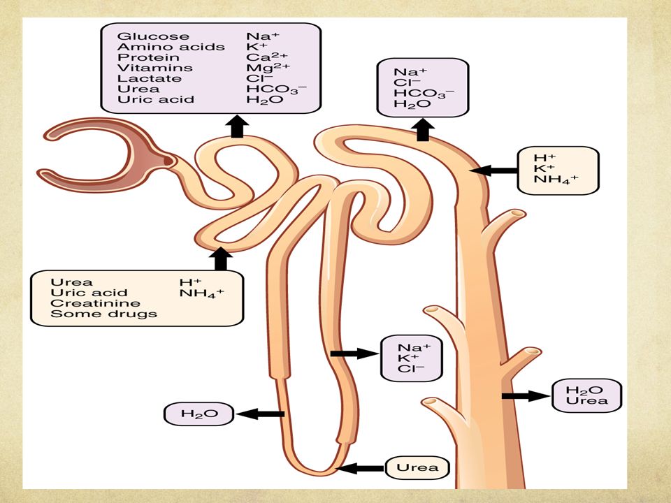
A duct system transporting urine from the Bowmans Capsule to the Loop of Henle. 65-80% of the filtrate is reabsorbed in the PCT. IT IS NOT A PLACE FOR TIGHT REGULATION, THAT WILL COME LATER ON. MOST REABSORPTION IS COUPLED WITH NA ION MOVEMENT. BUT THERE IS ALSO ACTIVE TRANSPORT AND EVEN SIMPLE DIFFUSION TAKING PLACE HERE.

17
Promixal Tubule Continued…
Substance % of Filtrate Reabsorbed Na & H2O ~66% Glucose& amino acids ~100% Potassium ~65% Urea ~50% Phosphate ~80%

18
A summary of the reabsorption along the proximal tubule:
Na: Diffusion through ion channels followed by water and Cl or Co- transported with glucose or amino acids. K: Potassium is absorbed mainly by the paracellular route with water via osmosis. Na/K ATPase is pumping 2 K back into the epithelial cells & K is then cleared from the cells using a co-transporter with chlorine. Urea: 50% of filtered urea is reabsorbed in the proximal tubule. However the concentration of urea actually increases thanks to the reabsorption of 70% of the filtered water in the same portion of the nephron. Glucose: Reabsorption of glucose can only occur in the proximal tubule and occurs regardless of the concentration gradient as it is completed via secondary active transport. It is reabsorbed using a co-transported with sodium.secondary active transport. It is reabsorbed using Na. H Secretion/HCO3 Reabsorption: The secretion of H+ in this section of the nephron is mainly a result of the Na+/H+ exchanger. Energy for it provided by Na/K ATPase. The majority (70%) of sodium is reabsorbed in the proximal tubule. It is reabsorbed into the cytosol of the epithelial cells either alone by diffusion through ion channels followed by water and chloride or together with another product such as glucose or AA using a co-transporter by secondary active co-transport. odium/potassium ATPase Pump and is an example of primary active transport. This pump removes three sodium ions from the cell and pumps two potassium ions back in.
 of sodium is reabsorbed in the proximal tubule. It is reabsorbed into the cytosol of the epithelial cells either alone by diffusion through ion channels followed by water and chloride or together with another product such as glucose or AA using a co-transporter by secondary active co-transport. odium/potassium ATPase Pump and is an example of primary active transport. This pump removes three sodium ions from the cell and pumps two potassium ions back in.")
21
Loop of Henle Is the part of the nephron that connects the proximal convoluted tubule (PCT) to the distal convoluted tubule (DCT). The main function of the Loop of Henle is to maintain a concentration gradient. As water is osmotically driven from the descending limb into the interstitium, it readily enters the vasa recta.

22
Loop of Henle Continued…
Is divided into 4 parts. Thin descending limb of the loop of Henle. Thin ascending limb of loop of Henle Thick ascending limb Cortical thick ascending limb It can be divided into four parts: Thin descending limb of loop of Henle The thin descending limb has low permeability to ions and urea, while being highly permeable to water. The loop has a sharp bend in the renal medulla going from descending to ascending thin limb. Thin ascending limb of loop of Henle The thin ascending limb is impermeable to water, but it is permeable to ions. Thick ascending limb of loop of Henle Sodium (Na+), potassium (K+) and chloride (Cl-) ions are reabsorbed from the urine by secondary active transport by a Na-K-Cl cotransporter (NKCC2). The electrical and concentration gradient drives more reabsorption of Na+, as well as other cations such as magnesium (Mg2+) and calcium (Ca2+). Cortical thick ascending limb The cortical thick ascending limb drains urine into the distal convoluted tubule.[2]
, potassium (K+) and chloride (Cl-) ions are reabsorbed from the urine by secondary active transport by a Na-K-Cl cotransporter (NKCC2). The electrical and concentration gradient drives more reabsorption of Na+, as well as other cations such as magnesium (Mg2+) and calcium (Ca2+). Cortical thick ascending limb. The cortical thick ascending limb drains urine into the distal convoluted tubule.[2]")
23
Loop of Henle Continued…
The descending loop of Henle receives isotonic (300 mOsm/L) fluid from the proximal convoluted tubule (PCT). The descending portion of the loop of Henle is extremely permeable to water and is less permeable to ions, therefore water is easily reabsorbed here and solutes are not readily reabsorbed. The ascending limb of the loop of Henle receives an even lower volume of fluid and has different characteristics compared to the descending limb. In the ascending portion, the loop becomes impermeable to water and the cells of the loop actively reabsorb solutes from the luminal fluid; therefore water is not reabsorbed and ions are readily reabsorbed.
 fluid from the proximal convoluted tubule (PCT). The descending portion of the loop of Henle is extremely permeable to water and is less permeable to ions, therefore water is easily reabsorbed here and solutes are not readily reabsorbed. The ascending limb of the loop of Henle receives an even lower volume of fluid and has different characteristics compared to the descending limb. In the ascending portion, the loop becomes impermeable to water and the cells of the loop actively reabsorb solutes from the luminal fluid; therefore water is not reabsorbed and ions are readily reabsorbed.")
24
Loop of Henle Continued…
Overall the loop of Henle reabsorbs around 25% of filtered ions and 20% of the filtered water in a normal kidney. These ions are mostly Na Cl K Ca HCO3.

26
Distal Convoluted Tubule
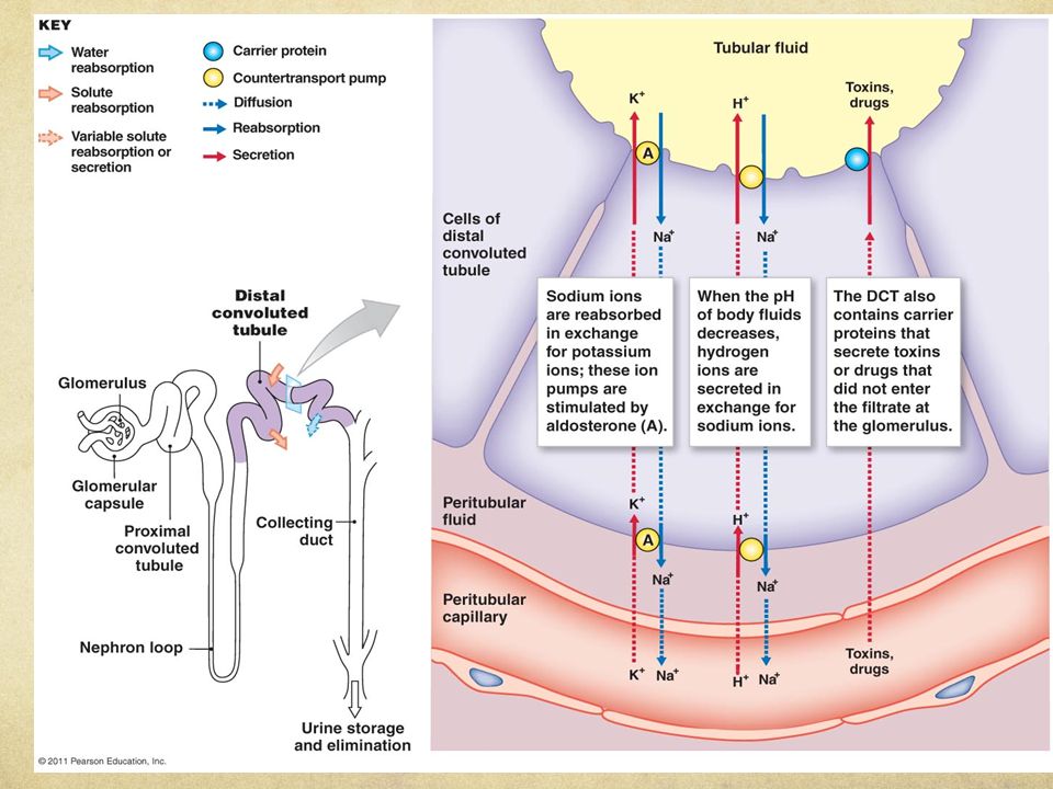
The distal convoluted tubule (DCT) is a portion of kidney nephron between the loop of Henle and the collecting duct system. Functions: Reabsorbs Na+ ions through coupled secretion of H+ or K+ ions into the tubular fluid via aldosterone. By acidifying the urine the distal convoluted tubule plays an important role in acid-base balance. Normally is relatively impermeable to water. However in the presence of antidiuretic hormone (ADH) its permeability to water increases permitting concentration of the urine. Secretes ammonium ions and some drugs. Forms part of the juxtaglomerular apparatus. Hemodynamic regulation via macula densa and renin Electrolyte homeostasis via Calcium reabsorption
 is a portion of kidney nephron between the loop of Henle and the collecting duct system. Functions: Reabsorbs Na+ ions through coupled secretion of H+ or K+ ions into the tubular fluid via aldosterone. By acidifying the urine the distal convoluted tubule plays an important role in acid-base balance. Normally is relatively impermeable to water. However in the presence of antidiuretic hormone (ADH) its permeability to water increases permitting concentration of the urine. Secretes ammonium ions and some drugs. Forms part of the juxtaglomerular apparatus. Hemodynamic regulation via macula densa and renin. Electrolyte homeostasis via Calcium reabsorption.")
27
Distal Convoluted Tubule cont…
On the apical surface (lumen side), the DCT cells have a Na/CL cotransporter and are permeable to Ca. On the basolateral surface (blood), there are several other pumps for Ca, Na & K.
, the DCT cells have a Na/CL cotransporter and are permeable to Ca. On the basolateral surface (blood), there are several other pumps for Ca, Na & K.")
28
Distal Convoluted Tubule cont…
Sodium and potassium levels are controlled by secreting K+ and absorbing Na+. Na absorption by the distal tubule is mediated by the hormone aldosterone. Aldosterone increases Na reabsorption. The DCT also participates in Calcium regulation by reabsorbing Ca in response to the Parathyroid hormone (PTH).
.")
31
Collecting Duct Is the final portion in the nephron.
The filtrate travels from the Distal Convoluted Tubule, through the collecting duct and into the ureter. It participates in electrolyte and fluid balance through reabsorption and excretion. These processes are regulated by the hormones aldosterone and antidiuretic hormone (ADH). In humans, the system accounts for 4–5% of the kidney's reabsorption of sodium and 5% of the kidneys reabsorption of water.
. In humans, the system accounts for 4–5% of the kidney s reabsorption of sodium and 5% of the kidneys reabsorption of water.")
32
Collecting duct cont… The wide variation in water reabsorption levels for the collecting duct system reflects its dependence on hormonal activation. The collecting ducts are largely impermeable to water without the presence of antidiuretic hormone (ADH or vasopressin). In the absence of ADH, water in the renal filtrate is left alone to enter the urine, promoting diuresis. When ADH is present, aquaporis allow for the absorption of water, thereby inhibiting diuresis. YOURE RUNNINGSWEATYLOSING SALT AND WATERBODY KNOWS YOU’RE DEHYDRATED-SIGNALS THE BRAINVASOPRESSIN/ADHFORCES AQUAPORINS TO OPEN IN THE COLLECTING DUCT URINE BECOMES CONCENTRATED.
. In the absence of ADH, water in the renal filtrate is left alone to enter the urine, promoting diuresis. When ADH is present, aquaporis allow for the absorption of water, thereby inhibiting diuresis. YOURE RUNNINGSWEATYLOSING SALT AND WATERBODY KNOWS YOU’RE DEHYDRATED-SIGNALS THE BRAINVASOPRESSIN/ADHFORCES AQUAPORINS TO OPEN IN THE COLLECTING DUCT URINE BECOMES CONCENTRATED.")
33
URINE Urine – is the sterile liquid of the body secreted by the kidneys through a process called urination and excreted through the urethra. Color, odor, pH, presence/absence of blood, turbidity & volume are all important aspects of the urine.

34
THE END! QUESTIONS??

Similar presentations


. Other excretory.>")


 System>")