Download presentation
Presentation is loading. Please wait.

1
Cardio-Vascular System
PHED1

2
Cardio-Vascular System
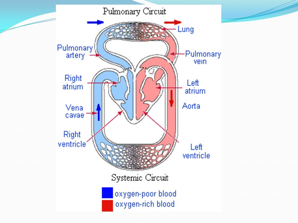
Is what determines our ability to participate in aerobic/endurance events Function: provide muscles with oxygen to sustain energy Components of c.v. system; - Heart (cardio) – pump - Blood vessels (vascular) – transportation network -Blood – transportation vehicle (carries oxygen)
 – pump. - Blood vessels (vascular) – transportation network. -Blood – transportation vehicle (carries oxygen)")
3
Aorta De-oxygentated Vein Septum Bicuspid x 2 Oxygenated Right Pressure Pulmonary Tricuspid x 2 Semi-lunar

6
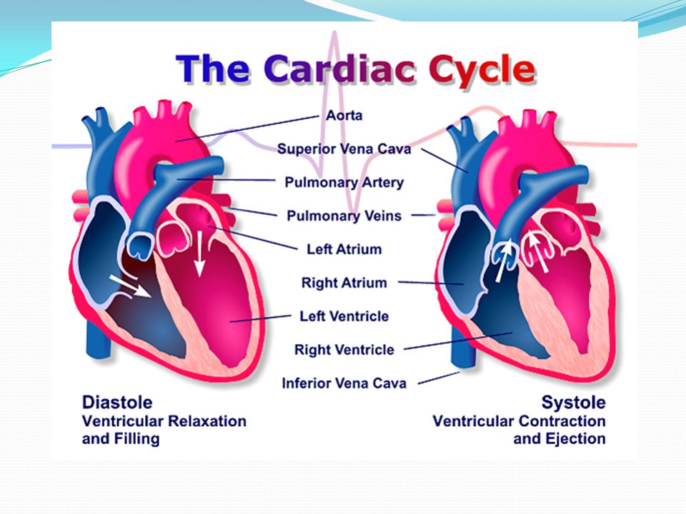
The Cardiac Cycle Cardiac Cycle - The events of one heart beat
One cycle – 0.8 second (72 cycles a minute) 2 main processes – contraction and relaxation of the heart muscle; Diastole – lasts 0.5 seconds, represents the relaxation phase, chambers fill with blood Systole – lasts 0.3 seconds, represents the contraction phase, blood pushed out of chambers/heart
 2 main processes – contraction and relaxation of the heart muscle; Diastole – lasts 0.5 seconds, represents the relaxation phase, chambers fill with blood. Systole – lasts 0.3 seconds, represents the contraction phase, blood pushed out of chambers/heart.")
8
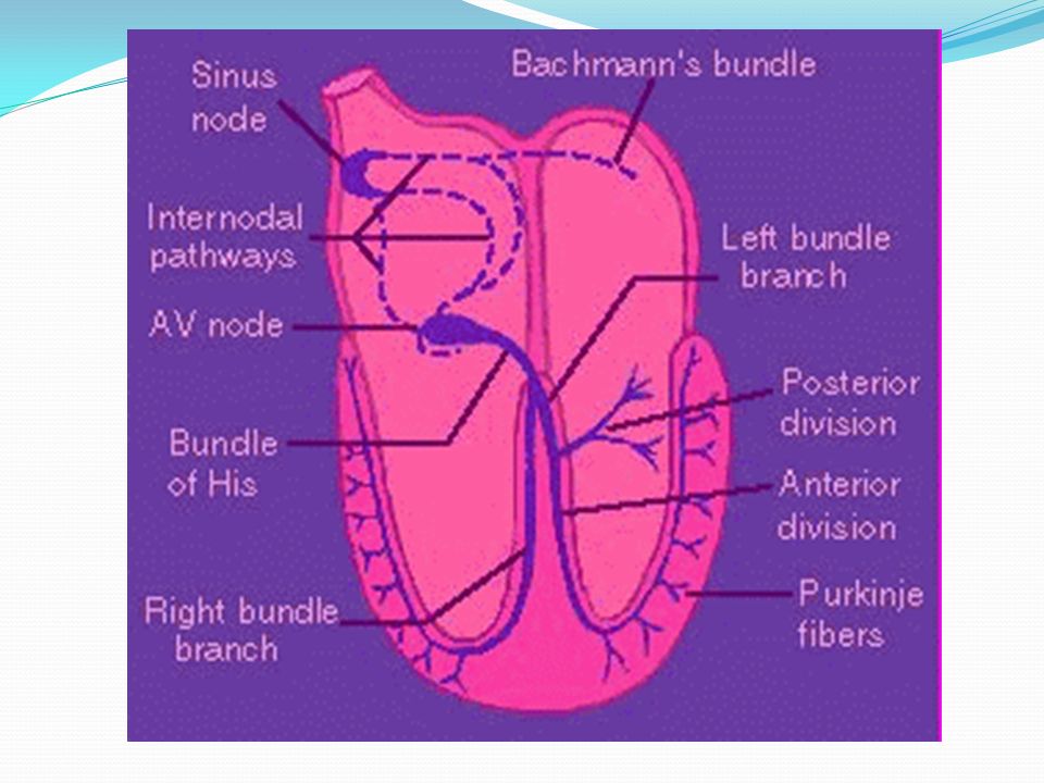
The Heart’s Conduction System
The heart is myogenic This means it generates its own electrical impulse The impulse that it generates is spread throughout the heart and causes it to contract This is known as the cardiac impulse

9
The Spread of the Electrical Impulse
The impulse starts in the SA Node (located at the top of the right atrium) Called the pacemaker
 Called the pacemaker.")
10
The impulse travels through the atria walls
This causes both atria to contract The cardiac impulse then reaches the AV node Also located in the right atrium The AV node helps delay the impulse to allow the atria to finish their contraction

11
It then spreads the impulse down the bundle of His
This is located in the Septum of the heart

12
The bundle of His splits into left and right branches
The impulse spreads around the ventricle walls through a network of purkinje fibres This causes both ventricles to contract

13
The ventricles then relax
The cycle is repeated with the next cardiac impulse

15
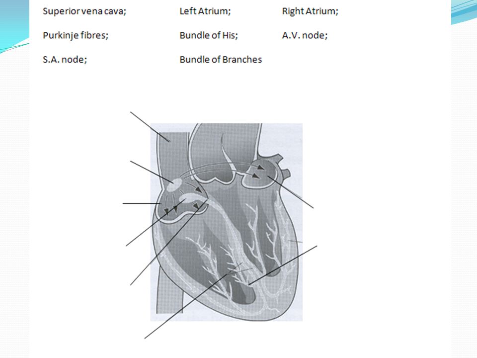
Cardiac Impulse

17
Q. Describe how the sinoatrial node (SAN) and the atrioventricular node (AVN) control the increase in heart rate during exercise SAN initiates heart beat/sends impulses; intrinsic/myogenic/pacemaker; spread of impulses through atria; atria contracts/systole; impulse reaches AV Node; Reduced delay of spread of impulses; Bundle of His; Purkinje fibres conducting impulses; ventricular systole/contraction; period of diastole/relaxation for filling;

18
Cardiac Dynamics

19
Make sure you have included these points:
Cardiac output = heart rate x stroke volume Ejection fraction = the percentage of blood forced out of the heart per beat Cardiac hypertrophy – as the muscle walls have increased in size the contraction of the heart will be stronger, therefore pushing more blood out of the heart per beat Bradycardia – the term means “slow heart” (resting heart rate below 60 bpm) Stroke volume measured in ml per beat (average at rest 70 ml per beat) Heart Rate beats per minute (average at rest 72 bpm) Cardiac output in ml per minute or litres per minute (average at rest 5 litres per minute – which would be 5000ml per minute)
 Stroke volume measured in ml per beat (average at rest 70 ml per beat) Heart Rate beats per minute (average at rest 72 bpm) Cardiac output in ml per minute or litres per minute (average at rest 5 litres per minute – which would be 5000ml per minute)")
20
Changes to the Values During Exercise

21
Types of Exercise Sub-maximal Exercise – Maximal Exercise -

22
Stroke Volume Increases during exercise – why?
At a linear rate to the speed/intensity of the exercise (up to about 40-60% of maximum intensity exercise) Once 40-60% of maximum intensity is reached stroke volume plateaus. Therefore stroke volume reaches its maximum during sub-maximal exercise
 Once 40-60% of maximum intensity is reached stroke volume plateaus. Therefore stroke volume reaches its maximum during sub-maximal exercise.")
23
What causes stroke volume (and therefore Q) to increase?
More blood is being returned to the heart – this is called venous return Less blood left in heart (End Systolic Volume) Increased diastolic filling occurs, this increases the pressure and stretches the walls of the ventricles, which means that a more forceful contraction is produced, This is known as Starling’s Law (more stretch = more forceful contraction)
 Increased diastolic filling occurs, this increases the pressure and stretches the walls of the ventricles, which means that a more forceful contraction is produced, This is known as Starling’s Law (more stretch = more forceful contraction)")
24
During maximal exercise the cardiac output will need to be increased, however stroke volume has already reached its maximum – what happens to allow Q to increase? Heart rate increases As a result of this stroke volume starts to decrease – the increase in hr means that there is not as much time for the ventricles to fill up with blood, so there is less to eject (causes the hr to increase even more)
")
25
Changes in stroke volume in response to increasing exercise intensity;

26
Heart Rate Before Exercise
Increases above resting hr before exercise has begun – known as Anticipatory Rise, is as a result of the release of adrenalin which stimulates SA node Maximal Exercise Increases dramatically once exercise starts, continues to increase as intensity increases Decreases as exercise intensity decreases Reaches its maximum at ________________?

27
Changes in heart rate in response to increasing exercise intensity;

28
Heart Rate and Sub-Maximal Exercise
Plateaus during sub-maximal exercise, called Steady State – this means that the oxygen demand is being meet After Exercise After exercise – drops dramatically Then gradually decreases

29
Cardiac Output Increases directly in line with intensity from resting up to maximum Plateaus during sub-maximal exercise

30
Changes in cardiac output in response to increasing exercise intensity;

31
Summary When exercise starts Q is increased by an increase in both HR and SV. When the intensity of exercise increases above 40-60% of maximum intensity, SV plateaus and any further increases in Q come about as a result of an increase in HR.

32
Extension Questions Recap
What happens to stroke volume during sub-maximal exercise? What happens to heart rate during sub-maximal exercise and what is this called? What happens to heart rate during maximal exercise? What happens to heart rate during sub-maximal exercise in warmer conditions?

33
Adaptations to the Heart as a Result of Training

34
Stroke Volume Heart muscle increases in size, known as . . Cardiac Hypertrophy AND Athlete’s heart The left ventricle increases in size – why this ventricle? Thicker walls of the heart allow a more forceful contractions, there more blood can be pumped per beat, resulting in an increase in. . .

35
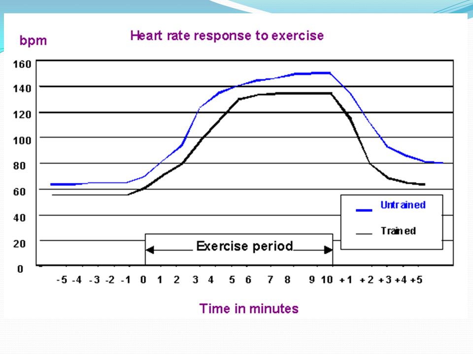
Heart Rate Due to an increase in SV the heart will not have to pump as many times (both at rest and during exercise), resulting in a decrease in. . . Heart rate (at rest and during exercise) When an athlete’s resting heart rate falls below 60bpm it is known as . . . Bradycardia During sub-maximal exercise, a trained athlete’s heart rate would not rise as much It would reach steady state sooner And recover faster Greater heart rate range – resting heart rate is lower so there is more room for an increase when exercising Maximum heart rate stays the same (220-age)
 When an athlete’s resting heart rate falls below 60bpm it is known as Bradycardia. During sub-maximal exercise, a trained athlete’s heart rate would not rise as much. It would reach steady state sooner. And recover faster. Greater heart rate range – resting heart rate is lower so there is more room for an increase when exercising. Maximum heart rate stays the same (220-age)")
36
Cardiac Output The volume of cardiac output at rest. . . Stays the same (lower resting heart rate but increase in stroke volume) The maximum cardiac output of an individual. . . Increases, so a trained athlete can deliver oxygen to the muscles for a longer period of time)
")
37
Other Factors The percentage of blood that the heart pumps out per beat is known as ejection fraction A trained athlete experiences an increase in ejection fraction because their heart will pump more forcefully each beat Even though resistance training (strength) does not work the CV system, an athlete will still will experience an increase in the size of their heart muscle (myocardium) and therefore their stroke volume will increase (more forceful contraction) The heart itself will experience capillarisation – this will increase the blood supply to the heart and ensure it continues to work for longer
 does not work the CV system, an athlete will still will experience an increase in the size of their heart muscle (myocardium) and therefore their stroke volume will increase (more forceful contraction) The heart itself will experience capillarisation – this will increase the blood supply to the heart and ensure it continues to work for longer.")
39
Summary of Changes Heart Rate Stroke Volume Cardiac Output Rest
Decreases (below 60bpm = bradycardia) Increases -can contract more forcefully (as does ejection fraction) Stays the same Exercise Lowers during sub-maximal exercise Greater heart rate range (starts lower so has more room for increase) Maximum stays same (220-age) Increases as heart muscle is stronger – can contract more forcefully (as does ejection fraction) Stays the same during sub-maximal exercise Maximum cardiac output increases (athlete can last longer)
 Increases -can contract more forcefully (as does ejection fraction) Stays the same. Exercise. Lowers during sub-maximal exercise. Greater heart rate range (starts lower so has more room for increase) Maximum stays same (220-age) Increases as heart muscle is stronger – can contract more forcefully (as does ejection fraction) Stays the same during sub-maximal exercise. Maximum cardiac output increases (athlete can last longer)")
Similar presentations