Download presentation
Presentation is loading. Please wait.

1
An orthopaedic overview

2
Characteristic Hip Pains: ◦ Dull ache- OA, degenerative, tendinitis/ bursitis ◦ Sharp – Impingement, acute sprain, labrum tear, subluxation/dislocation, fracture Pain frequently noted in groin and medial thigh Symptoms: pain, weakness, numbness, clicking, giving way Referred Pain from: Back, Abdomen, Pubic symphysis Refers Pain to: knee

8
Mechanism: High energy: ◦ Motor vehicle crash (50-60%) ◦ Motorcycle crash (10-20%) ◦ Pedestrian versus car (10-20%) ◦ Falls (8-10%) ◦ Crush (3-6%) Physical examination is specific for pelvic instability, but it has a low sensitivity: high level of suspicion Pain, swelling, WB/NWB, deformity, crepitus, Consider Blood loss and signs of shock GU exam: rectal tone, bladder control, perineum, boggy prostate, scrotal hematoma, hematuria digital rectal examination has a very low sensitivity for diagnosing pelvic fractures
 ◦ Motorcycle crash (10-20%) ◦ Pedestrian versus car (10-20%) ◦ Falls (8-10%) ◦ Crush (3-6%) Physical examination is specific for pelvic instability, but it has a low sensitivity: high level of suspicion Pain, swelling, WB/NWB, deformity, crepitus, Consider Blood loss and signs of shock GU exam: rectal tone, bladder control, perineum, boggy prostate, scrotal hematoma, hematuria digital rectal examination has a very low sensitivity for diagnosing pelvic fractures")
9
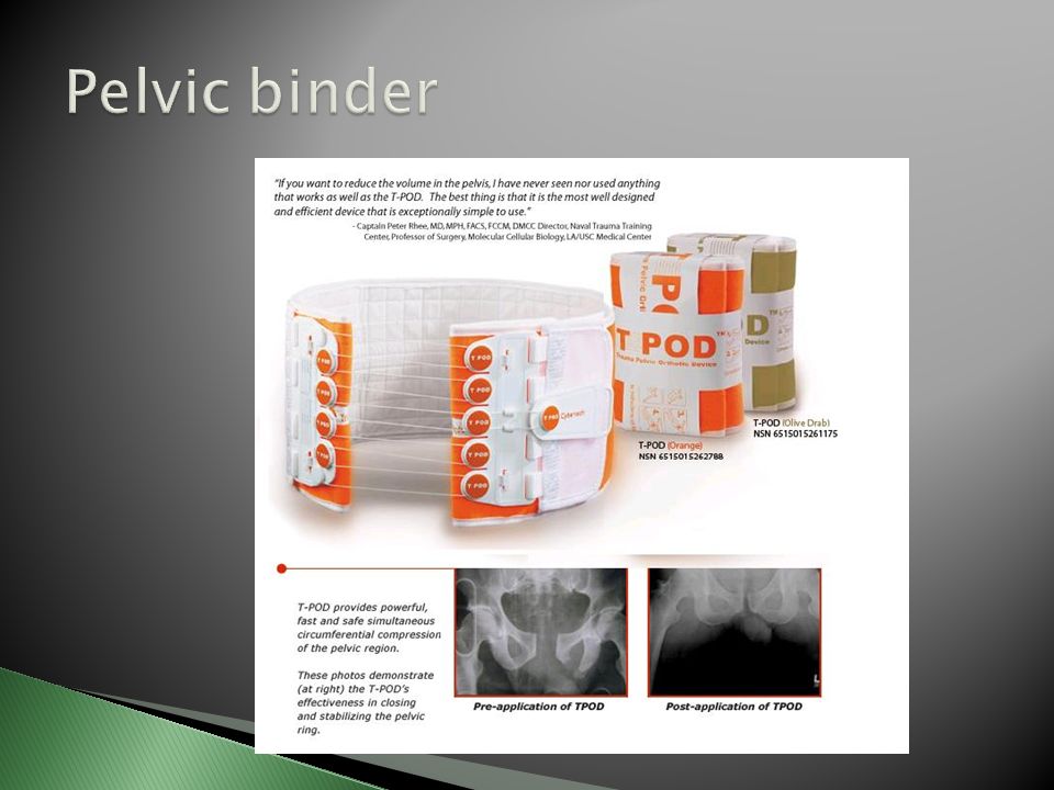
Management: pelvic binder (T-pod), IV, analgesia, Blood, Evacuation for surgical assessment X-ray: pelvic ring- usually disrupted in 2 places Tile classification: based on the integrity of the posterior sacroiliac complex Young classification system is based on mechanism of injury Death most commonly due to hemorrhage or multiple injuries
, IV, analgesia, Blood, Evacuation for surgical assessment X-ray: pelvic ring- usually disrupted in 2 places Tile classification: based on the integrity of the posterior sacroiliac complex Young classification system is based on mechanism of injury Death most commonly due to hemorrhage or multiple injuries")
11
Mechanism: high velocity trauma, MVA, falls from height Multiple fracture patterns: MOI Pain, non WB, presentations of hip, Neurovascular exam, abdominal exam, LLD, position of lower limb Stabilize, IV, analgesic, Evacuation for X-ray, surgical assessment 20% concomitant pelvic fracture

12
“ People come into this world under the brim of the pelvis and leave it by the neck of the femur. ”

14
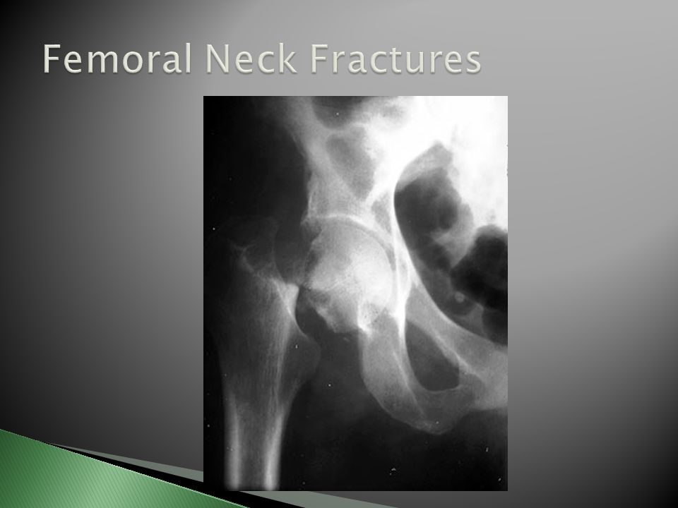
MOI: Young- MVA, fall from height ◦ Older: simple fall, Osteoporosis: abrupt step, Runners: stress fractures Acute onset hip pain, unable to WB O/E: shortened leg, external rotation, painful ROM, crepitus Neurovascular exam Stabilize, IV, analgesia Evacuation for X-ray and surgical assessment

16
Garden Classification: 1-4 Treatment: ◦ Young: internal fixation (+/- reduction) ◦ Older: internal fixation non displaced, hemi- arthroplasty
 ◦ Older: internal fixation non displaced, hemi- arthroplasty")
18
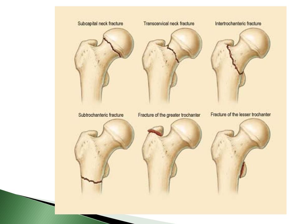
Extra-capsular fracture including the greater and lesser trochanter (b/w neck and shaft) Traumatic force to trochanteric area Acute pain, unable to WB, shortened, ER Stabilize, IV, analgesic Evacuation for X-ray, surgical assessment Treatment: Dynamic Hip Screw fixation
 Traumatic force to trochanteric area Acute pain, unable to WB, shortened, ER Stabilize, IV, analgesic Evacuation for X-ray, surgical assessment Treatment: Dynamic Hip Screw fixation")
19
Mechanism: high energy trauma Pain, deformity, Non WB Neurovascular status: urgent reduction? Procedural sedation, blood loss into fracture site…1000mL Reduction, immobilize, IV, analgesia, Blood products, +/- antibiotics Evacuation to surgical capability Surgery: internal fixation- IM nail/ plate

20
Complications: ◦ Haemorrhage requiring transfusion ◦ Fat embolism – ARDS ◦ Increased risk of open fracture ◦ Nerve injury ◦ Infection

21
Supracondylar: above condyles Condylar, Inter-condylar= intra-articular involvement Mechanism: high energy force, axial load Pain, hemarthrosis, non WB, ER, shortened Immobilize, IV, analgesia Evacuation for surgical fixation Complication: femoral artery tear

22
A.Anterior B.Posterior

25
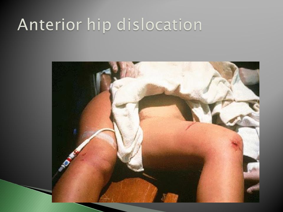
***Orthopaedic Emergency Mechanism: blow to knee in hip abduction Shortened, abducted, ER limb Neurovascular exam Stabilize, IV, analgesia, Urgent Evacuation for X-ray, reduction under sedation/GA Complications: as per posterior

26
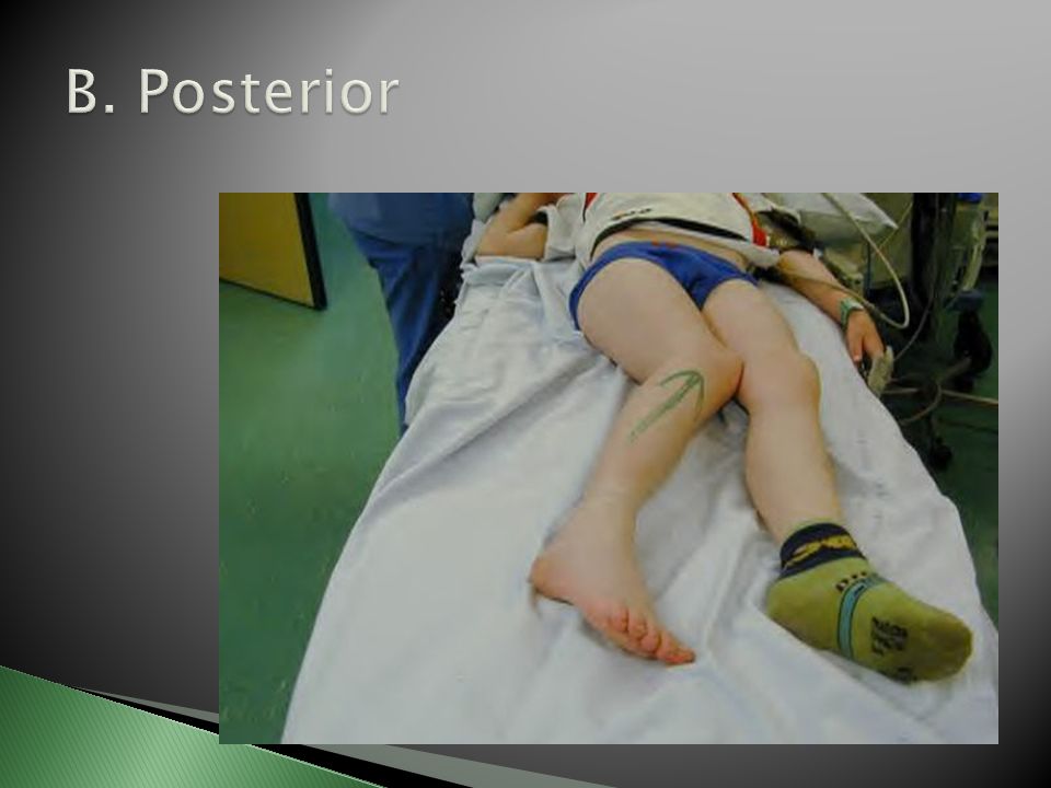
***Orthopaedic Emergency Mechanism: high force through femur with hip in flexion and adduction (dashboard ) Pain, Shortened, Add and IR of hip
 Pain, Shortened, Add and IR of hip")
27
Stabilize, IV, analgesia, Urgent Evacuation for X-ray- r/o fracture, reduction under sedation/ GA, ORIF risk of AVN with delayed reduction (>6 hrs)
")
28
Slow onset degenerative change often following injury or prolonged exposure to impact, poor biomechanics, congenital hip disorder Pain into groin and medial thigh worse with activity, intermittent flares with acute pain and swelling

29
O/E: trendelenberg gait, decreased ROM, strength deficit, ligament laxity X-ray: decreased joint space, osteophyte formation, sclerosis of femoral head, subchondral cysts Treatment: NSAIDS for acute flare, Tylenol/NSAID for long-term analgesia Physiotherapy: ROM, strengthening, gait aids Partial/Total hip replacement

30
Etiology: Loss of vascular supply to femoral head Primarily distal to proximal intra-osseous blood supply Predisposing factors: systemic steroid, dislocation of femur, fracture of femoral neck, chronic alcohol use, sickle cell, septic arthritis, “the Bends”

31
Symptoms: Pain in groin, worse with WB O/E: abnormal range of motion if collapse of cartilage on femoral head Normal strength on manual muscle testing Pain on compression testing X-ray may show crescent sign Treatment: Non WB until new bone formation

32
Etiology: trauma to hip, abnormal gait mechanics, muscle tightness, over-training Rule out cellulitis or infection Pain at lateral aspect of hip, worse with weight bearing/ walking/ direct pressure O/E: pain on palpation over greater trochanter, +/- tight ilio-tibial band, muscle imbalance, pain on single leg stance

33
Treatment: Rest, Ice, NSAIDS Physiotherapy for stretching, muscle imbalance Consider corticosteroid injection for refractive conditions

34
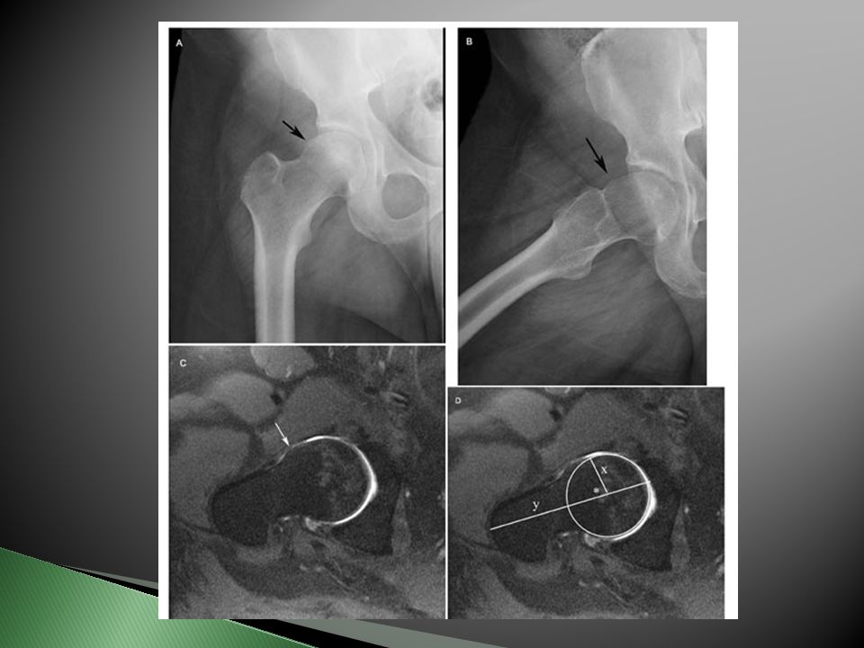
Abnormal contact between the acetabulum and femoral head-neck junction Primarily an impingement issue Groin pain with activity or extreme ROM Usually younger active people Can lead to labral tears

38
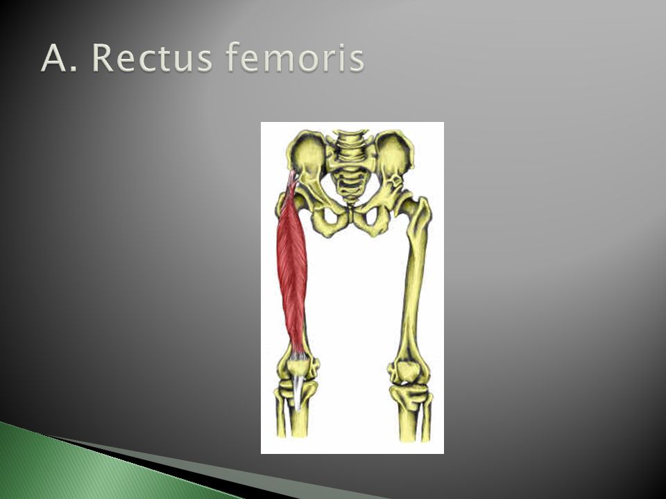
A.Rectus femoris B.Vastus lateralis

40
Adductors: groin pull Hip flexors: Rectus femoris strain Snapping hip: iliopsoas Piriformis syndrome Iliotibial band syndrome Gluteal strain

41
Let’s take a break.

Similar presentations


, twist or hyperextension.>")


