Download presentation
Presentation is loading. Please wait.

1
Generation and reading of the 12 lead ECG
AWC Chow

2
The 12 lead ECG Advantages Common clinical tool
Independent marker of cardiac disease Non-invasive Rapid information acquisition Cheap Gold standard for arrhythmia management

3
The 12 lead ECG Disadvantages Average of potentials Limited resolution
Snapshot of activity Electrical and not haemodynamic data

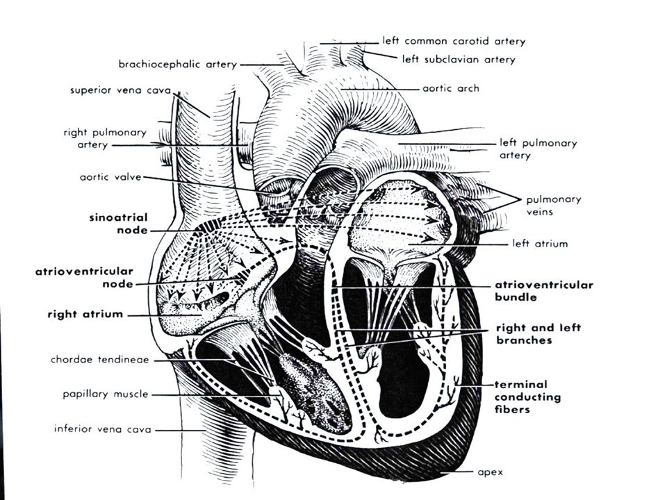
5
Non-specialised atrial tissue
Left bundle Anterior superior fascicle Right bundle Posterior inferior fascicle

6
- +/- +

7
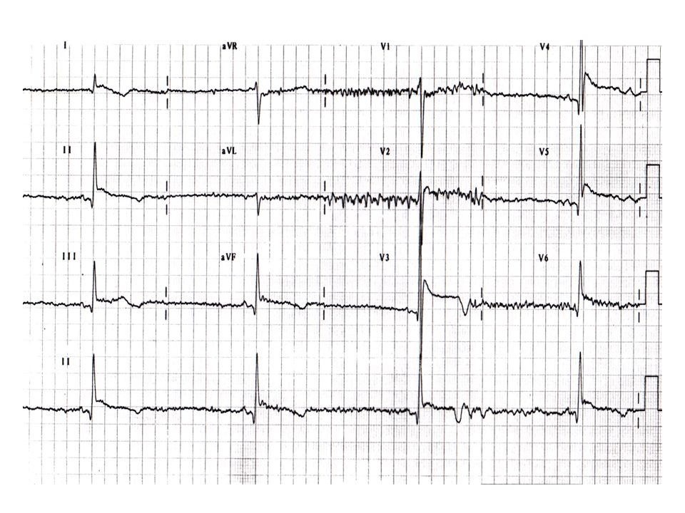
History of the ECG 1842 -Carlo Matteucci shows that an electric current accompanies each heart beat. Sanderson and Page record the heart's electrical current with a capillary electrometer British physiologist Augustus D. Waller publishes the first human electrocardiogram. Einthoven develops the string galvometer 1910 – Eithoven’s triangle

8
Theoretical consideration
Myocytes have a resting potential Transmembrane flux create voltage difference - activation Cellular coupling cause rapid deploarisation Ionic flux seen ECG deflections

9
Theoretical considerations
Resting state - no potential/field change Depolarisation - boundary potential change Represented as a dipole/vector Restitution of polarity: repolarisation

10
Theoretical considerations
Greater muscle mass Larger potential change Larger voltage changes of ECG Direction of activation dependent on Site of initiation Specialised conduction system distribution Anatomical considerations Barriers (scar, valves) Muscle mass
 Muscle mass.")
11
RA LA I II III RL LL

12
aVR +210 aVL -30 I 0 II +60 III +120 aVF +90

13
PR T P QT QRS

14
Diagnostic criteria for LVH
There are many different criteria for LVH. Sokolow + Lyon (Am Heart J, 1949;37:161) S V1+ R V5 or V6 > 35 mm Cornell criteria (Circulation, 1987;3: ) SV3 + R avl > 28 mm in men SV3 + R avl > 20 mm in women Framingham criteria (Circulation,1990; 81: ) R avl > 11mm, R V4-6 > 25mm S V1-3 > 25 mm, S V1 or V2 + R V5 or V6 > 35 mm, R I + S III > 25 mm Romhilt + Estes (Am Heart J, 1986:75:752-58) Point score system
 S V1+ R V5 or V6 > 35 mm. Cornell criteria (Circulation, 1987;3: ) SV3 + R avl > 28 mm in men. SV3 + R avl > 20 mm in women. Framingham criteria (Circulation,1990; 81: ) R avl > 11mm, R V4-6 > 25mm. S V1-3 > 25 mm, S V1 or V2 + R V5 or V6 > 35 mm, R I + S III > 25 mm. Romhilt + Estes (Am Heart J, 1986:75:752-58) Point score system.")
15
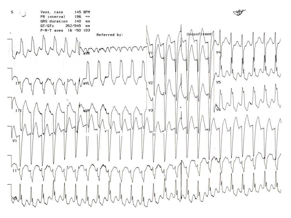
Causes of RBBB normal finding in children and tall thin adults
right ventricular hypertrophy chronic lung disease even without pulmonary hypertension anterolateral myocardial infarction left posterior hemiblock pulmonary embolus Wolff-Parkinson-White syndrome - left sided accessory pathway atrial septal defect ventricular septal defect

16
Causes of LBBB left anterior hemiblock
Q waves of inferior myocardial infarction artificial cardiac pacing emphysema hyperkalaemia Wolff-Parkinson-White syndrome - right sided accessory pathway tricuspid atresia ostium primum ASD

17
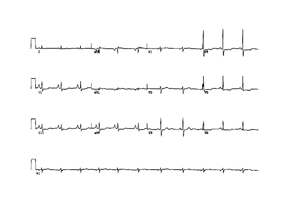
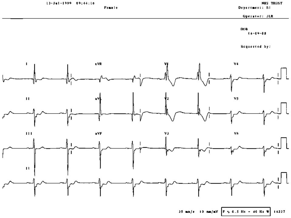
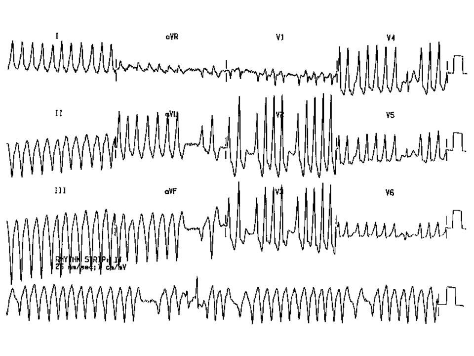
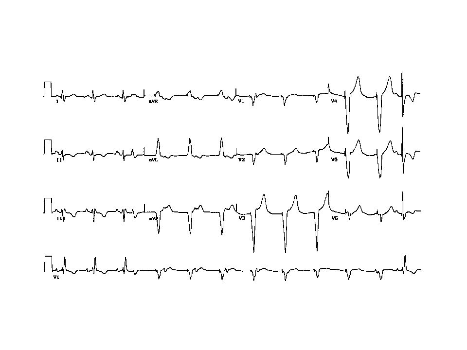
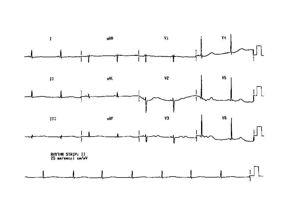
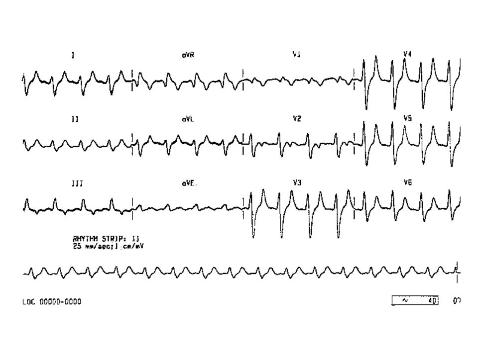
ECG Analysis Rate 60-100b/min Rhythm SR PR <200ms QRS <120ms
Axis to +120 QT interval <500ms ST segment

Similar presentations


>")


>")


