Download presentation
Presentation is loading. Please wait.

1
Brain asymetry

2
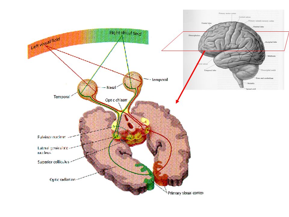
Visual pathway

4
Image courtesy of Dr. Paul Wellman V1 (Striate Cortex)
")
5
Modularity: structures for faces, places, and bodies

6
Figure 4.18 (a) Monkey brain showing location of the inferotemporal cortex (IT) in the lower part of the temporal lobe. (b) Human brain showing location of the fusiform face area (FFA) in the fusiform gyrus, which is located under the temporal lobe.
 Human brain showing location of the fusiform face area (FFA) in the fusiform gyrus, which is located under the temporal lobe..")
7
Brain Asymmetry experiment You see a sequence of faces (old/young, or young/old) You judge which face, the one shown first or the other shown second, looks younger. Young

8
Brain asymmetry explanation The two faces were always mirror images of each other. So they had identical age information. For a right-handed person, the right hemisphere is more strongly involved in judgment about faces.

9
The information to the left of fixation goes first to the right hemisphere. As a result, the younger half on the left will look younger than the mirror image (with the younger half on the right). Young
. Young.")
Similar presentations


 Prosopagnosia usually results from localized.>")


 Ch 4-6, 9.>")
