Download presentation
Presentation is loading. Please wait.

1
Dr Rim Braham

2
Located within the sella tursica Contiguous to vascular and neurologic structures Cavernous sinuses Cranial nerves Optic chiasm Hypothalamic neural cells synthesize specific releasing and inhibiting hormones Secreted directly into the portal vessels of the pituitary stalk Blood supply derived from the superior and inferior hypophyseal arteries Pituitary Gland

3
Hypothalamic–Pituitary Axis

5
Anterior pituitary gland Secrete various trophic hormones Disease in this region may result in syndromes of hormone excess or deficiency Posterior pituitary gland More of a terminus of axons of neurons in the supraoptic and paraventricular nuclei of the hypothalamus Storehouse for the hormones The main consequence of disease in this area is disordered water homeostasis Pituitary Gland

6
Production of six major hormones: Prolactin (PRL) Growth hormone (GH) Adrenocorticotropin hormone (ACTH) Luteinizing hormone (LH) Follicle-stimulating hormone (FSH) Thyroid-stimulating hormone (TSH) Anterior Pituitary Gland
 Growth hormone (GH) Adrenocorticotropin hormone (ACTH) Luteinizing hormone (LH) Follicle-stimulating hormone (FSH) Thyroid-stimulating hormone (TSH) Anterior Pituitary Gland")
8
Secreted in a pulsatile manner Feedback control at the level of the hypothalamus and pituitary to modulate pituitary function exerted by the hormonal products of the peripheral target glands Tumors cause characteristic hormone excess syndromes Hormone deficiency may be inherited or acquired Anterior Pituitary Gland

9
Hypopituitarism

10
Gonadotropin Deficiency Women Oligomenorrhea or amenorrhea Loss of libido Vaginal dryness or dyspareunia Loss of secondary sex characteristics (estrogen deficiency) Men Loss of libido Erectile dysfunction Infertility Loss of secondary sex characteristics (testosterone deficiency) Atrophy of the testes Gynecomastia (testosterone deficiency)
 Men Loss of libido Erectile dysfunction Infertility Loss of secondary sex characteristics (testosterone deficiency) Atrophy of the testes Gynecomastia (testosterone deficiency)")
11
Results in hypocortisolism Malaise Anorexia Weight-loss Gastrointestinal disturbances Hyponatremia Pale complexion Unable to tan or maintain a tan No features of mineralocorticoid deficiency Aldosterone secretion unaffected ACTH Deficiency

12
Hypothyroidism Atrophic thyroid gland TSH Deficiency

13
Inability to lactate postpartum Often 1 st manifestation of Sheehan syndrome (usually secondary to post partum hemorrhage ) Prolactin Deficiency
 Prolactin Deficiency")
14
Adults Often asymptomatic May complain of Fatigue Degrees exercise tolerance Abdominal obesity Loss of muscle mass Children GH Deficiency Constitutional growth delay Growth Hormone Deficiency

15
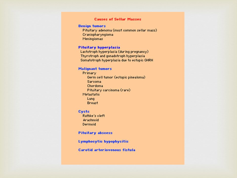
Etiology Anterior pituitary diseases Deficiency one or more or all anterior pituitary hormones Common causes: Primary pituitary disease Hypothalamic disease Interruption of the pituitary stalk Extrasellar disorders Hypopituitarism

16
Primary pituitary disease Tumors Pituitary surgery Radiation treatment Hypothalamic disease Functional suppression of axis Exogenous steroid use Extreme weight loss Exercise Systemic Illness Interruption of the pituitary stalk Extrasellar disorders Craniopharyngioma Rathke pouch

17
Hypopituitarism Developmental and genetic causes Dysplasia Septo-Optic dysplasia Developmental hypothalamic dysfunction Kallman Syndrome Laurence-Moon-Bardet-Biedl Syndrome Frohlich Syndrome (Adipose Genital Dystrophy)
")
18
Acquired causes: Infiltrative disorders Cranial irradiation Lymphocytic hypophysitis Pituitary Apoplexy Empty Sella syndrome Sheehan syndrome Hypopituitarism

19
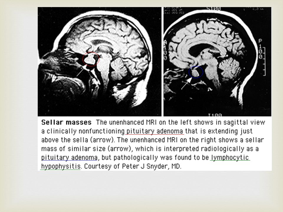
Lymphocytic Hypophysitis Etiology Presumed to be autoimmune Clinical Presentation Women, during postpartum period Mass effect (sellar mass) Deficiency of one or more anterior pituitary hormones ACTH deficiency is the most common Diagnosis MRI - may be indistinguishable from pituitary adenoma Treatment Corticosteroids – often not effective Hormone replacement Hypopituitarism: Acquired
 Deficiency of one or more anterior pituitary hormones ACTH deficiency is the most common Diagnosis MRI - may be indistinguishable from pituitary adenoma Treatment Corticosteroids – often not effective Hormone replacement Hypopituitarism: Acquired")
21
Pituitary Apoplexy Hemorrhagic infarction of a pituitary adenoma/tumor Considered a neurosurgical emergency Presentation: Variable onset of severe headache Nausea and vomiting Meningismus Vertigo +/ - Visual defects +/ - Altered consciousness Symptoms may occur immediately or may develop over 1-2 days Hypopituitarism: Acquired

22
Pituitary Apoplexy Transient or permanent hypopituitarism is possible undiagnosed acute adrenal insufficiency Diagnose with CT/MRI Differentiate from leaking aneurysm Treatment: Surgical - Transsphenoid decompression Visual defects and altered consciousness Medical therapy – if symptoms are mild Corticosteroids Hypopituitarism: Acquired

23
Empty Sella Syndrome Often an incidental MRI finding Usually have normal pituitary function Implying that the surrounding rim of pituitary tissue is fully functional Hypopituitarism may develop insidiously Pituitary masses may undergo clinically silent infarction with development of a partial or totally empty sella by cerebrospinal fluid (CSF) filling the dural herniation. Rarely, functional pituitary adenomas may arise within the rim of pituitary tissue, and these are not always visible on MRI Hypopituitarism: Acquired

24
Clinical Presentation Can present with features of deficiency of one or more anterior pituitary hormones Clinical presentation depends on: Age at onset Hormone effected, extent Speed of onset Duration of the deficiency Hypopituitarism

25
Diagnosis Biochemical diagnosis of pituitary insufficiency Demonstrating low levels of trophic hormones in the setting of low target hormone levels Provocative tests may be required to assess pituitary reserve Hypopituitarism

26
Treatment Hormone replacement therapy usually free of complications Treatment regimens that mimic physiologic hormone production allow for maintenance of satisfactory clinical homeostasis Hypopituitarism

27
Hormone Replacement Trophic Hormone DeficitHormone Replacement ACTHHydrocortisone (10-20 mg A.M.; 10 mg P.M.) Cortisone acetate (25 mg A.M.; 12.5 mg P.M.) Prednisone (5 mg A.M.; 2.5 mg P.M.) TSHL-Thyroxine (0.075-0.15 mg daily) FSH/LHMales Testosterone enanthate (200 mg IM every 2 wks) Testosterone skin patch (5 mg/d) Females Conjugated estrogen (0.65-1.25 mg qd for 25days) Progesterone (5-10 mg qd) on days 16-25 Estradiol skin patch (0.5 mg, every other day) For fertility: Menopausal gonadotropins, human chorionic gonadotropins GHAdults: Somatotropin (0.3-1.0 mg SC qd) Children: Somatotropin [0.02-0.05 (mg/kg per day)] VasopressinIntranasal desmopressin (5-20 ug twice daily) Oral 300-600 ug qd
![ Hormone Replacement Trophic Hormone DeficitHormone Replacement ACTHHydrocortisone (10-20 mg A.M.; 10 mg P.M.) Cortisone acetate (25 mg A.M.; 12.5 mg P.M.) Prednisone (5 mg A.M.; 2.5 mg P.M.) TSHL-Thyroxine ( mg daily) FSH/LHMales Testosterone enanthate (200 mg IM every 2 wks) Testosterone skin patch (5 mg/d) Females Conjugated estrogen ( mg qd for 25days) Progesterone (5-10 mg qd) on days Estradiol skin patch (0.5 mg, every other day) For fertility: Menopausal gonadotropins, human chorionic gonadotropins GHAdults: Somatotropin ( mg SC qd) Children: Somatotropin [ (mg/kg per day)] VasopressinIntranasal desmopressin (5-20 ug twice daily) Oral ug qd](http://images.slideplayer.com/24/7332900/slides/slide_27.jpg " Hormone Replacement Trophic Hormone DeficitHormone Replacement ACTHHydrocortisone (10-20 mg A.M.; 10 mg P.M.) Cortisone acetate (25 mg A.M.; 12.5 mg P.M.) Prednisone (5 mg A.M.; 2.5 mg P.M.) TSHL-Thyroxine ( mg daily) FSH/LHMales Testosterone enanthate (200 mg IM every 2 wks) Testosterone skin patch (5 mg/d) Females Conjugated estrogen ( mg qd for 25days) Progesterone (5-10 mg qd) on days Estradiol skin patch (0.5 mg, every other day) For fertility: Menopausal gonadotropins, human chorionic gonadotropins GHAdults: Somatotropin ( mg SC qd) Children: Somatotropin [ (mg/kg per day)] VasopressinIntranasal desmopressin (5-20 ug twice daily) Oral ug qd")
28
Pituitary Tumors

30
Microadenoma < 1 cm Macroadenoma > 1 cm Is the tumor causing local mass effect? Is hypopituitarism present? Is there evidence of hormone excess? Clinical presentation: Mass effect Superior extension May compromise optic pathways – leading to impaired visual acuity and visual field defects May produce hypothalamic syndrome – disturbed thirst, satiety, sleep, and temperature regulation Lateral extension May compress cranial nerves III, IV, V, and VI – leaning to diplopia Inferior extension May lead to cerebrospinal fluid rhinorrhea Pituitary Tumors

31
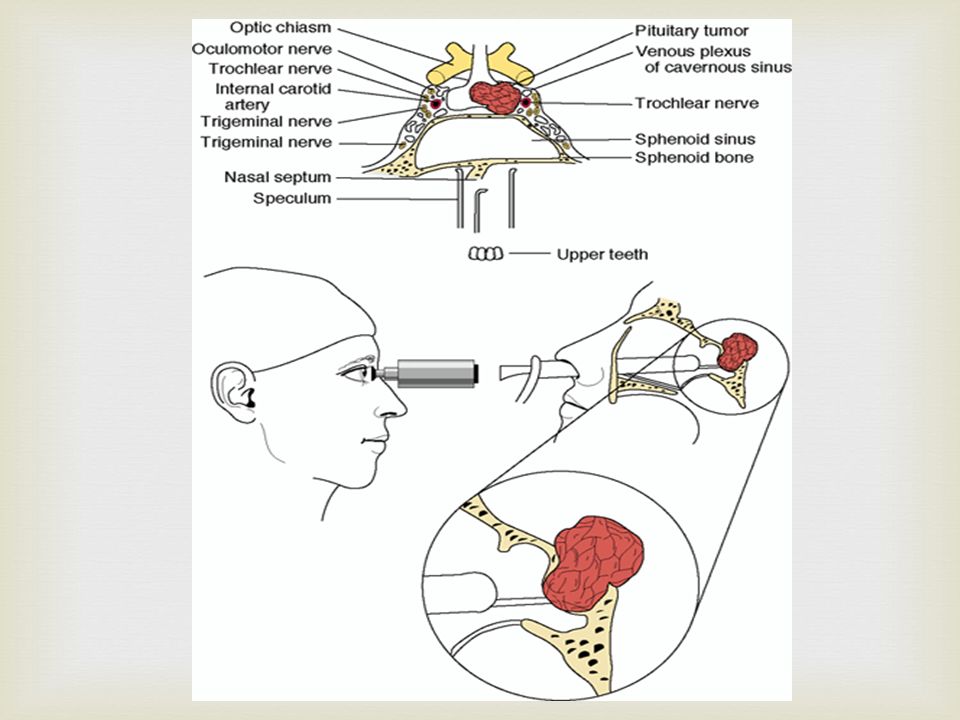
Diagnosis Check levels of all hormones produced Check levels of target organ products Treatment Surgical excision, radiation, or medical therapy Generally, first-line treatment surgical excision Drug therapy available for some functional tumors Simple observation Option if the tumor is small, does not have local mass effect, and is nonfunctional Not associated with clinical features that affect quality of life Pituitary Tumors

34
Most common functional pituitary tumor Usually a micro adenoma Can be a space occupying macroadenoma – often with visual field defects Although many women with hyperprolactinemia will have galactorrhea and/ or amenorrhea The absence these the two signs do not excluded the diagnosis GnRH release is decreased in direct response to elevated prolactin, leading to decreased production of LH and FSH Prolactinoma

35
Women Amenorrhea – this symptom causes women to present earlier Hirsutism Men Impotence – often ignored Tend to present later Larger tumors Signs of mass effect Prolactinoma

36
Essential to rule out secondary causes!! Drugs which decrease dopamine stores Phenothiazines Amitriptyline Metoclopramide Factors inhibiting dopamine outflow Estrogen Pregnancy Exogenous sources Hypothyroidism If prolactin level > 200, almost always a prolactinoma (even in a nursing mom) Prolactin levels correlate with tumor size in the macroadenomas Suspect another tumor if prolactin low with a large tumor Prolactinoma
 Prolactin levels correlate with tumor size in the macroadenomas Suspect another tumor if prolactin low with a large tumor Prolactinoma.")
37
Diagnosis Assess hypersecretion Basal, fasting morning PRL levels (normally <20 ug/L) Multiple measurements may be necessary Pulsatile hormone secretion levels vary widely in some individuals with hyperprolactinemia Both false-positive and false-negative results may be encountered May be falsely lowered with markedly elevated PRL levels (>1000 ug/L) assay artifacts; sample dilution is required to measure these high values accurately May be falsely elevated by aggregated forms of circulating PRL, which are biologically inactive (macroprolactinemia) Hypothyroidism should be excluded by measuring TSH and T4 levels Prolactinoma
 Multiple measurements may be necessary Pulsatile hormone secretion levels vary widely in some individuals with hyperprolactinemia Both false-positive and false-negative results may be encountered May be falsely lowered with markedly elevated PRL levels (>1000 ug/L) assay artifacts; sample dilution is required to measure these high values accurately May be falsely elevated by aggregated forms of circulating PRL, which are biologically inactive (macroprolactinemia) Hypothyroidism should be excluded by measuring TSH and T4 levels Prolactinoma")
38
Treatment Medical Cabergoline – dopamine receptor agonist Bromocriptine - dopamine agonist Safe in pregnancy Will restore menses Decreases both prolactin and tumor size (80%) Surgical Transsphenoidal surgery – irridation (if pt cannot tolerate rx) Prolactinoma
 Surgical Transsphenoidal surgery – irridation (if pt cannot tolerate rx) Prolactinoma")
40
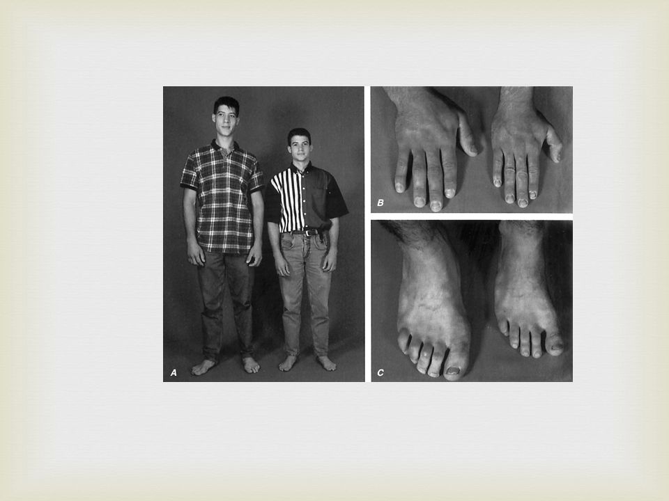
Gigantism GH excess before closure of epipheseal growth plates of long bones Acromegaly GH excess after closure of epipheseal growth plates of long bones Insidious onset Usually diagnosed late Growth Hormone Tumors or somatotrophic adenomas

41
May have DM or glucose intolerance Hypogonadism Large hands and feet Large head with a lowering brow and coarsening features Hypertensive – 25% Colon polyps 3-6 more likely than general population Multiple skin tags Growth Hormone Tumors

42
Diagnosis Screen: Check for high IGF-I levels (>3 U/ml) Remember, levels very high during puberty Confirm: 100gm glucose load Positive: GH levels do not increase to <5ng/ml Treatment Surgical Radiation Bromocriptine - temporizing measure May decrease GH by 50% Somatostatin analogue, Octreotide For suboptimal response to other treatment Growth Hormone Tumors
 Remember, levels very high during puberty Confirm: 100gm glucose load Positive: GH levels do not increase to <5ng/ml Treatment Surgical Radiation Bromocriptine - temporizing measure May decrease GH by 50% Somatostatin analogue, Octreotide For suboptimal response to other treatment Growth Hormone Tumors")
44
corticotrophic adenomas: secrete adenocorticotropic hormone (ACTH), Cushing's disease gonadotrophic adenomas: secrete luteinizing hormone (LH), follicle-stimulating hormone (FSH) and their subunits, usually doesn't cause symptoms thyrotrophic adenomas (rare) secrete thyroid- stimulating hormone (TSH), occasionally hyperthyroidism, usually doesn't cause symptoms 25% of pituitary adenomas are nonsecretive: Non functioning pituitary adenomas Other pituitary Tumors
, Cushing s disease gonadotrophic adenomas: secrete luteinizing hormone (LH), follicle-stimulating hormone (FSH) and their subunits, usually doesn t cause symptoms thyrotrophic adenomas (rare) secrete thyroid- stimulating hormone (TSH), occasionally hyperthyroidism, usually doesn t cause symptoms 25% of pituitary adenomas are nonsecretive: Non functioning pituitary adenomas Other pituitary Tumors")
45
A pituitary incidentaloma is a previously unsuspected sellar mass that is detected on an imaging study performed for reasons other than pituitary symptoms or disease. Pituitary incidentalomas are common. In imaging studies, the frequency of incidentally discovered pituitary lesions is 4 to 20 % by computed tomography (CT) scan and 10 to 38 % by magnetic resonance imaging (MRI). evaluation for hormonal hyper- and hyposecretion (hypopituitarism). Pituitary Incidentalomas
 scan and 10 to 38 % by magnetic resonance imaging (MRI). evaluation for hormonal hyper- and hyposecretion (hypopituitarism). Pituitary Incidentalomas.")
Similar presentations


. The posterior pituitary is part of the brain; the anterior pituitary is not In embryonic development, anterior.>")


>")
