Download presentation
Presentation is loading. Please wait.

1
Techniques of Mandibular and Maxillary Anesthesia
Dr. Mumena C.H

2
Introduction Choice of anesthesia Mandibular technique
Maxillary technique

3
Choice of LA 3 Important factors; Specific nerve to be blocked,
Onset of action, Duration action,

4
Duration of action The duration of action of LA may vary from 30min to 180 minutes or longer, Duration is related to dosage, increasing dosage increases duration, Duration can be increased by adding Epinephrine (vasoconstrictor) to the local anaesthesia, Duration of action differs for different agents NB. Read the properties of each agent.
 to the local anaesthesia, Duration of action differs for different agents. NB. Read the properties of each agent.")
5
Nerve to be blocked Small nerves are in general easier to be block than larger onces, Nerve endings and cutaneous nerves are easily and quickly blocked by low concentration of drug given by infiltration,

6
Onset of action Depending on the type of operation, e.g acute pain then rapid onset required,

7
Review Anat. Trigeminal nerve
5th cranial nerve. Major sensory of the face, mouth and nasal cavity. Motor and proprioceptive innervation to muscles of mastication. Origin- Sensory neurons-upper part of pons, Motor neurons-inferior surface of pons 3 divisions Ophthalmic, maxillary, and mandibular). Successful practice of dentistry is based on blockade of the various branches of these nerves.
. Successful practice of dentistry is based on blockade of the various branches of these nerves.")
8
Mandibular nerve 3rd and largest branch of trigeminal nerve.
Composed of motor and sensory roots. Exits cranium foramen ovale. Motor innervation to the muscles of mastication.

9
Mandibular nerve Sensory to mandibular teeth and gingiva, lower lip, cheek, anterior two thirds of the tongue, auricle and skin over temporal region. Braches – Inferior alveolar nerve, Lingual nerve Buccal nerve Mental nerve Incisive nerve

10
Inferior alveolar nerve block in Dental
Inferior alveolar nerve together with lingual nerve are anesthetized by the same or different injections. Lingual nerve mucous membrane of the floor of mouth, anterior 2/3 of tongue and lingual gingiva. I.A.n mandibular teeth and surrounding hard and soft tissue up to the midline, exception buccal soft tissues in the molar area.

11
Inferior alveolar nerve block in Dental
Anatomical landmarks anterior border of ramus, external and internal oblique ridges, the coronoid notch, retromolar triangle and pterygomandibular ligament. Target area for injection mandibular foramen located in the mid-portion of ramus and 1 cm above the occlusal plane. Sometimes found in the area between mid-portion of ramus and posterior 1/3 of ramus.

12
Inferior alveolar nerve block in Dental cont…
Several technique elaborated difficulty of locating mandibular foramen hence LA failure. There is two techniques: Traditional technique, Direct technique Indirect technique Alternative technique; High ramus –neck of condyle approach (Gow-gates technique) and Tuberosity approach (Akinos technique)-Tresmus.
 and. Tuberosity approach (Akinos technique)-Tresmus.")
13
Inferior alveolar Nerve Block Anesthesia
Lower success rate than Maxillary anesthesia - approx % Related to bone density Less access to nerve trunks Most commonly performed technique Has highest failure rate (15-20%) Success depends on depositing solution within 1 mm of nerve trunk
 Success depends on depositing solution within 1 mm of nerve trunk.")
14
Inferior Alveolar Nerve Block
Not a complete mandibular nerve block. Requires supplemental buccal nerve block May require infiltration of incisors or mesial root of first molar

15
Inferior Alveolar Nerve Block
Nerves anesthetized Inferior Alveolar Mental Incisive Lingual

16
Inferior Alveolar Nerve Block
Areas Anesthetized Mandibular teeth to midline Body of mandible, inferior ramus Buccal mucosa anterior to mental foramen Anterior 2/3 tongue & floor of mouth Lingual soft tissue and periosteum

17
Inferior Alveolar Nerve Block
Indications Multiple mandibular teeth Buccal anterior soft tissue Lingual anesthesia

18
Inferior Alveolar Nerve Block
Contraindications Infection/inflammation at injection site Patients at risk for self injury (eg. children)
")
19
Inferior Alveolar Nerve Block
10%-15% positive aspiration

20
Inferior Alveolar Nerve Block
Supplemental injection Periodontal ligament injection (PDL) Intraseptal
 Intraseptal.")
21
Inferior Alveolar Nerve Block
Target Area Inferior alveolar nerve, near mandibular foramen Landmarks Coronoid notch Pterygomandibular raphe Occlusal plane of mandibular posterior teeth (molars).
.")
22
Inferior Alveolar Nerve Block
Precautions Do not inject if bone not contacted Avoid forceful bone contact Avoid use of cold agents

23
Inferior Alveolar Nerve Block
Failure of Anesthesia Injection too low Injection too anterior Accessory innervation -Mylohyoid nerve -contralateral Incisive nerve innervation

24
Inferior Alveolar Nerve Block
Complications Hematoma Trismus Facial paralysis (Inject into parotid gland).
.")
25
Inferior Alveolar Nerve Block
General hints: Technique Apply topical Area of insertion: medial ramus, mid-coronoid notch, level with occlusal plane (1 cm above), 3/4 posterior from coronoid notch to pterygomandibular raphe advance to bone (20-25 mm)
, 3/4 posterior from coronoid notch to pterygomandibular raphe. advance to bone (20-25 mm)")
26
Traditional technique of inferior alveolar nerve block-Direct technique
Palpate the oblique ridge in the oral vestibule with index finger. Follow it posteriorly to where it ascends as sharp anterior border of the ramus of mandible. Move the finger to the temporal crest (internal oblique line) of the mandible and is left in this position. The finger is now in the retromolar fossa with the finger nail backwards.
 of the mandible and is left in this position. The finger is now in the retromolar fossa with the finger nail backwards.")
27
Traditional technique of inferior alveolar nerve block-Direct technique
Draw an imaginary line from point between the occlusal surfaces of the two premolars in the opposite quadrant to the midpoint of the fingernail. This imaginary line when extended posteriorly ends just above the mandibular foramen. Therefore the syringe needle will be directed along this line.

28
Traditional technique of inferior alveolar nerve block-Indirect technique
After locating the injection site the syringe is held parallel to the mandibular occlusal plane on the same side as the tooth to be blocked. The needle is directed approximately 1 cm (with the syringe) above the mandibular arch. Aspirate the needle and inject 0.5 ml of LA for lingual nerve. Then move the syringe to the other side of the arch over the opposite premolars teeth.
 above the mandibular arch. Aspirate the needle and inject 0.5 ml of LA for lingual nerve. Then move the syringe to the other side of the arch over the opposite premolars teeth.")
29
Alternative technique-
More reliable. Gow gates technique; Also known as high ramus- neck of condyle approach. Penetration into the oral mucosa is along the medial border of the mandibular ramus lateral to the pterygomandibular depression, but medial to the temporalis muscle tendon. The needle inserted along a line extending from the corner of the mouth opposite the site of injection The needle should be 25G and usually 1in long. The needle should advanced until bone is contacted and then withdrawn 1 mm. Anesthetic solution deposited after negative aspiration

36
Alternative technique- cont…
Akinos technique; Also known as Tuberosity approach. This technique is useful where patient has intense trismus. Mucosa is penetrated along the medial surface of the mandibular ramus. The mouth is kept closed (teeth in occlusion) with the cheek and muscles of mastication relaxed. The same needle side. The depth of penetration is 1 ½ inches.
 with the cheek and muscles of mastication relaxed. The same needle side. The depth of penetration is 1 ½ inches.")
37
The Akinosi technique

38
The Akinosi technique

39
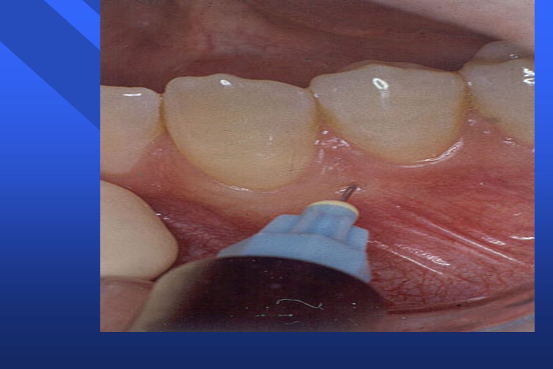
Avoiding self-inflicted trauma
reduce area of soft tissue anaesthesia intraligamentary anaesthesia intra-osseous anaesthesia palatal approaches to pulp

43
Intraligamentary anaesthesia
injection dentine gingiva pulp pdl alveolus

44
Intraligamentary anaesthesia
injection dentine gingiva pulp alveolus

45
Intraligamentary anaesthesia
injection dentine gingiva pulp alveolus

46
Intraligamentary anaesthesia
injection dentine gingiva pulp alveolus

47
Intraligamentary and intra-osseous anaesthesia
injection dentine gingiva pulp intra-osseous injection alveolus

48
Intraligamentary and intra-osseous anaesthesia
injection dentine gingiva pulp intra-osseous injection alveolus

49
Success of intraligamentary anaesthesia depends on:
tooth least successful with lower incisors solution dependent upon vasoconstrictor concentration

51
self-aspirating } aspirating non-aspirating

53
aspiration force diaphragm deformation basal diaphragm recoils

54
Force Time initial aspiration force subsequent force

56
Buccal nerve block Anterior branch of Mandibular nerve (V3)
Provides buccal soft tissue anesthesia adjacent to mandibular molars Not required for most restorative procedures Buccal is blocked by the injection in the buccal mucosa right to the 3rd molar just above the occlusal plane.

57
Buccal Nerve Block Indications Contraindications
Anesthesia required - mucoperiosteum buccal to mandibular molars Contraindications Infection/inflammation at injection site

58
Buccal Nerve Block Advantages Technically easy High success rate
Disadvantages Discomfort

59
Buccal Nerve Block Alternatives Buccal infiltration PDL Intraseptal

60
Buccal Nerve Block Landmarks Mandibular molars Mucobuccal fold

61
Buccal Nerve Block Complications Hematoma (unusual)
Positive aspiration 0.7 %

62
Long Buccal Nerve Block
Technique; Apply topical Retract the cheek Insert the needle distal to the tooth with the syringe horizontal into the buccal fold. About 0.5 ml of LA solution is injected. The barrel of the needle should be facing downwards.

65
Mental nerve block Mental nerve exits mandible through mental foramen.
The foramen is located between root apices of 1st and 2nd premolrs. Technique; Insert the needle at an angle to the bony canal towards the mental foramen. Inject ml LA solution, another injection is required in the lingual gingiva and mucosa.

66
Mental Nerve Block Provides sensory innervation to buccal soft tissue anterior to mental foramen, lip and chin

67
Mental Nerve Block Indication Need for anesthesia in innervated area
Contraindication Infection/inflammation at injection site

68
Mental Nerve Block Advantages Easy, high success rate
Usually atraumatic Disadvantage Hematoma

69
Mental Nerve Block Alternatives Local infiltration PDL Intraseptal
Inferior alveolar nerve block Gow Gates

70
Mental Nerve Block Complications Few Hematoma Positive aspiration
5.7 %

75
Incisive Nerve Block Terminal branch of IAN
Originates in mental foramen and proceeds anteriorly Good for bilateral anterior anesthesia Not effective for anterior lingual anesthesia

76
Incisive Nerve Block Nerves anesthetized Incisive Mental

77
Incisive Nerve Block Areas Anesthetized
Mandibular labial mucous membranes Lower lip / skin of chin Incisor, cuspid and bicuspid teeth

78
Incisive Nerve Block Indication
Anesthesia of pulp or tissue required anterior to mental foramen Contraindication Infection/inflammation at injection site

79
Incisive Nerve Block Advantages High success rate
Pulpal anesthesia w/o lingual anesthesia Disadvantages Lack of lingual or midline anesthesia

80
Incisive Nerve Block Complications Hematoma Positive aspiration 5.7 %

81
Maxillary nerve Foramen rotundum Courses
pterygopalatine fossa & infraorbital canal. Exit the face through infraorbital foramen. Supply sensory innervation-maxillary teeth, gingiva, maxillary sinus, hard & soft palate, lower eyelid, side of the nose, upper lip, mucous membrane of nasopharynx, and skin over the anterior temporal region. Necessary to block one or more of nerve or branches listed above. 3 branches: Posterior, middle & anterior superior alveolar nerves

82
Maxillary nerve cont… Posterior superior alveolar nerve-all molars except mesiobuccal root of 1st molar. Technique- Dry area with gauze, Apply topical anesthesia Insert palpating finger into muccobuccal fold of maxilla till contacts the zygomatic arch at it juncture with maxillary bone. Cheek retracted gentle laterally, Mouth partially opened. Long needle 1 5/8 inches inserted through maxillary mucosa at 450 angle to all 3 planes of orientation. Advance needle slowly in this orientation. This ensure deposition of LA soln in close proximity to the nerve as it enters its foramina.

83
Maxillary nerve cont… Avoids contact with the ptrygoid plexus of veins. Needle should be advanced to a depth of one half to two thirds of the dental needle. Inject slowly two thirds of LA soln after negative aspiration. Withdraw needle the remaining one third may be used to block mesiobuccal root of 1st molar. Middle superior alveolar nerve-located within the maxilla. Innervate mesiobuccal root of 1st molar, premolars, antral lining corresponding to these teeth and buccal alveolar bone and soft tissues in this area. Technique; Retract cheek mucosa after preparation as described previously. 1 to 1 5/8 inches needle should be used. Orient needle so that it conforms to the maxilla curvature in this area.

84
Maxillary nerve cont… Advance slowly to a depth where whereby its tip approximates the level of the apices of the premolar teeeth. Inject slowly following negative aspiration. LA diffuses through the maxilla blocking the MSAn. Anterior superior alveolar nerve (ASAn)- supply canine, lateral and central incisors, buccal alveolar bone, and soft tissues in this region. Technique; 1 to 1 5/8 inches needle. Needle inserted and advanced slowly along the long canine eminence, Until tip approximates the level of the apex of canine tooth. Inject slowly after negative aspiration.
- supply canine, lateral and central incisors, buccal alveolar bone, and soft tissues in this region. Technique; 1 to 1 5/8 inches needle. Needle inserted and advanced slowly along the long canine eminence, Until tip approximates the level of the apex of canine tooth. Inject slowly after negative aspiration.")
85
Maxillary nerve cont… Greater palatine nerve (GPn)- exit into the hard palate though greater palatine foramen located at level of 1 cm above margin of palatal gingiva and between 2nd and 3rd molar. Technique; Insert the needle slightly anterior to the greater palatine (depression btn 2nd & 3rd Molars) foramen and perpendicular to the hard palate. The needle advanced to contact the bone. Withdraw 2mm and approximately one fourth of a dental cartilage (0.5 ml) injected. Nasopalatine nerve (NPn)- enters oral cavity through incisive foramen, located in the midline of the anterior hard palate directly beneath incisive papilla. Technique; insert a needle into the incisive papilla at an angle
- exit into the hard palate though greater palatine foramen located at level of 1 cm above margin of palatal gingiva and between 2nd and 3rd molar. Technique; Insert the needle slightly anterior to the greater palatine (depression btn 2nd & 3rd Molars) foramen and perpendicular to the hard palate. The needle advanced to contact the bone. Withdraw 2mm and approximately one fourth of a dental cartilage (0.5 ml) injected. Nasopalatine nerve (NPn)- enters oral cavity through incisive foramen, located in the midline of the anterior hard palate directly beneath incisive papilla. Technique; insert a needle into the incisive papilla at an angle.")
86
Maxillary nerve cont… To the midline and advanced until the rim of the foramen is contacted. Needle withdrawn 1-2 mm and one fourth of a dental cartilage injected following negative aspiration. Infra-orbital block; exits via infra orbital firamen, Technique; foramen located in vertical line with the pt’s pupil as he looks straight ahead. Palpate the foramen, retract the cheek to expose the buccal vestibule adjacent to the bicuspids. Insert the needle between bicuspids and parallel to their long axes. Penetrate far to enough to reach infra orbital foramen. Following negative aspiration deposit LA. It is possible to produce unilateral analgesia of I, C, P, and mesiobuccal root of 1st molar.

87
Palatal access

88
I hope L.A is the bridge to Effective surgical procedures.
BEST OF LUCKY

Similar presentations


>")


 nerve block is a local anaisthisia that anesthetizes the maxillary canine, the central and lateral incisors, and.>")


