Download presentation
Presentation is loading. Please wait.

1
Obstructive Pulmonary Disease

2
Characterized by airway obstruction that is increased with expiration.
More force is required to expire a given volume of air, or emptying of lungs is slowed, or both. The most common obstructive diseases are asthma, chronic bronchitis, and emphysema.

3
COPD Asthma Emphysema Chronic Bronchitis

4
Many people have both chronic bronchitis and emphysema, and together these are often called chronic obstructive pulmonary disease - COPD Major symptom of obstructive pulmonary disease is dyspnea, and wheezing.

5
COPD Asthma Emphysema Pathologic Diagnosis Chronic Bronchitis
Physiologic Diagnosis Chronic Bronchitis Clinical Diagnosis

6
1- Bronchial Asthma More intermittent and acute than COPD.
It is a reversible disorder It occurs at all ages. Runs in families, so evidence genetics plays a role. Environmental factors interact with inherited factors to increase the risk of asthma.

7
Childhood exposure to high levels of allergens, smoking and/or respiratory viruses increases chances of developing asthma. Major events in acute asthma attack are bronchiolar constriction, mucus hyper-secretion inflammatory swelling.

8
Pathogenesis Smooth muscle spasm in bronchioles. Vascular congestion. Edema formation. Production of thick mucus. Impaired muco-ciliary function. Thickening of airway walls.

9
Untreated, this can lead to airway damage that is irreversible.
Obstruction increases resistance to air flow and decreases flow rates Impaired expiration causes hyperinflation of alveoli, and increases the work of breathing

10
Clinical manifestations
Dyspnea Sometimes, cough Wheezing Attacks may continue from few hours to days or even weeks. During remission individual is asymptomatic and pulmonary function tests are normal

11
2- Chronic Bronchitis Hyper-secretion of mucus and chronic productive cough for at least 3 months of the year for at least two consecutive years. Incidence may be increased up to 20 times in persons who smoke and more in persons exposed to air pollution.

13
Pathophysiology Inspired irritants result in inflammatory cell infiltration into the bronchial wall. Causes bronchial edema and increases size and number of mucus glands and goblet cells. Mucus is thick, and can’t be cleared because of impaired ciliary function. Increases susceptibility to infection and injury

14
Initially affects only larger bronchi, but eventually all airways involved.
Airways collapse in early expiration, blocked by mucus leads to hypo-ventilation Hypoxemia Air trapping prevents respiratory muscles from functioning efficiently (barrel chest).
.")
15
Barrel Chest

17
Treatment Best treatment is PREVENTION.
Stop smoking halts progression of the disease Bronchodilators, expectorants, and chest physical therapy. Acute attacks may require antibiotics, and steroids. Oxygen therapy may be required

18
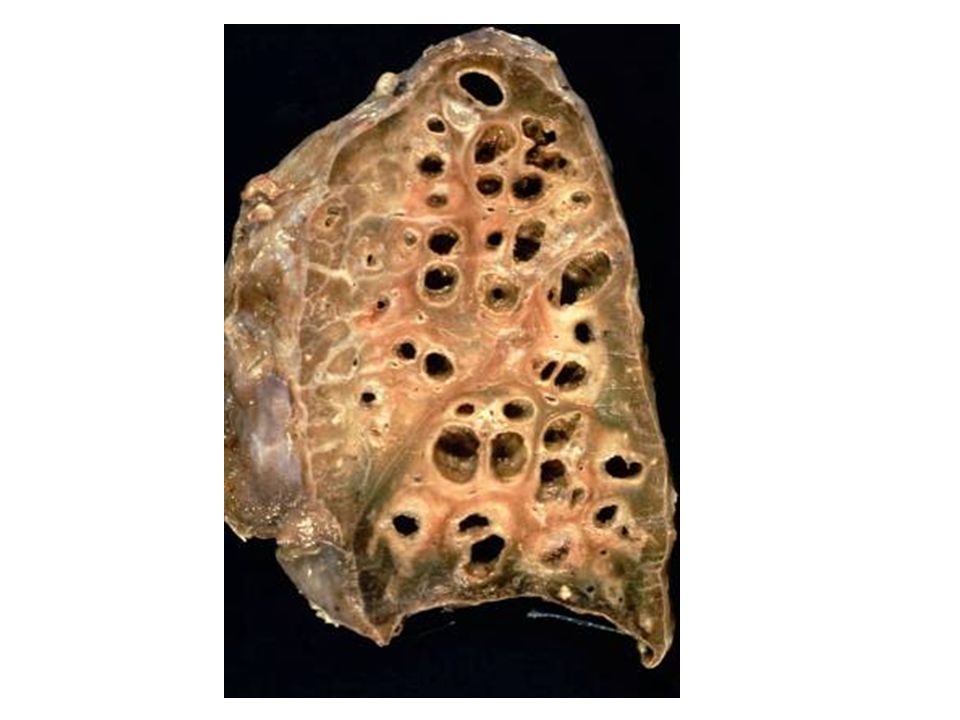
3- Emphysema Abnormal, permanent enlargement of the gas-exchange airways and destruction of the alveolar walls. Obstruction results from changes in lung tissue rather than mucus production and inflammation (pathological changes). Major mechanism is loss of elastic recoil
. Major mechanism is loss of elastic recoil.")
19
Major cause is smoking Other causes are air pollution and recurrent respiratory infections Primary emphysema linked to an inherited deficiency of the enzyme alpha 1- antitrypsin which can affect lung tissue.

20
Normal Lung versus Emphysema

21
Normal Lung versus Emphysema

22
Pathophysiology Begins with the destruction of the alveolar septa, which eliminates portions of the capillary bed, and increases the volume of air in the alveolus. Continued alveolar loss and loss of elastic recoil Expiration becomes difficult, causing hyperexpansion of the chest These are not effective in gas exchange and result in hypoxia.

23
Clinical manifestations
Dyspnea at rest Barrel chest Minimal wheezing Prolonged expiration Hypoventilation and hypoxia

24
Pursed Lip Breathing

25
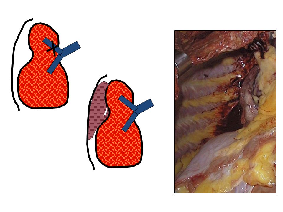
Atelectasis Collapse or incomplete expansion of part or all of the lung Types: Resorption (obstruction of airway). Compressive (pleural effusion or pneumothorax)
")
27
Bronchiectasis Dilatation of bronchi and bronchioles secondary to chronic inflammation Associated conditions Obstruction Cystic fibrosis Immotile cilia syndromes Necrotizing pneumonia

29
Respiratory Failure 29

30
Respiratory Failure (RF)
The inability of the lungs to adequately oxygenate the blood and to clear it of carbon dioxide. RF could be :- Acute RF: Acute Respiratory Distress Syndrome (ARDS) Pulmonary embolism Direct injury to the lungs, airways or chest wall Indirect due to injury of another body system, as brain. 30
 Pulmonary embolism. Direct injury to the lungs, airways or chest wall. Indirect due to injury of another body system, as brain. 30.")
31
Chronic respiratory failure
Due to progressive hypoventilation from airway obstruction or restrictive disease 31

32
Respiratory failure always presents with:-
Dysnpea always present, but may be difficult to detect a change in a chronic patient Since nervous tissue is highly oxygen-dependent --- Drowsiness, Memory and visual impairment, Headache due to increased intracranial pressure due to cerebral vasodilatation

33
Two principal patterns:
1- Hypoxic Respiratory Failure 2- Hypoxic-Hypercapnic Respiratory Failure

34
1- Hypoxic Respiratory Failure:
Seen when pO2 {oxygen partial pressure} falls to or below 60 mm Hg Typically seen in:- chronic bronchitis and emphysema. lung consolidation due to bacterial infection (pneumonia). in lung collapse, pulmonary hypertension, pulmonary embolism and ARDS. 34
. in lung collapse, pulmonary hypertension, pulmonary embolism and ARDS. 34.")
35
With a progressive lowering of pO2, more widespread tissue damage and loss of consciousness can occur.

36
2- Hypoxic-Hypercapnic Respiratory Failure
When arterial pCO2 {Carbon dioxide partial pressure} (normally 40 mm Hg) exceeds 45 mm HG, condition is called hypercapnia 36
 exceeds 45 mm HG, condition is called hypercapnia. 36.")
37
Most often caused by airway obstructive conditions, and hypoventilation from CNS problem, thoracic cage or neuromuscular abnormalities.

38
CNS effects produce muscular tremors, drowsiness and coma.
Attempts to compensate include increased heart rate and vasodilatation, which produces warm, moist skin. CNS effects produce muscular tremors, drowsiness and coma. Hypercapnia also produces acidosis. 38

39
ARDS Mortality in persons < 60 is 40%
Those over 65 and immunocompromised still have mortality over 60 % Most survivors have almost normal lung function 1 year after acute illness. 39

40
Clinical manifestations:
Symptoms developed progressively: Hyperventilation → respiratory alkalosis → dyspnea and hypoxemia → metabolic acidosis → respiratory acidosis → further hypoxemia → hypotension, decreased cardiac output, death 40

41
Evaluation and Treatment
Diagnosis based on physical examination, blood gases and imaging Treatment is based on early detection, supportive therapy and prevention of complications, esp. infection Often requires mechanical ventilation. 41

42
Tumors of the Lung

43
Carcinoma of the Lung Causes 7% of all deaths
More common in males than females Causes 85-95% smoking 1% asbestos + smoking (estimate) Rare arsenic, chromium, nickel, 43
 Rare arsenic, chromium, nickel, 43.")
44
44

45
Classification of Lung Carcinoma (Major Types)
Squamous cell carcinoma 35% Adenocarcinoma 30% Small cell carcinoma 25% Large cell carcinoma 10% 45

46
Squamous cell carcinoma
Frequency: 35% Smoking: X 25 (increased risk) Males > females Survival (5 years): % Arises in bronchial squamous metaplasia Centrally located May cavitate 46
 Males > females. Survival (5 years): % Arises in bronchial squamous metaplasia. Centrally located. May cavitate. 46.")
47
Squamous cell carcinoma

48
Adenocarcinoma Frequency: 30% Smoking: X 3 (increased risk)
Males < females Survival (5 years): % Peripheral
: % Peripheral.")
49
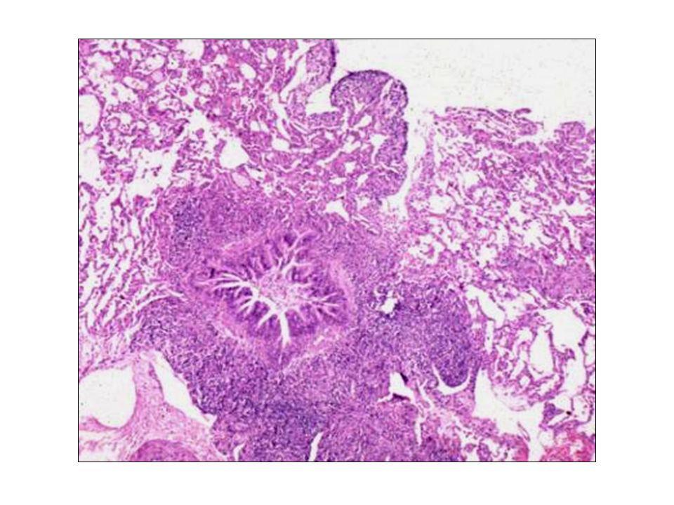
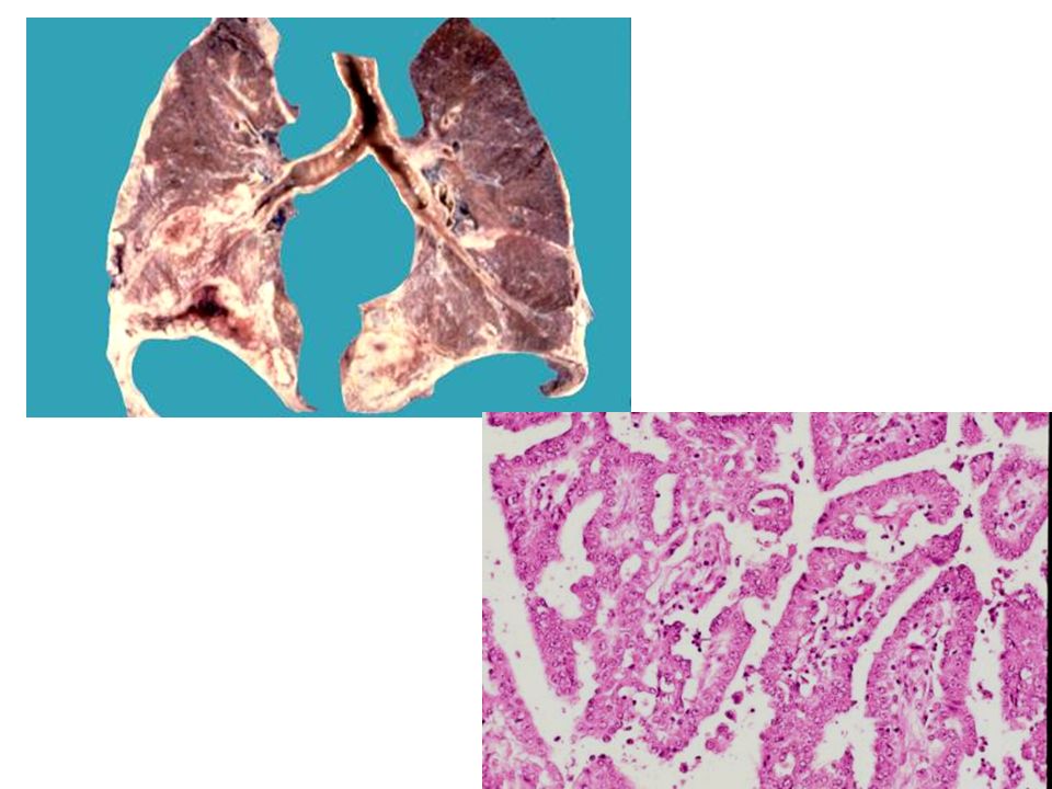
Bronchiolo-alveolar carcinoma
Frequency: 2 % Smoking Males = females Survival (5 years): %. Single or multiple tumor nodules
: %. Single or multiple tumor nodules.")
50
Small cell carcinoma Frequency: 25 % Smoking: 95% of patients
Males >> females Survival (5 years): %.
: %.")
51
Small cell carcinoma

52
Large Cell Carcinoma Frequency: 10 % Gross picture: Peripheral lesion
Microscopic Wastebasket group of tumors that do not fit the criteria of a squamous cell carcinoma, adenocarcinoma, or small cell carcinoma Prognosis: Similar to adenocarcinoma

53
Large Cell Carcinoma

54
Mesothelioma Malignant tumor of mesothelial cells Highly malignant neoplasm with short survival Most patients (70%) have an asbestos exposure history Asbestos exposure also increases the risk of pulmonary cancer Smoking is not related to mesothelioma
 have an asbestos exposure history. Asbestos exposure also increases the risk of pulmonary cancer. Smoking is not related to mesothelioma.")
Similar presentations


>")


 With atria and blood vessels on for.>")


