Download presentation
Presentation is loading. Please wait.

1
Branchial Cleft Cyst By: Dr. Waleed Alhajii

2
What is a Branchial Cyst?
Definition: Congenital epithelial cysts, which arise on the lateral part of the neck due to failure of obliteration of the second branchial cleft in embryonic development.

3
Commonest cysts to arise in the neck.
Branchila cleft cyst = Lymphoepithelial cyst “Lymphatic origin, Modern Theory” The name branchial means in Greek “Gill” , Those structures are responsible of development of Gills in fish. “Classic Theory” Commonest cysts to arise in the neck. Previously, the Branchila cleft cysts were explained based on embryological origin only, lately many theories are trying to explain the etiology The ‘Branchial theory’ is now less accepted and the most appropriate hypothesis explaining the aetiology of these cysts is the "Lymph node inclusion theory" with he palatine tonsils as the most likely source of the enclosed epithelium.

4
Classical location Anterior to the sternocleidomastoid muscle.
However there have been a number of case reports describing cysts which were found in areas other than the classical position.

5
Etiology Many theories They can be grouped into two categories:
the congenital The cervical lymph nodes cystic transformation theories. There are many theories for the etiology of lateral cervical cysts. They can be grouped into two categories: the congenital and the cervical lymph nodes cystic transformation theories.

6
Easier approach to Embryology
Structures between the developing head and the heart (i.e., the face, neck, oropharynx, and the larynx) develop from the branchial apparatus. There are six branchial arches; the last two are rudimentary. Each arch has a bar of mesoderm.
 develop from the branchial apparatus. There are six branchial arches; the last two are rudimentary. Each arch has a bar of mesoderm.")
7
Caudal to each of the four arches is an internal pouch lined with entoderm.
Externally is branchial cleft, lined with ectoderm. Between each bar, a branchial plate, composed of entoderm and ectoderm, separates the branchial cleft from the branchial pouch.

8
The relationship of pouches to clefts can result in remnants that are sinuses internally to the pharynx, externally to the skin, or cysts with or without connection to either surface. Cleft I remnants are rare (< 1% of branchial cleft remnants) and either involve the parotid gland (and CN VII) or emerge below the ear. These may present as a painless swelling of the parotid and may be associated with the external ear canal. These are different from pre-auricular inclusion cysts. Cleft II remnants are more common and are located anterior to the sternocleidomastoid. The tract may run between the internal and external carotid and may have an internal opening at the superior pole of the tonsil. The tract must be completely excised. Remnants of the third cleft lie deep to the carotids and may have an internal opening into the pyriform sinus (see larynx anatomy).
 and either involve the parotid gland (and CN VII) or emerge below the ear. These may present as a painless swelling of the parotid and may be associated with the external ear canal. These are different from pre-auricular inclusion cysts. Cleft II remnants are more common and are located anterior to the sternocleidomastoid. The tract may run between the internal and external carotid and may have an internal opening at the superior pole of the tonsil. The tract must be completely excised. Remnants of the third cleft lie deep to the carotids and may have an internal opening into the pyriform sinus (see larynx anatomy). .")
9
Anatomical Considerations
The second arch grows downwards and ultimately covers the third and fourth arches. The buried clefts normally disappear around the seventh week of development. If a portion of the cleft remains entrapped and fails to disappear, its remnants form a cyst.

10
Second cleft Cyst with tract extending up to Pharynx
Note tract goes between internal & external carotid arteries and close to cranial nerves IX, X, XII which control among other functions tongue movement and swallowing. This diagram is a subject of debate Most authors believe it can never have a cord or tract attached leading to the skin or the pharynx Some believe it can occur rarely, I couldn’t get any solid proof “operative images” to confirm or deny.

11
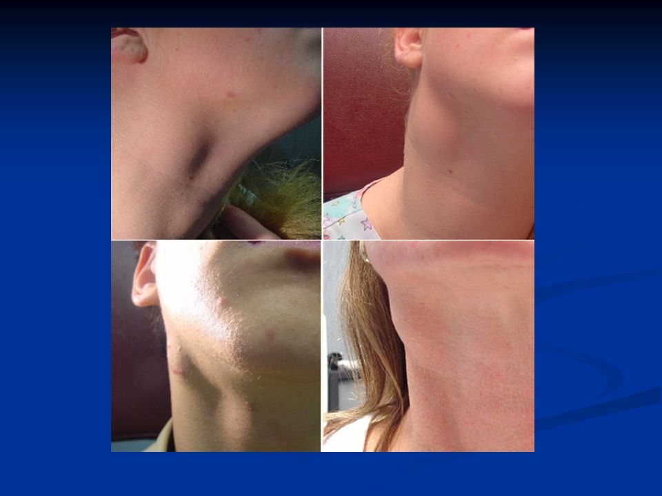
Clinical Presentation

12
History Solitary, painless mass in the neck of a child or a young adult. History of intermittent swelling and tenderness of the lesion during upper respiratory tract infection may exist. Discharge if associated with a sinus tract. May present with locally compressive symptoms. + family history. A branchial cyst commonly presents as a solitary, painless mass in the neck of a child or a young adult. A history of intermittent swelling and tenderness of the lesion during upper respiratory tract infection may exist. Discharge may be reported if the lesion is associated with a sinus tract. In some instances, patients may present with locally compressive symptoms. A family history may be present.

13
Physical Examination Primary lesion: Branchial cysts are smooth, nontender, fluctuant masses, which occur along the lower one third of the anteromedial border of the sternocleidomastoid muscle between the muscle and the overlying skin. Secondary lesion: The lesion may be tender if secondarily inflamed or infected. When associated with a sinus tract, mucoid or purulent discharge onto the skin or into the pharynx may be present.

16
Diagnosis Cyst arising off midline of the neck and having lymphoepithelial characteristics should be regarded as a branchial cyst. Usually occur in the 2nd or 3rd decade of life. Most commonly found in the anterior triangle of the neck anterior to the upper third of the sternomastoid. A cyst occupying the posterior triangle is extremely rare. Hence they should be suspected in all the cystic swellings of the neck except the median ones. As per King’s criteria any cyst arising outside the midline of the neck and having lymphoepithelial characteristics should be regarded as a branchial cyst. Usually occur in the 2nd or 3rd decade of life. They are most commonly found in the anterior triangle of the neck anterior to the upper third of the sternomastoid. A cyst occupying the posterior triangle is extremely rare. However these cysts have been reported to occur in all the regions of the neck, and even in the mediastinum and the abdomen. Hence they should be suspected in all the cystic swellings of the neck except the median ones.

17
Imaging On general principle it’s less helpful than expected
Although you can always tell where is the lesion, but differentiating between other causes of cystic neck masses is not always easy.

18
Ultrasound Well defined, echogenic mass usually anterior to the carotid artery, draped anterior to the sternocleidomastoid muscle

19
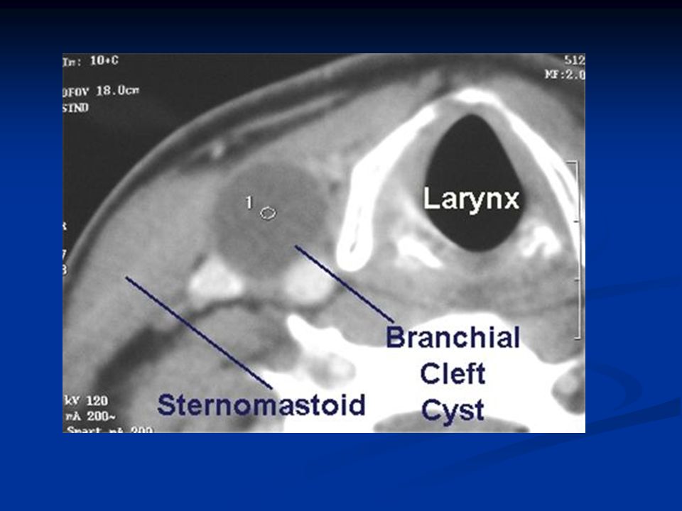
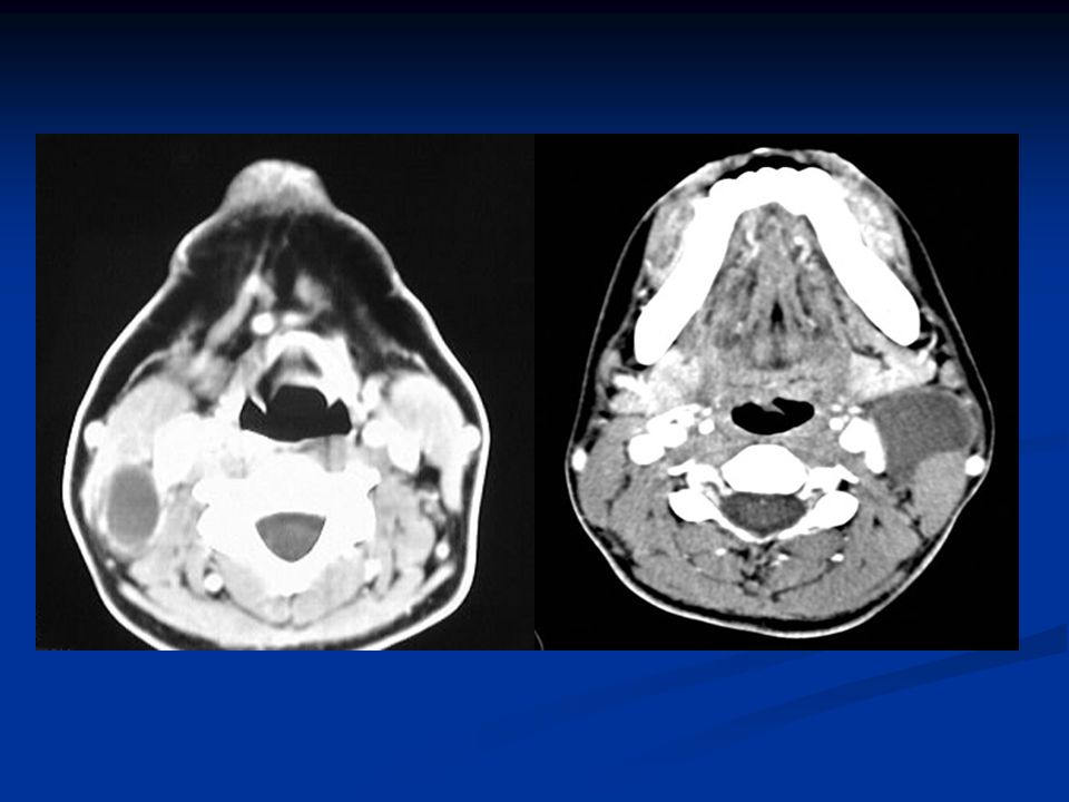
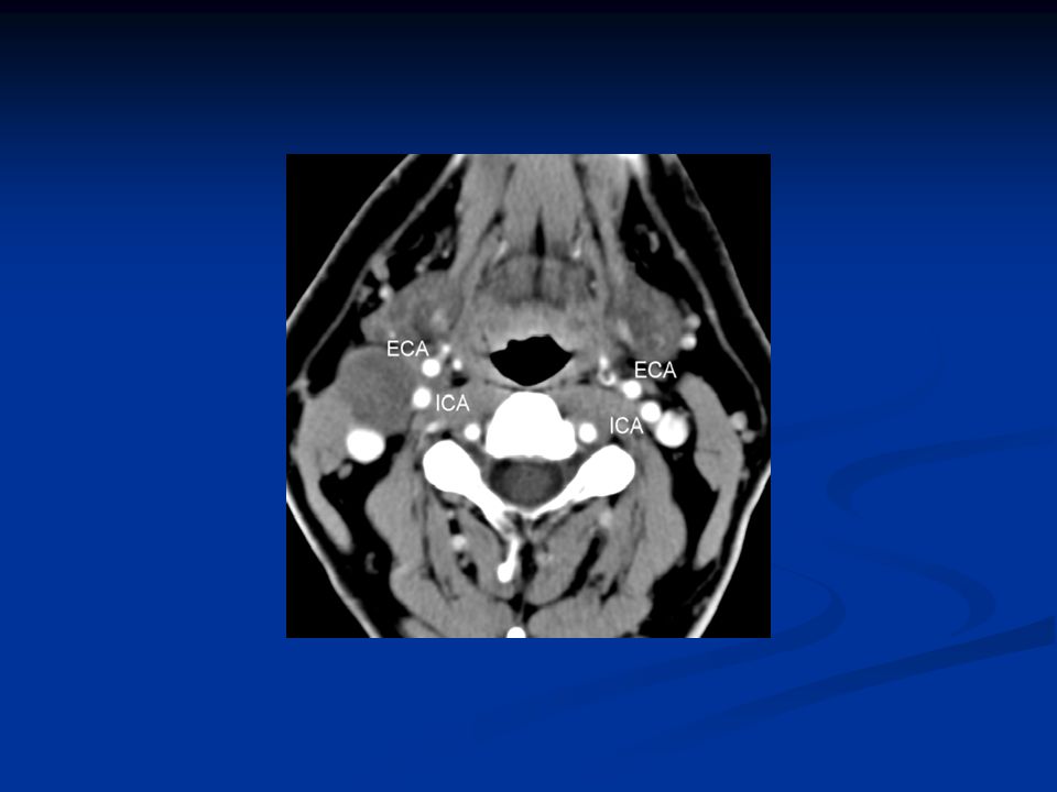
CT Well defined, low density unilocular mass with a thin uniformly enhancing rim

24
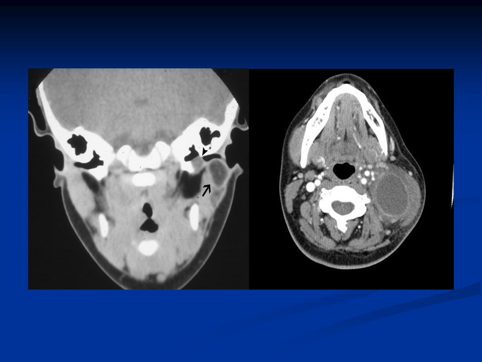
Axial contrast-enhanced CT scan shows a left-sided cyst with a thick, enhancing rim. This cyst is behind the submandibular gland, lateral to the carotid sheath structures, and deep to the anterior margin of the sternocleidomastoid muscle. There is an enhancing tract (arrow) extending from the cyst toward the left palatine tonsil. This was an infected second branchial cleft cyst with an internal tract. Such a tract typically passes between the internal and external carotid arteries and ends in the palatine tonsil. Axial contrast-enhanced CT scan shows a left-sided cyst with a thick, enhancing rim. This cyst is behind the submandibular gland, lateral to the carotid sheath structures, and deep to the anterior margin of the sternocleidomastoid muscle. There is an enhancing tract (arrow) extending from the cyst toward the left palatine tonsil. This was an infected second branchial cleft cyst with an internal tract. Such a tract typically passes between the internal and external carotid arteries and ends in the palatine tonsil.
 extending from the cyst toward the left palatine tonsil. This was an infected second branchial cleft cyst with an internal tract. Such a tract typically passes between the internal and external carotid arteries and ends in the palatine tonsil.")
25
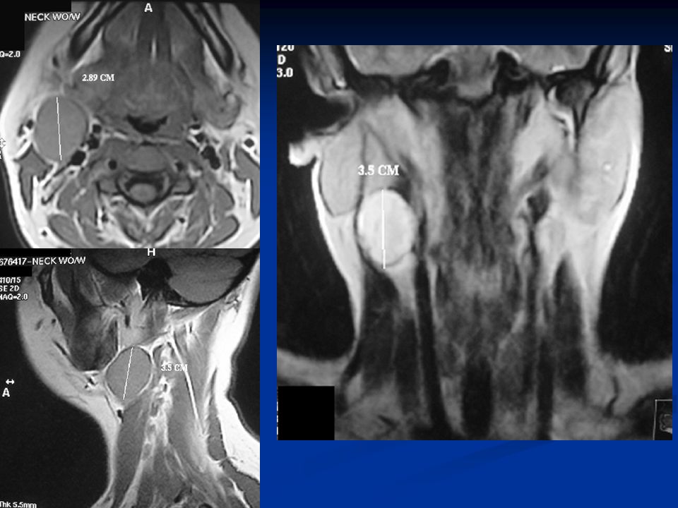
MRI MRI allows for finer resolution during preoperative planning. The wall may be enhancing on gadolinium scans.

27
Differential Diagnosis
Branchiogenic carcinoma Tuberculous adenitis Lipoma Metastatic malignant neoplasms (SCCA from a primary site in the aerodigestive tract) Cystic hygroma (lymphangioma) Carotid body tumors Lymphomas Hemangiomas Thyroid cysts Ectopic thyroid Cervical thymic cysts Thyroglossal duct cyst Parotid cystic tumors Before we discuss imaging features, let’s take a look on the DD of a branchial cyst
 Cystic hygroma (lymphangioma) Carotid body tumors. Lymphomas. Hemangiomas. Thyroid cysts. Ectopic thyroid. Cervical thymic cysts. Thyroglossal duct cyst. Parotid cystic tumors. Before we discuss imaging features, let’s take a look on the DD of a branchial cyst.")
28
Axial contrast-enhanced CT scan shows a well-delineated fatty mass in the subcutaneous tissues of the back of the neck. Branchial cleft cyst

29
Cystic hygroma Cystic lymphangioma Branchial cleft cyst

30
Parotid, malignant tumors
Parotid, malignant tumors. Axial T1-weighted MRI with fat saturation and contrast enhancement shows an enhancing mass extending into the superficial and deep lobes of the right parotid gland. Pathology indicated a squamous cell carcinoma. Branchial cleft cyst

31
T1-Weighted MRI. A well defined mass is present along the anterior triangle of the neck on the right side. There are low signal regions within this mass suggesting the presence of calcifications and flow-voids (arrows). T2-Weighted Axial Images Through the Submandi- bular Region. The mass is bright on T2-weighted images and again exhibits focal lucencies compatible with flow voids. Contrast Enhanced MR in the Axial Plane. There is bright enhancement of the mass. Flow voids produce a "salt and pepper" appearance. Paragangliomas

32
Axial contrast-enhanced CT scan shows a well-delineated irregular mass lesion taking early KM enhancment Branchial cleft cyst Hemangioma

33
Treatment The treatment of branchial cleft cysts is surgical excision.

34
This branchial cleft cyst was followed superiorly to the region of the oropharynx, but no communication was found. The picture below shows the anatomy of the carotid triangle after removal of the cyst.

36
Marina Waves, Salmiya Waterfront, Kuwait
Thank You Marina Waves, Salmiya Waterfront, Kuwait

Similar presentations