Download presentation
Presentation is loading. Please wait.

1
Structure, Function, and Diseases
The Skeletal System: Structure, Function, and Diseases of the bones and joints

2
The Skeletal System Parts of the skeletal system
Bones (skeleton) Joints Cartilages Ligaments (bone to bone) (tendon=bone to muscle) Divided into two divisions Axial skeleton- skull, spinal column Appendicular skeleton – limbs and girdle Copyright © 2003 Pearson Education, Inc. publishing as Benjamin Cummings
 Joints. Cartilages. Ligaments (bone to bone) (tendon=bone to muscle) Divided into two divisions. Axial skeleton- skull, spinal column. Appendicular skeleton – limbs and girdle. Copyright © 2003 Pearson Education, Inc. publishing as Benjamin Cummings.")
3
Functions of Bones Support of the body Protection of soft organs
Movement due to attached skeletal muscles Storage of minerals and fats Blood cell formation Copyright © 2003 Pearson Education, Inc. publishing as Benjamin Cummings

4
Bones of the Human Body The skeleton has 206 bones
Two basic types of bone tissue Compact bone Homogeneous Spongy bone Small needle-like pieces of bone Many open spaces Figure 5.2b Copyright © 2003 Pearson Education, Inc. publishing as Benjamin Cummings

5
Bones are classified by their shape: LONG
bones are longer than they are wide 2. SHORT usually square in shape, cube like 3. FLAT flat , curved 4. IRREGULAR odd shapes Change this on notes!

6
Classification of Bones on the Basis of Shape
Figure 5.1 Copyright © 2003 Pearson Education, Inc. publishing as Benjamin Cummings

7
Types of Bone Cells Osteocytes Osteoblasts Osteoclasts
Mature bone cells Osteoblasts Bone-forming cells Osteoclasts Bone-destroying cells Break down bone matrix for remodeling and release of calcium Bone remodeling is a process by both osteoblasts and osteoclasts Copyright © 2003 Pearson Education, Inc. publishing as Benjamin Cummings

8
Changes in the Human Skeleton
In embryos, the skeleton is primarily hyaline cartilage During development, much of this cartilage is replaced by bone Cartilage remains in isolated areas Bridge of the nose Ears Parts of ribs Joints Copyright © 2003 Pearson Education, Inc. publishing as Benjamin Cummings

9
K J k hyoid Vertebral column carpals metacarpals Cranium Face Clavicle
Scapula Sternum Humerus Ribs Pelvis Radius Ulna Femur Patella Tibia Fibula Tarsals Metatarsals Phalanges J hyoid Vertebral column carpals metacarpals

10
Axial Appendicular

11
The Axial Skeleton Forms the longitudinal part of the body
Divided into three parts Skull Vertebral Column Rib Cage Slide 5.20a Copyright © 2003 Pearson Education, Inc. publishing as Benjamin Cummings

12
The Appendicular Skeleton
Includes the limbs and girdles Divided into two parts PECTORAL GIRDLE Clavicle + scapula + arms & hands PELVIC GIRDLE Pelvis + legs & feet Slide 5.20a Copyright © 2003 Pearson Education, Inc. publishing as Benjamin Cummings

13
The Skull AXIAL SKELETON 8 sutured bones in cranium
Facial bones: 13 sutured bones mandible Cranium encases brain attachments for muscles sinuses

14
= sutures Bones of the Skull = bone markings

15
Bones of the Skull Figure 5.11
Copyright © 2003 Pearson Education, Inc. publishing as Benjamin Cummings

16
Allows for growth

17
Paranasal Sinuses Hollow portions of bones surrounding the nasal cavity Figure 5.10 Slide 5.25a Copyright © 2003 Pearson Education, Inc. publishing as Benjamin Cummings

18
The Vertebral Column Vertebrae separated by intervertebral discs
The spine has a normal curvature Each vertebrae is given a name according to its location Figure 5.14 Slide 5.28 Copyright © 2003 Pearson Education, Inc. publishing as Benjamin Cummings

19
C2 = axis C1 = atlas C1 - C7 T1 – T12 5 fused L1 – L5 4 fused

20
The Hyoid Bone The only bone that does not articulate with another bone Serves as a moveable base for the tongue Figure 5.12 Slide 5.26 Copyright © 2003 Pearson Education, Inc. publishing as Benjamin Cummings

21
Vertebrae

22
Ribs 1 - 7 8 -12

23
Common Types of Fractures
Table 5.2

24
Stages in the Healing of a Bone Fracture
Hematoma External callus Bony callus of spongy bone Healed fracture New blood vessels Internal callus (fibrous tissue and cartilage) Spongy bone trabecula Hematoma formation Fibrocartilage callus formation Bony callus formation Bone remodeling Figure 5.5
 Spongy bone trabecula. Hematoma formation. Fibrocartilage callus formation. Bony callus formation. Bone remodeling. Figure 5.5.")
25
Stages in the Healing of a Bone Fracture
Hematoma Hematoma formation Figure 5.5, step 1

26
Stages in the Healing of a Bone Fracture
Hematoma External callus New blood vessels Internal callus (fibrous tissue and cartilage) Spongy bone trabecula Hematoma formation Fibrocartilage callus formation Figure 5.5, step 2
 Spongy bone trabecula. Hematoma formation. Fibrocartilage callus formation. Figure 5.5, step 2.")
27
Stages in the Healing of a Bone Fracture
Hematoma External callus Bony callus of spongy bone New blood vessels Internal callus (fibrous tissue and cartilage) Spongy bone trabecula Hematoma formation Fibrocartilage callus formation Bony callus formation Figure 5.5, step 3
 Spongy bone trabecula. Hematoma formation. Fibrocartilage callus formation. Bony callus formation. Figure 5.5, step 3.")
28
Stages in the Healing of a Bone Fracture
Hematoma External callus Bony callus of spongy bone Healed fracture New blood vessels Internal callus (fibrous tissue and cartilage) Spongy bone trabecula Hematoma formation Fibrocartilage callus formation Bony callus formation Bone remodeling Figure 5.5, step 4
 Spongy bone trabecula. Hematoma formation. Fibrocartilage callus formation. Bony callus formation. Bone remodeling. Figure 5.5, step 4.")
29
Bone Fractures A break in a bone Types of bone fractures
Closed (simple) fracture – break that does not penetrate the skin Open (compound) fracture – broken bone penetrates through the skin Greenstick- frays, hard to repair, breaks like a green twig Bone fractures are treated by reduction and immobilization Realignment of the bone Copyright © 2003 Pearson Education, Inc. publishing as Benjamin Cummings
 fracture – break that does not penetrate the skin. Open (compound) fracture – broken bone penetrates through the skin. Greenstick- frays, hard to repair, breaks like a green twig. Bone fractures are treated by reduction and immobilization. Realignment of the bone. Copyright © 2003 Pearson Education, Inc. publishing as Benjamin Cummings.")
30
Joints A joint, or articulation, is the place where two bones come together. Fibrous- Immovable:connect bones, no movement. (skull and pelvis). Cartilaginous- slightly movable, bones are attached by cartilage, a little movement (spine or ribs). Synovial- freely movable, much more movement than cartilaginous joints. Cavities between bones are filled with synovial fluid. This fluid helps lubricate and protect the bones.
. Synovial- freely movable, much more movement than cartilaginous joints. Cavities between bones are filled with synovial fluid. This fluid helps lubricate and protect the bones.")
31
The Synovial Joint Slide 5.51 Figure 5.28
Copyright © 2003 Pearson Education, Inc. publishing as Benjamin Cummings

32
Types of Joints Hinge-
A hinge joint allows extension and retraction of an appendage. (Elbow, Knee)
 .")
33
Ball and Socket-
A ball and socket joint allows for radial movement in almost any direction. They are found in the hips and shoulders. (Hip, Shoulder)
.")
34
Gliding - In a gliding or plane joint bones slide past each other
Gliding -
In a gliding or plane joint bones slide past each other. Mid-carpal and mid-tarsal joints are gliding joints. (Hands, Feet)
")
35
Saddle - This type of joint occurs when the touching surfaces of two bones have both concave and convex regions with the shapes of the two bones complementing one other and allowing a wide range of movement. (Thumb)
.")
36
Fixed joint

37
Diseases and Conditions
of the Skeletal System

38
Arthritis

42
Bursitis Inflammation of the Bursa (fluid filled sac surrounding the joint). A bursa can become inflamed from injury, infection (rare in the shoulder), or due to an underlying rheumatic condition. Bursitis is typically identified by localized pain or swelling, tenderness, and pain with motion of the tissues in the affected area.
, or due to an underlying rheumatic condition. Bursitis is typically identified by localized pain or swelling, tenderness, and pain with motion of the tissues in the affected area.")
45
Tendonitis Sometimes the tendons become inflamed for a variety of reasons, and the action of pulling the muscle becomes irritating. If the normal smooth gliding motion of your tendon is impaired, the tendon will become inflamed and movement will become painful. This is called tendonitis, and literally means inflammation of the tendon. The most common cause of tendonitis is overuse.

47
Carpal Tunnel Syndrome
Any condition that causes swelling or a change in position of the tissue within the carpal tunnel can squeeze and irritate the median nerve. Irritation of the median nerve in this manner causes tingling and numbness of the thumb, index, and the middle fingers, a condition known as "carpal tunnel syndrome."

50
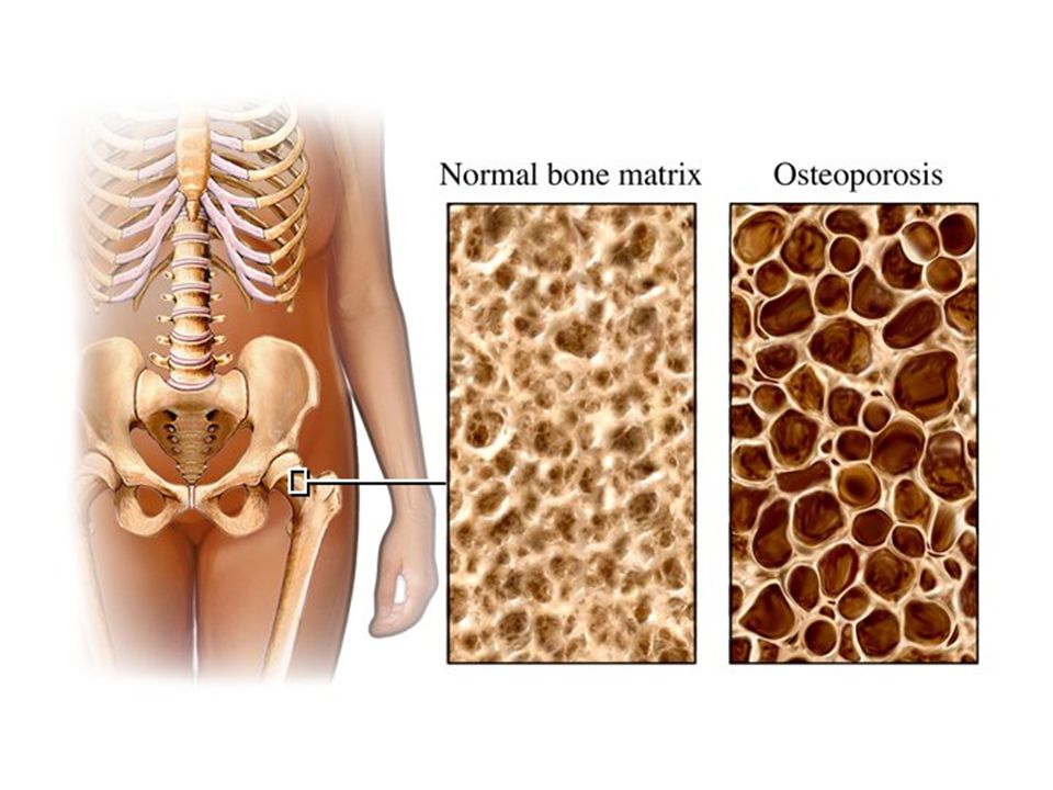
Osteoporosis Osteoporosis is a term that means "porous bones." It is a skeletal disease affecting women and men. Osteoporosis is a condition in which bones have lost minerals especially calcium - making them weaker, more brittle, and susceptible to fractures (broken bones). Any bone in the body can be affected by osteoporosis, but the most common places where fractures occur are the back (spine), hips, and wrists.
. Any bone in the body can be affected by osteoporosis, but the most common places where fractures occur are the back (spine), hips, and wrists.")
53
Scoliosis Scoliosis is an abnormal curvature of the spine. If your child has scoliosis, the view from behind may reveal one or more abnormal curves. Scoliosis runs in families, but doctors often don't know the cause. More girls than boys have severe scoliosis. Adult scoliosis may be a worsening of a condition that began in childhood, but wasn't diagnosed or treated. In other cases, scoliosis may result from a degenerative joint condition in the spine.

59
Kyphosis With kyphosis, your spine may look normal or you may develop a hump. Kyphosis can occur as a result of developmental problems; degenerative diseases, such as arthritis of the spine; osteoporosis with compression fractures of the vertebrae; or trauma to the spine. It can affect children, adolescents and adults.

62
Lordosis A normal spine, when viewed from behind appears straight. However, a spine affected by lordosis shows evidence of a curvature of the back bones (vertebrae) in the lower back area, giving the child a "swayback" appearance.
 in the lower back area, giving the child a swayback appearance.")
65
Rickets Rickets is the softening and weakening of bones in children, usually because of an extreme and prolonged vitamin D deficiency. Some skeletal deformities caused by rickets may need corrective surgery.

69
Scurvy The human body lacks the ability to synthesize and make vitamin C and therefore depends on exogenous dietary sources to meet vitamin C needs. Consumption of fruits and vegetables or diets fortified with vitamin C are essential to avoid ascorbic acid deficiency. Even though scurvy is uncommon, it still occurs and can affect adults and children who have chronic dietary vitamin C deficiency.

72
Gout Gout is a disease that results from an overload of uric acid in the body. This overload of uric acid leads to the formation of tiny crystals of urate that deposit in tissues of the body, especially the joints. When crystals form in the joints it causes recurring attacks of joint inflammation. Chronic gout can also lead to deposits of hard lumps of uric acid in and around the joints and may cause joint destruction, decreased kidney function, and kidney stones.

76
Acromegaly Acromegaly is a serious condition that occurs when the body produces too much of the hormones that control growth. ・The hormone most often affected is called growth hormone, or GH. It is produced by the pituitary gland, a tiny organ at the base of the brain. Growth hormone promotes growth of bone, cartilage, muscle, organs, and other tissues. When there is too much growth hormone in the body, these tissues grow larger than normal. This excessive growth can cause serious disease and even premature death.

81
Spina Bifida Spina bifida is a birth defect that involves the incomplete development of the spinal cord or its coverings. The term spina bifida comes from Latin and literally means "split" or "open" spine. Spina bifida occurs at the end of the first month of pregnancy when the two sides of the embryo's spine fail to join together, leaving an open area. In some cases, the spinal cord or other membranes may push through this opening in the back. The condition is usually detected before a baby is born and treated right away.

85
Sarcoma Osteosarcoma is the most common type of bone cancer. It arises in bone and is most commonly found in children and adolescents but a rare form occurs in adults, particularly in patients who have been cured of other cancers with radiation therapy.

90
Leukemia Leukemia is cancer of the blood cells. It starts in the bone marrow, the soft tissue inside most bones. Bone marrow is where blood cells are made. When you are healthy, your bone marrow makes:・White blood cells, which help your body fight infection.・Red blood cells, which carry oxygen to all parts of your body.・Platelets, which help your blood clot. When you have leukemia, the bone marrow starts to make a lot of abnormal white blood cells, called leukemia cells. They don't do the work of normal white blood cells, they grow faster than normal cells, and they don't stop growing when they should.

93
Bone Marrow Biopsy

94
Copy this chart in your notebook:
Homeostatic Imbalances of the SKELETAL SYSTEM Chapter & page Disorder Description 1 Chapter 6 2 Chapter 7 3 Chapter 7 4 Chapter 8 5 Chapter 8 See pages : 6 Chapter 8 Sprains, dislocations, bursitis, tendonitis Use Chapters 6,7,8 to complete the chart. Remember to look for the ORANGE “Homeotatic Imbalance” headings

Similar presentations


 Joints Cartilages Ligaments.>")


. Cartilaginous->")

