Download presentation
Presentation is loading. Please wait.

2
Fertilization and Pregnancy

3
Fertilization Pregnancy is the presence of developing offspring in the uterus, an event resulting from fertilization – the fusion of male and female gametes. For this fusion to occur in humans, the sperm needs to penetrate the egg cell, which is surrounded by a zona pellucida and a corona radiata.

4
Fertilization The acrosome reaction A protective covering (the acrosome) surrounds the head of the sperm; it contains enzymes that break down the cells that protect the egg. As the sperm touches the cells surrounding the egg (the corona radiate), the acrosome releases these enzymes, and they digest a path into the egg.
, the acrosome releases these enzymes, and they digest a path into the egg..")
5
Fertilization Role of the zona pellucida When the sperm cell reaches the zona pellucida, a thick, jelly-like layer surrounding the secondary oocyte, re- ceptors bind the sperm cell so it can pass through.

6
Fertilization The cortical reaction The head of the sperm cell fuses with the plasma membrane of the oocyte, triggering the cort- ical granules to release enzymes to thicken the zona pellucida so that it becomes a fertilization envelope Then no other sperm cells can fertilize the egg.

7
Fertilization The journey of the sperm cell: Sperm cells must reach the upper one-third of the uterine tubes for fertilization to occur. Under the influence of estrogen during the first half of the menstrual cycle, uterine se- cretions are thin, allowing sperm cells to swim easily to their destination. Fertilization occurs in the fallopian tube when one sperm penetrates the egg. A diploid zygote results.

8
Process of in vitro fertilization Fertilization that is done outside the body (in glass).
.")
9
Early embryo development After fertilization: the haploid sets of chromosomes from the male and female gametes line up at the equator and prepare for the first mitotic division. This division, within 24 hours after fertilization, is not followed by cell growth, and is, therefore, called a cleavage division. During the first 5 days several cleavage divisions occur within the fallopian tube. Even though cells are dividing, embryo doesn’t grow At the 8 cell stage compaction occurs – the cells squish together into a small ball called the morula.

10
Early embryo development The morula stage, up to 32 cells

11
Early embryo development Morula: A compact mass of 16 to 32 cells. The morula then leaves the fallopian tube and enters the uterus. The inside of the morula hollows out, and the cavity becomes filled with fluid, becoming the blastocyst.

12
Early embryo development The blastocyst begins to differentiate. Trophoblast: tissue around the outside of the blastocyst will become the extraembryonic membranes and placenta, after implantation in the uterus. Inner cell mass: a ball of cells inside the trophoblast will become the embryo proper. Blastocoel: the fluid-filled cavity.

13
Early Embryonic Development The blastocyst begins to differentiate. The rate of cell division increases. By time blastocyst reaches the uterine cavity there are ~100 cells, and it is ready to plant itself in the endometrium. (lining of uterus)
.")
14
Early embryo development Passage of the fertilized egg through the fallopian tube.

15
Early embryonic development http://www.youtube.com/watch?v=vslaD-mWaNA http://www.youtube.com/watch?v=28GTvrNvRRE

16
The placenta Uterus Day 6 Day 7 Day 18 Day 30 Development of the placenta from cells of the chorion (in green).
.")
17
The placenta Structure: Disk-shaped organ, contains embryonic and maternal blood vessels About the size of a dinner plate, and weighing about 1 kg allowing exchange of materials by diffusion. Two embryonic blood vessels contained with- in the umbilical cord.

18
Placenta Structure of the placental villus Maternal blood flows in the inter-villous space around the villi. Cells that separate maternal and fetal blood from the placental barrier must be selectively permeable for exchange of materials.ace area for processes. Microvilli increases Surface Area Fetal blood that flows through the placental villi is oxygenated and recharged with nutrients Waste products removed Distance between mater- nal and fetal blood is as small as 5 micrometers.

19
The placenta Function: produces progesterone/estrogen Estrogen maintains endometrium and inhibits menstrual cycle from 2nd trimester onwards Materials are exchanged between the maternal and fetal blood in the placenta. Nutrients: oxygen, glucose, amino acids, vitamins, antibodies. Waste products: carbon dioxide, urea transports glucose, amino acids, lipids, oxygen, carbon dioxide, antibodies, urea

20
HCG in early pregnancy Human Chorionic Gonadotropin Secreted by embryonic cells that form the placenta after implantation. Enters the bloodstream, binds to LHCGR (Luteinizing Hormone /Choriogonadotropin Receptor in Uterus and Ovary. This preserves the corpus luteum beyond 14 days so it can con- tinue producing progesterone to maintain the thickening & vascularization of the endometrial layer in the uterus. Triggers the hypothalamus to mobilize the mothers stored fat. HCG levels will double every 72 hours Level will reach its peak in the first 8 - 11 weeks of pregnancy and then will decline and level off for the remainder of the pregnancy.

21
Pregnancy Tests Human Chorionic Gonadotropin Can be detected 11 days after conception in the blood. More accurate. Can be detected 12-14 days after conception in the urine. A pregnancy test binds the HCG hormone to an indicator and antibody; the indicator is a pigment molecule (ex. a line on the home pregnancy test).
..")
22
The amnion The fetus is supported and protected by the amniotic sac and amniotic fluid.

23
The amnion Amniotic sac is an extra-embrionic membrane that extends all the way around the fetus. Provides a cushioning effect Provides thermal stability Provides an environment for free movement of fetus Development of muscles and skeleton of fetus

25
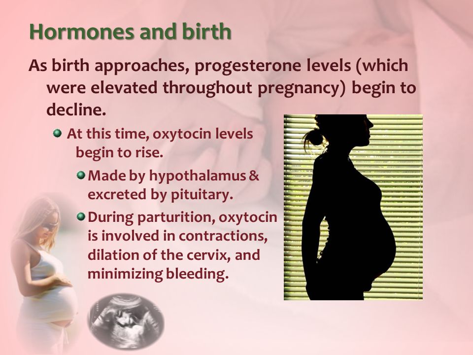
Hormones and birth As birth approaches, progesterone levels (which were elevated throughout pregnancy) begin to decline. At this time, oxytocin levels begin to rise. Made by hypothalamus & excreted by pituitary. During parturition, oxytocin is involved in contractions, dilation of the cervix, and minimizing bleeding.

26
Hormones and birth When a baby is ready to be delivered, it drops lower in the uterus and puts pressure on the cervix, causing the cervix to stretch.

27
Hormones and birth The stretching of the cervix further stimulates the release of the hormone oxytocin, which induces the uterus to contract more and push the baby further against the cervix. In turn, this causes the cervix to further stretch, which releases more oxy- tocin, and thus the cycle continues = positive feedback.

28
Childbirth Stretching of the cervix further stimulates the release of the hormone oxytocin, which induces the uterus to contract.

Similar presentations


>")


 in her ovaries by the time she is 7 years old, only approx. 300.>")
