Download presentation
Presentation is loading. Please wait.

1
Human Anatomy, First Edition McKinley & O'Loughlin
Chapter 26 : Digestive System

2
General Structure and Functions of the Digestive System
Organs of the Digestive System to: Ingest the food. Transport the food. Digest the food into smaller usable components. Absorb the necessary nutrients into the bloodstream. Expel the waste products from the body.

3
General Structure and Functions of the Digestive System
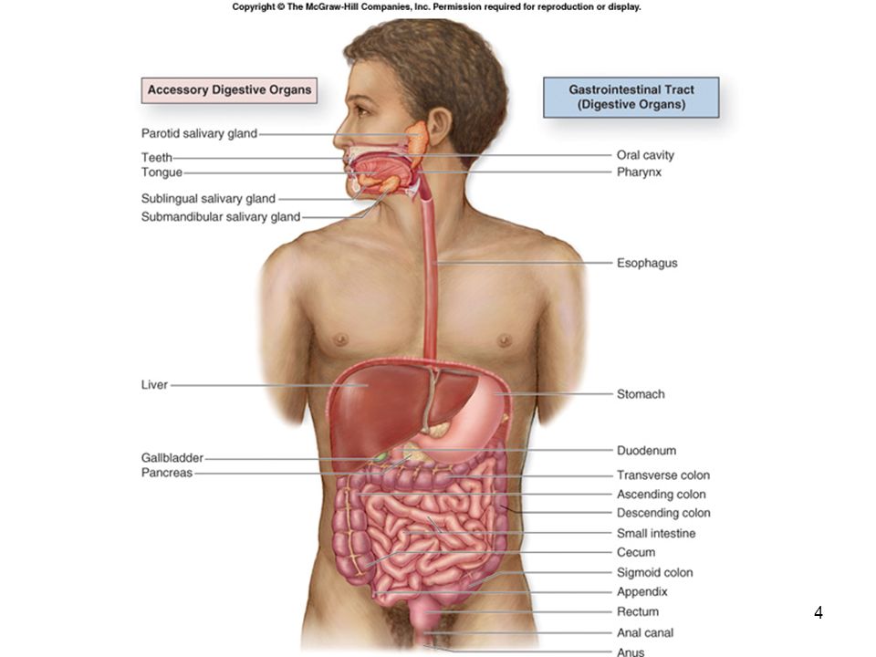
Composed of two separate categories of organs: digestive organs accessory digestive organs. Digestive organs collectively make up the: gastrointestinal (GI) tract. Also called: the digestive tract alimentary canal.
 tract. Also called: the digestive tract. alimentary canal.")
5
General Structure and Functions of the Digestive System
The GI tract organs: oral cavity pharynx esophagus stomach small intestine large intestine continuous tube about 30 feet (9–10 meters) from mouth to anus. Smooth muscle in the wall responsible for motility pushes materials from one end to the other.
 from mouth to anus. Smooth muscle in the wall. responsible for motility. pushes materials from one end to the other.")
6
General Structure and Functions of the Digestive System
Accessory digestive organs: do not form the GI tube can develop as outgrowths are connected to the GI tract (some by ducts) Assist the GI tract in the digestion of food. Include: Teeth Tongue Salivary glands Liver Gallbladder Pancreas
 Assist the GI tract in the digestion of food. Include: Teeth. Tongue. Salivary glands. Liver. Gallbladder. Pancreas.")
7
Digestive System Functions
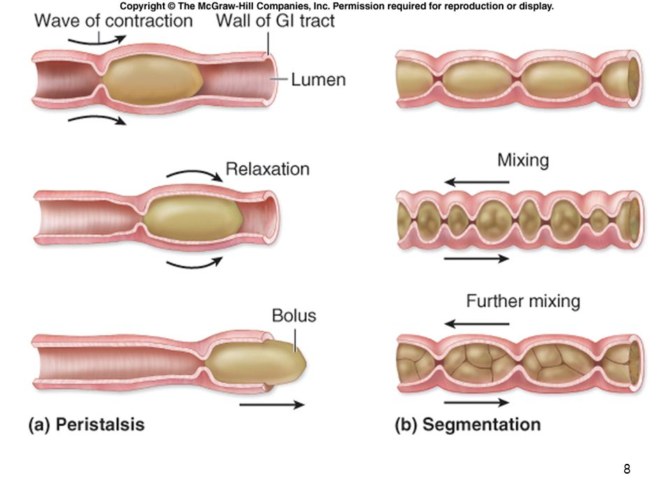
Ingestion Digestion: break down of large particles of food mechanical digestion chemical digestion Propulsion peristalsis segmentation Secretion: digestive enzymes hormones Absorption: from external environment into internal environment across mucosa Elimination of wastes (defecation)
")
9
Oral Cavity (mouth) Entrance to the GI tract.
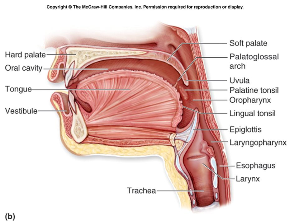
Initial site of digestion: mechanical digestion (via mastication) chemical digestion (via enzymes in saliva). Bounded anteriorly by the teeth and lips Bounded posteriorly by the oropharynx. Superior boundary is formed by the hard and soft palates. Floor, or inferior surface, of the oral cavity the tongue the mylohyoid muscle covered with mucosa.
 chemical digestion (via enzymes in saliva). Bounded anteriorly by the teeth and lips. Bounded posteriorly by the oropharynx. Superior boundary is formed by the hard and soft palates. Floor, or inferior surface, of the oral cavity. the tongue. the mylohyoid muscle covered with mucosa.")
11
Oral Cavity (mouth) Two regions of the oral cavity
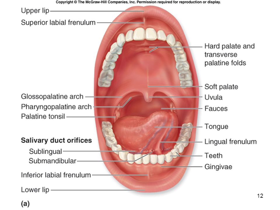
Vestibule is the space between the cheeks or lips and the gums. Oral cavity proper. The lateral walls are formed by the cheeks. Contain buccinator muscles Lips (labia). Orbicularis oris muscle Keratinized stratified squamous ET Gingivae, or gums. Dense regular CT Nonkeratinized ET Labial frenulum.
. Orbicularis oris muscle. Keratinized stratified squamous ET. Gingivae, or gums. Dense regular CT. Nonkeratinized ET. Labial frenulum.")
14
Palate Hard palate Soft palate
Anterior two-thirds of the palate hard and bony Soft palate Posterior one-third soft and muscular primarily composed of skeletal muscle. Extending inferiorly from the posterior part of the soft palate is the uvula. When swallowing, the soft palate and the uvula elevate to close off the opening of the nasopharynx.

15
Palate Fauces represent the opening between the oral cavity and the oropharynx. Fauces are bounded by paired muscular folds: glossopalatine arch (anterior fold) pharyngopalatine arch (posterior fold) Palatine tonsils are housed between the arches.
 pharyngopalatine arch (posterior fold) Palatine tonsils are housed between the arches.")
16
Tongue An accessory digestive organ Formed from:
skeletal muscle covered with lightly keratinized stratified squamous epithelium. Manipulates and mixes ingested materials during chewing Forms the bolus. a globular mass of partially digested material Performs important functions in swallowing.

17
Tongue Inferior surface of the tongue
attaches to the floor of the oral cavity By the lingual frenulum. Numerous small projections (papillae) cover the superior (dorsal) surface. Posterior surface contains lingual tonsils. Skeletal muscles move the tongue.
 cover the superior (dorsal) surface. Posterior surface contains lingual tonsils. Skeletal muscles move the tongue.")
18
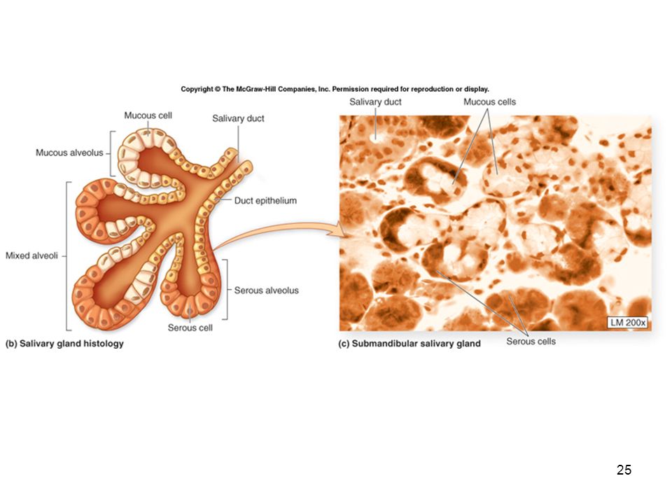
Salivary Glands Collectively produce and secrete saliva.
a fluid that assists in the initial activities of digestion Volume of saliva secreted daily ranges between 1.0 and 1.5 L. Most is produced during mealtime Smaller amounts are produced continuously to ensure that the oral cavity remains moist.

19
Salivary Glands Components of saliva Functions Water: makes up 99%
Amylase: first step of chemical digestion Lysozyme: antimicrobial Functions Moisten food Food molecules into solution: taste Form bolus: for swallowing Cleanse oral cavity.

20
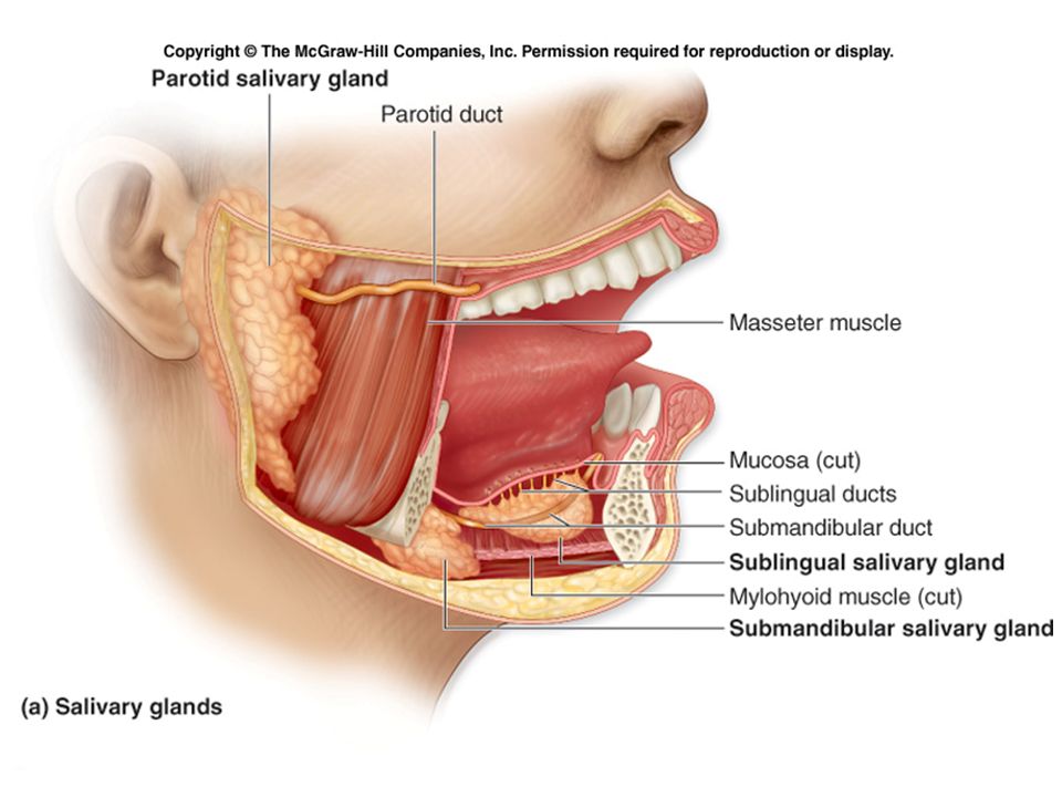
Salivary Glands Three pairs of large, multicellular salivary glands:
parotid glands submandibular glands sublingual glands

21
The Parotid Glands Largest salivary glands.
located anterior and inferior to the ear partially overlying the masseter muscle. Produce about 25–30% of saliva conducted through the parotid duct to the oral cavity.

22
The Submandibular Glands
Inferior to the body of the mandible. Produce most of the saliva (about 60–70%). ducts opens through a papilla in the floor of the mouth lateral to the the lingual frenulum.
. ducts opens through a papilla in the floor of the mouth. lateral to the the lingual frenulum.")
23
The Sublingual Glands Inferior to the tongue
internal to the oral cavity mucosa. Each gland has multiple tiny sublingual ducts open onto the inferior surface of the oral cavity posterior to the submandibular duct papilla. Contribute only about 3–5% of the total saliva.

26
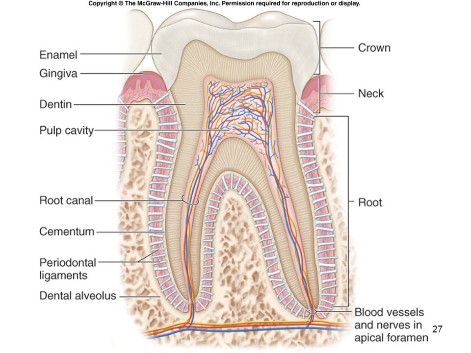
Teeth Collectively known as the dentition. Responsible for mastication
first part of the mechanical digestion. A tooth has: exposed crown constricted neck one or more roots Roots of the teeth fit into dental alveoli are sockets within the alveolar processes on both the maxillae and the mandible. Collectively, the roots, the dental alveoli, and the periodontal ligament that binds the roots to the alveolar processes form a gomphosis joint.

30
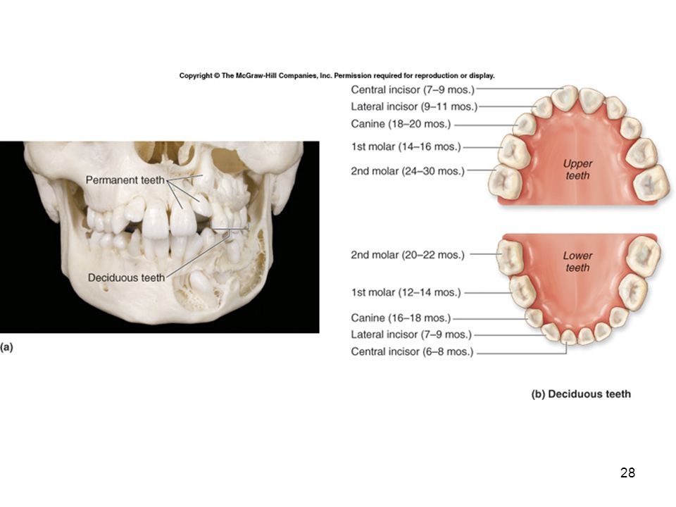
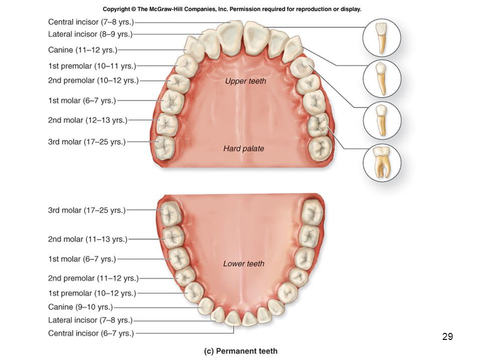
Teeth Two sets of teeth 20 deciduous teeth, also called “milk teeth,” erupt between 6 months and 30 months after birth. These teeth are eventually lost and replaced by 32 permanent teeth. The more anteriorly placed permanent teeth tend to appear first, followed by the posteriorly placed teeth. The last teeth to erupt are the third molars, often called “wisdom teeth,” in the late teens or early 20’s. Often the jaw lacks space to accommodate these final molars, and they may either emerge only partially or grow at an angle and become impacted. Impacted teeth cannot erupt properly because of the angle of their growth.

31
Pharynx Review Pharyngeal constrictors Innervated by the vagus nerves

32
General arrangement of abdominal GI organs
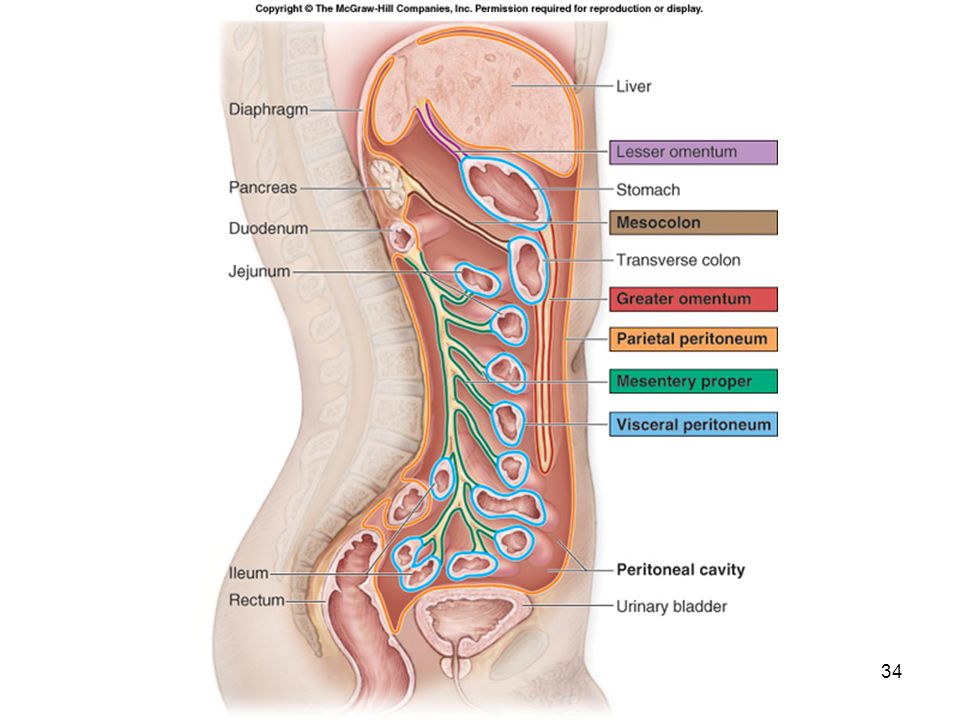
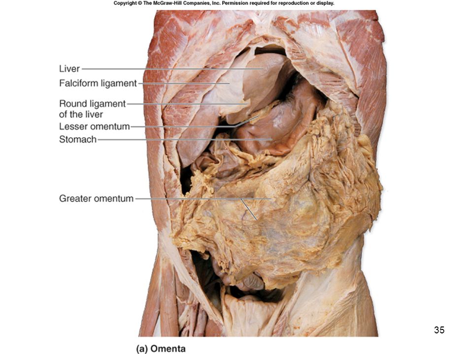
Peritoneum Parietal peritoneum Visceral peritoneum Peritoneal cavity Intraperitoneal organs Retroperitoneal organs

33
General arrangement of abdominal GI organs
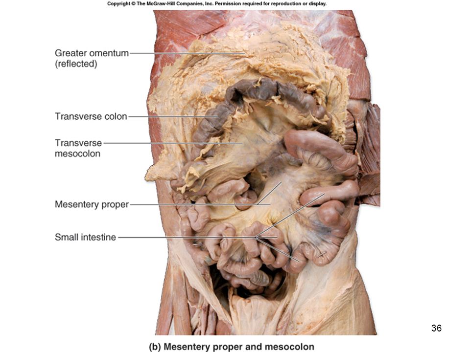
Mesentaries Double layered folds of peritoneum Greater omentum Lesser omentum Mesentery proper Suspends small intestine from posterior wall of abdomen Mesocolon Suspends large intestine Peritoneal ligament Peritoneum that attaches one organ to another

37
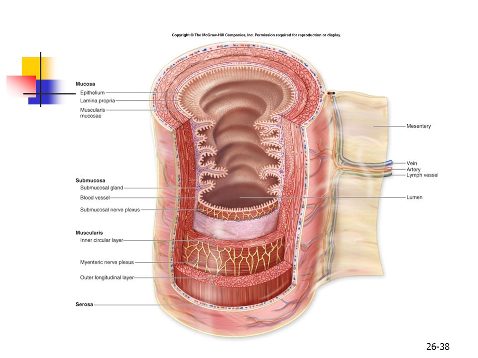
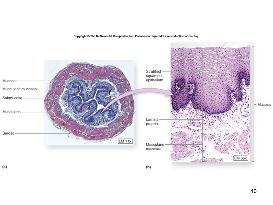
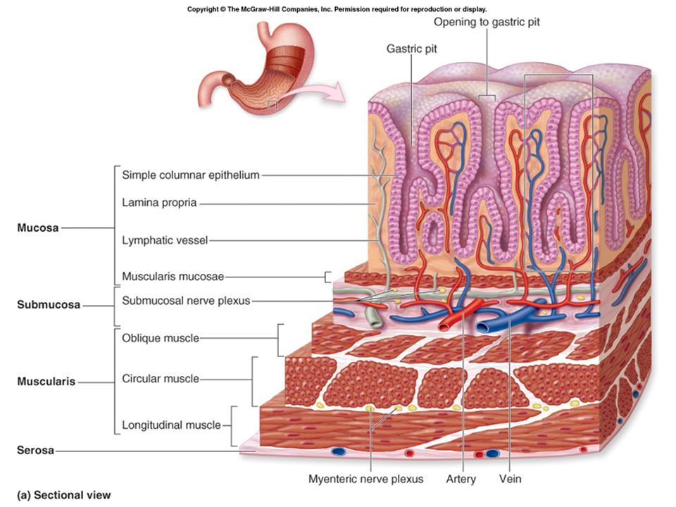
General Histology of GI Organs
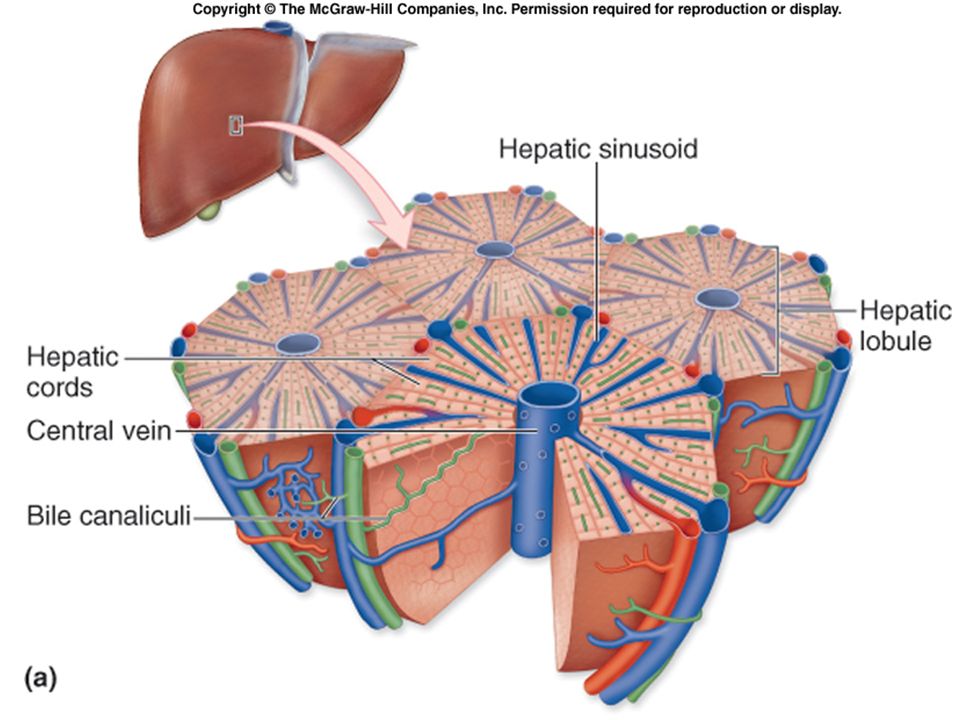
from the esophagus through the large intestine a tube composed of four concentric layers called tunics. From deep to superficial, these tunics are: the mucosa the submucosa submucosal nerve plexus (Meissner plexus) the muscularis myenteric plexus (Auerbach plexus) the adventitia or serosa
 the muscularis. myenteric plexus (Auerbach plexus) the adventitia or serosa.")
39
Esophagus Tubular passageway Histology Pharynx to stomach Bolus
About 25 cm in adult Esophageal hiatus: through diaphragm Histology Mucosa: nonkeritinized stratified squamous ep. Submucosa: thick, elastic fibers, mucous glands Muscularis: inner circular, outer longitudinal Both skeletal and smooth Adventitia

41
Esophagus Superior esophageal sphincter: Inferior esophageal sphincter
Skeletal muscle Where pharynx and esophagus meet Inferior esophageal sphincter Also cardiac sphincter Circular smooth muscle Orifice between esophagus and stomach

42
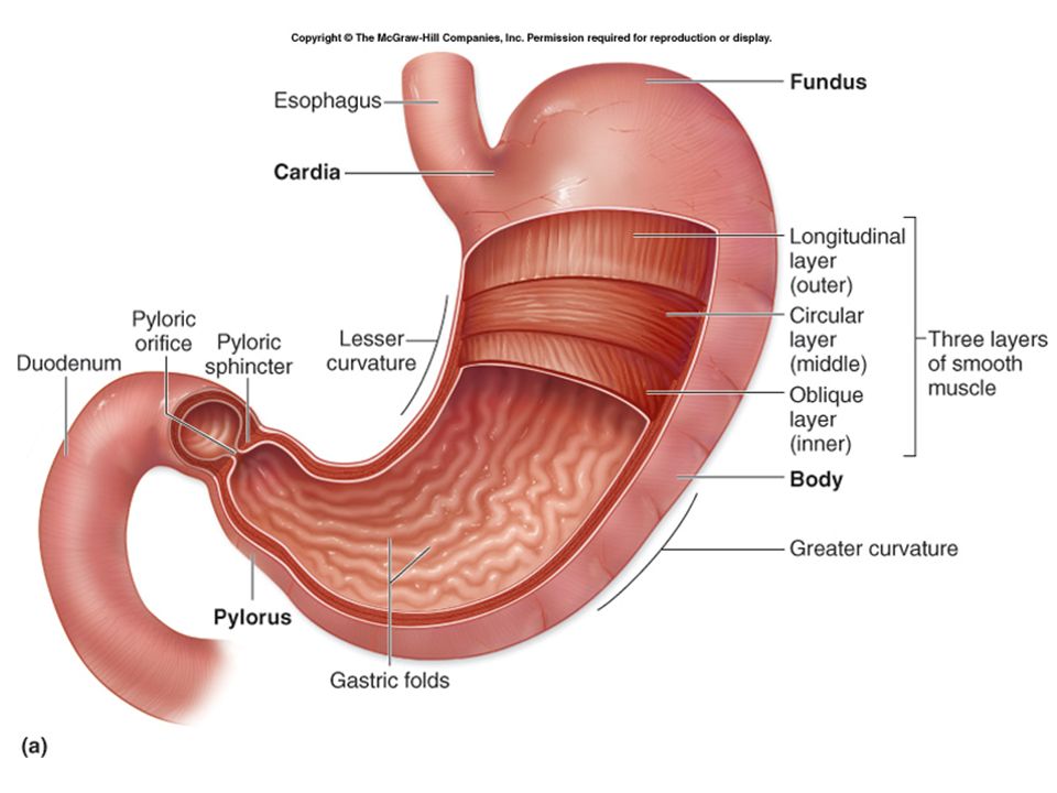
Stomach General J-shaped Functions Digestion Results in chyme
Chemical Mechanical Results in chyme Limited absorption

43
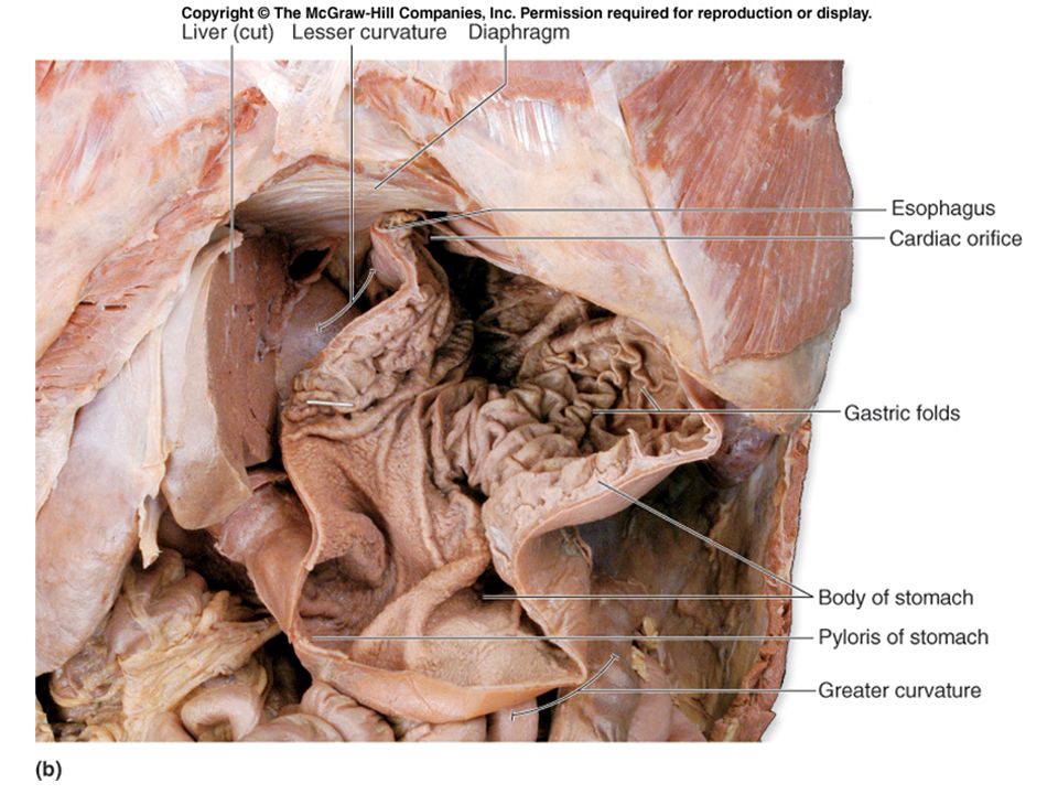
Stomach Gross anatomy Cardia Fundus Body Pylorus Greater curvature
Cardiac orifice Fundus Body Pylorus Pyloric sphincter Pyloric orifice Greater curvature Greater omentum Lesser curvature Lesser omemtum Gastric folds (rugae)
")
46
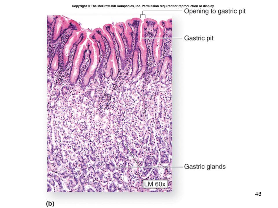
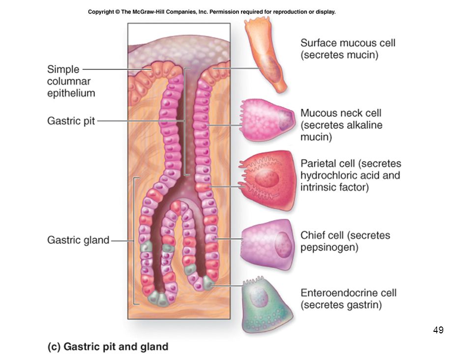
Stomach Histology Mucosa: simple columnar Muscularis Gastric pits
Gastric glands Muscularis 3 layers Inner oblique Middle circular Outer longitudinal

50
Small Intestine Finishes chemical digestion
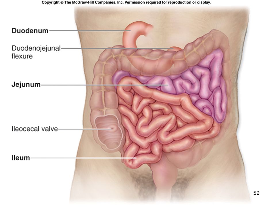
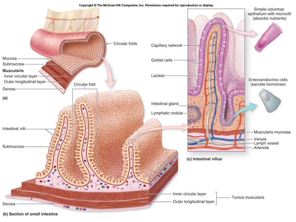
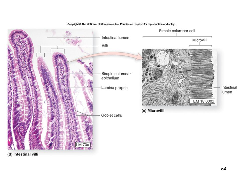
Responsible for absorbing most of the nutrients. Ingested nutrients spend at least 12 hours in the small intestine. thin-walled tube about 6 meters (20 feet) in length. coiled Extends from the pylorus of the stomach to the cecum of the large intestine occupies a significant portion of the abdominal cavity.
 in length. coiled. Extends from the pylorus of the stomach to the cecum of the large intestine. occupies a significant portion of the abdominal cavity.")
51
Small Intestine The duodenum The jejunum The ileum
first segment of the small intestine. approximately 25 centimeters (10 inches) long originates at the pyloric sphincter major duodenal papilla The jejunum middle region of the small intestine. approximately 2.5 meters (7.5 feet) makes up approximately two-fifths of the small intestine’s total length. primary region for chemical digestion and nutrient absorption The ileum is the last region of the small intestine. about 3.6 meters (10.8 feet) in length forms approximately three-fifths of the small intestine. terminates at the ileocecal valve sphincter that controls the entry of materials into the large intestine.
 long. originates at the pyloric sphincter. major duodenal papilla. The jejunum. middle region of the small intestine. approximately 2.5 meters (7.5 feet) makes up approximately two-fifths of the small intestine’s total length. primary region for chemical digestion and nutrient absorption. The ileum. is the last region of the small intestine. about 3.6 meters (10.8 feet) in length. forms approximately three-fifths of the small intestine. terminates at the ileocecal valve. sphincter that controls the entry of materials into the large intestine.")
55
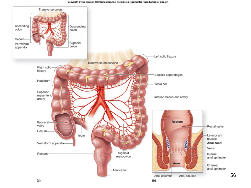
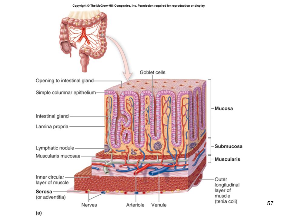
Large Intestine approximate length of 1.5 meters (5 feet)
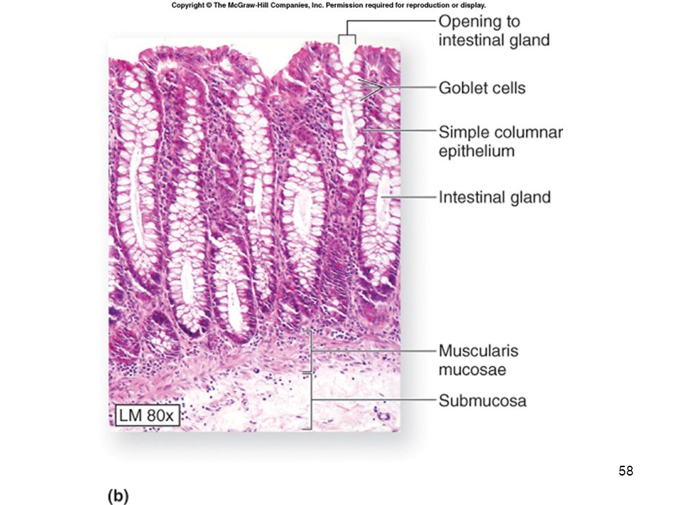
diameter of 6.5 centimeters (2.5 inches). Absorbs most of the water and electrolytes from the remaining digested material. Watery material that first enters the large intestine soon solidifies and becomes feces. Stores fecal material until the body is ready to defecate. Absorbs a very small percentage of nutrients still remaining in the digested material. Composed of four segments: the cecum, colon, rectum, anal canal
. Absorbs most of the water and electrolytes from the remaining digested material. Watery material that first enters the large intestine soon solidifies and becomes feces. Stores fecal material until the body is ready to defecate. Absorbs a very small percentage of nutrients still remaining in the digested material. Composed of four segments: the cecum, colon, rectum, anal canal.")
59
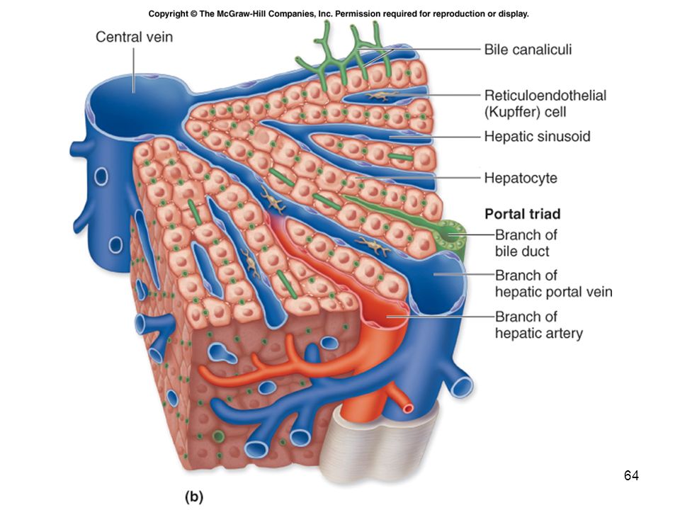
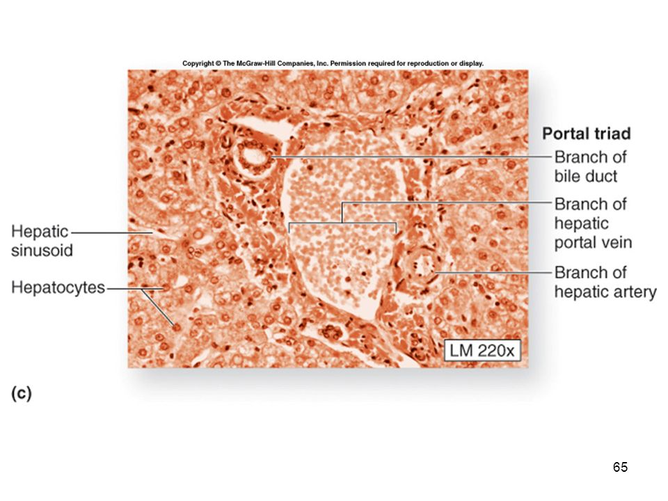
Accessory Digestive Organs
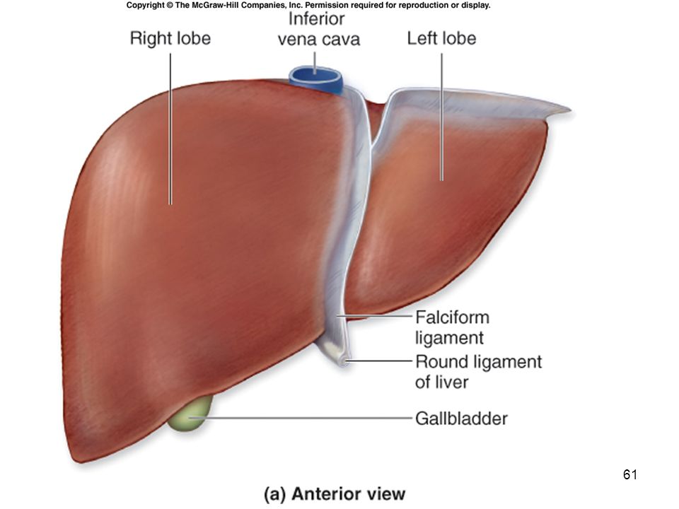
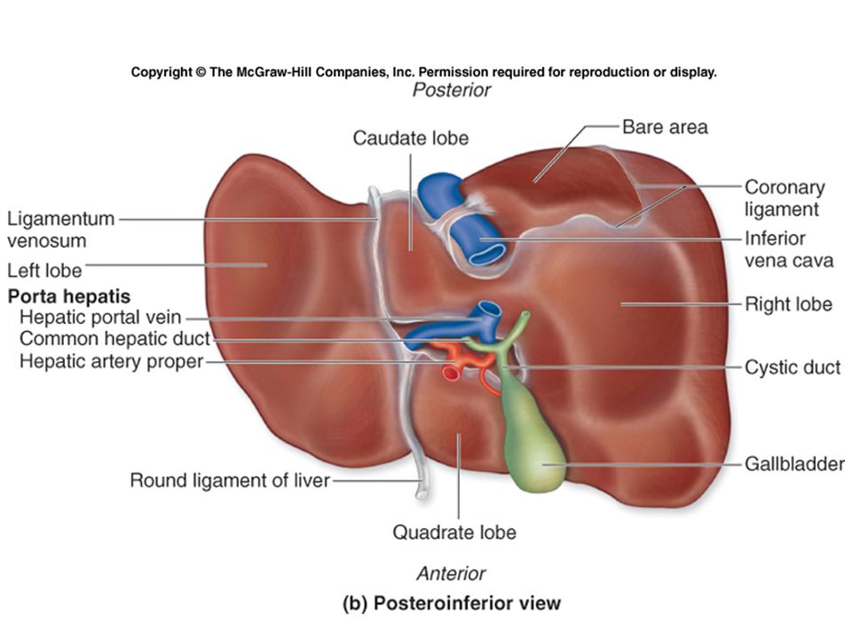
The liver composed of four incompletely separated lobes supported by two ligaments Right lobe Left lobe Falciform ligament Round ligament Caudate lobe Quadrate lobe

60
Functions of The Liver Produce bile.
a greenish fluid that breaks down fats into small droplets to assist in their chemical digestion Detoxify drugs, metabolites, and poisons. Store excess nutrients and vitamins and release them when they are needed. Synthesize blood plasma proteins such as albumins, globulins, and proteins required for blood clotting. Phagocytize debris in the blood. Help break down and recycle components of aged erythrocytes and damaged or worn-out formed elements.

66
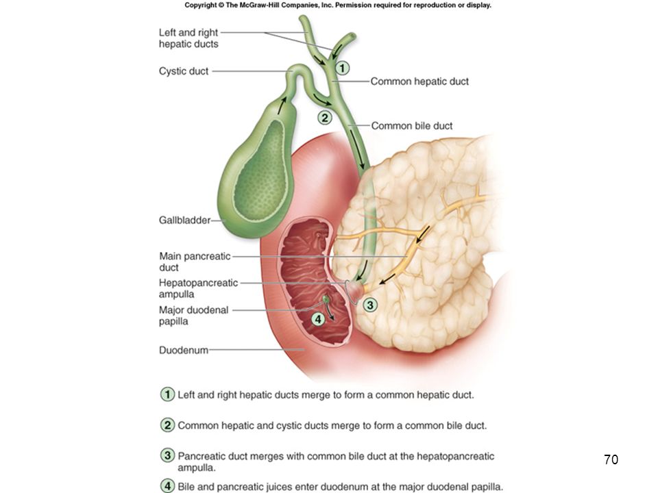
Accessory Digestive Organs
Gallbladder concentrates bile produced by the liver and stores this concentrate until it is needed for digestion cystic duct connects the gallbladder to the common bile duct can hold approximately 40 to 60 milliliters of concentrated bile

67
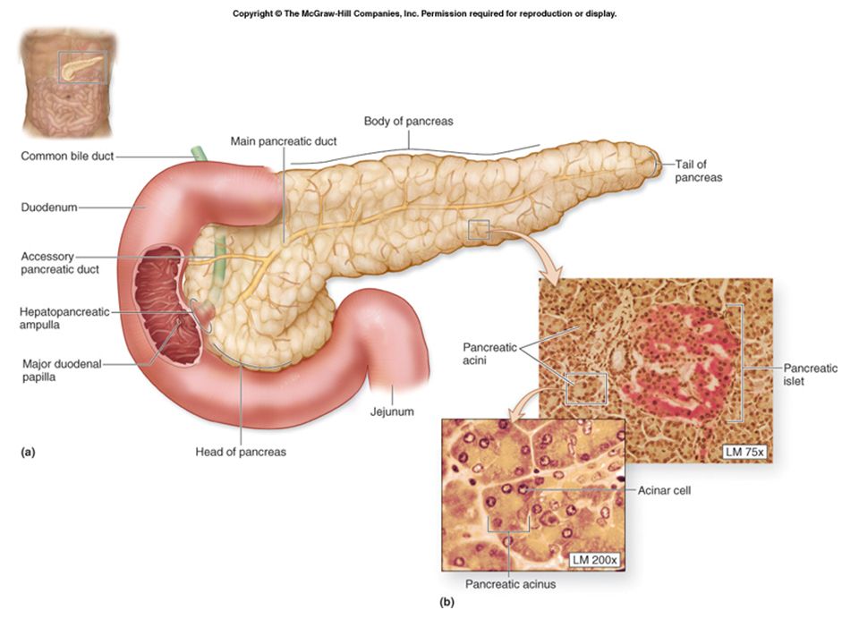
Accessory Digestive Organs
Pancreas mixed gland because it exhibits both endocrine and exocrine functions Endocrine functions are performed by the pancreatic islets. Exocrine activity results in the secretion of digestive enzymes, collectively called pancreatic juice, into the duodenum.

69
Accessory Digestive Organs
The biliary apparatus. network of thin ducts that carry bile from the liver and gallbladder to the duodenum the left and right lobes of the liver drain bile into the left and right hepatic ducts, respectively the left and right hepatic ducts merge to form a single common hepatic duct the cystic duct attaches to the common hepatic duct and carries bile to and from the gallbladder

Similar presentations


>")


![Anatomy Practical [PHL 212]](/14/4428258/big_thumb.jpg "Anatomy Practical [PHL 212]>")


