Download presentation
Presentation is loading. Please wait.

1
הגישה הקלינית להדמיית הריאה ד"ר צבי פרידלנדר מכון הריאה מרכז רפואי הדסה ע"כ

2
טכניקות להדמיית הריאה X-ray film Fluoroscopy ((שיקוף CT (HRCT, CT-Angio) PET & PET-CT MRI Lung scan (V/P) – מיפוי ריאות U/S
 PET & PET-CT MRI Lung scan (V/P) – מיפוי ריאות U/S")
3
צלום החזה התקין שם המצולם נכון... מיקום (סימטרי / א-סימטרי). חשיפה (חדור / לא-חדור). נשימה (ב-INS או EXP). PA לעומת AP.
. נשימה (ב-INS או EXP). PA לעומת AP..")
4
לבחור את שיטת הדימות המתאימה ! Screening – X-ray Diaphragmatic paralysis - Fluoroscopy Parenchyma – CT (HRCT) Mediastinum - CT (window), - MRI Chest wall – MRI
 Mediastinum - CT (window), - MRI Chest wall – MRI.")
5
לבחור את שיטת הדימות המתאימה ! Pleura – CT, U/S Tumor staging – PET-CT Split Function – Quantitative perfusion scan Pulmonary embolism CT-Angio Lung scan (מיפוי)
.")
6
Basic patterns in Lung Diseases Air bronchogram Silhouette sign Pulmonary opacity Increased transradiancy (translucency) of the lung
 of the lung")
7
Air bronchogram Silhouette sign Pulmonary opacity Increased transradiancy (translucency) of the lung Basic patterns in Lung Diseases
 of the lung Basic patterns in Lung Diseases")
8
הסננה / בצקת ברקמה סמוכה לברונכים הניגוד מדגיש את דרכי האוויר. מאפיין לרב תהליך זיהומי. Air bronchogram

10
אותו חולה – ב-CT

13
Air bronchogram Silhouette sign Pulmonary opacity Increased transradiancy (translucency) of the lung Basic patterns in Lung Diseases
 of the lung Basic patterns in Lung Diseases")
16
Air bronchogram Silhouette sign Pulmonary opacity Increased transradiancy (translucency) of the lung
 of the lung")
17
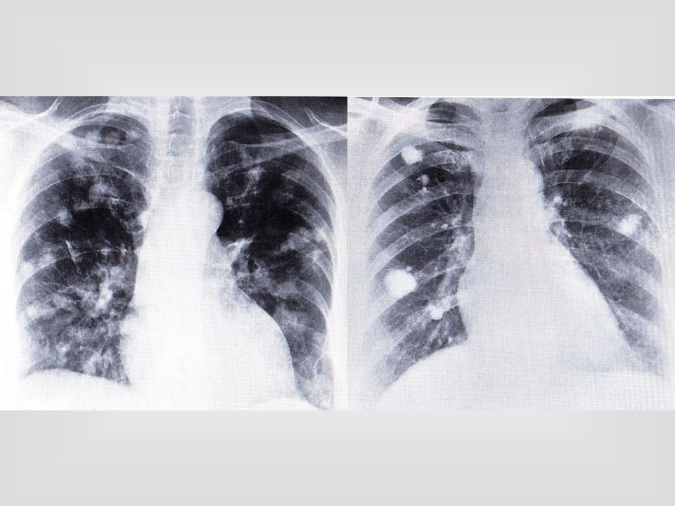
Atelectasis Pulmonary mass (nodule) Ring shadows and cysts Line shadows Air-space filling (alveolar pattern) Widespread nodular, reticulonodular and honeycomb shadowing (interstitial pattern) Pulmonary opacity
 Ring shadows and cysts Line shadows Air-space filling (alveolar pattern) Widespread nodular, reticulonodular and honeycomb shadowing (interstitial pattern) Pulmonary opacity")
24
Atelectasis Pulmonary mass (nodule) Ring shadows and cysts Line shadows Air-space filling (alveolar pattern) Widespread nodular, reticulonodular and honeycomb shadowing (interstitial pattern) Pulmonary opacity
 Ring shadows and cysts Line shadows Air-space filling (alveolar pattern) Widespread nodular, reticulonodular and honeycomb shadowing (interstitial pattern) Pulmonary opacity")
30
Atelectasis Pulmonary mass (nodule) Ring shadows and cysts Line shadows Air-space filling (alveolar pattern) Widespread nodular, reticulonodular and honeycomb shadowing (interstitial pattern) Pulmonary opacity
 Ring shadows and cysts Line shadows Air-space filling (alveolar pattern) Widespread nodular, reticulonodular and honeycomb shadowing (interstitial pattern) Pulmonary opacity")
31
Pneumatocele

33
Lung abscess Cavitating tumor

34
Lung abscess

35
Cavitating tumor

36
Cystic bronchiectases

37
Atelectasis Pulmonary mass (nodule) Ring shadows and cysts Line shadows Air-space filling (alveolar pattern) Widespread nodular, reticulonodular and honeycomb shadowing (interstitial pattern) Pulmonary opacity
 Ring shadows and cysts Line shadows Air-space filling (alveolar pattern) Widespread nodular, reticulonodular and honeycomb shadowing (interstitial pattern) Pulmonary opacity")
38
Kerley lines מזוהים בצל"ח. טיפוסיים למצב של בצקת ריאות. אלו עכירויות (opacities), אינטרסטיציאליות, לינאריות, שנגרמות ע"י נוזל או תאים ב-Interstitium.
, אינטרסטיציאליות, לינאריות, שנגרמות ע י נוזל או תאים ב-Interstitium..")
40
Kerley lines Kerley B – Short Parallel Peripheral right angles and in-contact with the pleura. Mostly at the bases. Represent interlobular septa – CHF and ILD

43
Atelectasis Pulmonary mass (nodule) Ring shadows and cysts Line shadows Air-space filling (alveolar pattern) Widespread nodular, reticulonodular and honeycomb shadowing (interstitial pattern) Pulmonary opacity
 Ring shadows and cysts Line shadows Air-space filling (alveolar pattern) Widespread nodular, reticulonodular and honeycomb shadowing (interstitial pattern) Pulmonary opacity")
44
Alveolar pattern - causes Water- Edema Pus- Pneumonia Blood- Trauma, Vasculitis Cells- Bronchioloalveolar Carcinoma Protein- Alveolar proteinosis Fat- Lipoid Pneumonia

45
Pulmonary edema

47
Pneumonia

48
Acinar shadows (TB)
")
49
Atelectasis Pulmonary mass (nodule) Ring shadows and cysts Line shadows Air-space filling (alveolar pattern) Widespread nodular, reticulonodular and honeycomb shadowing (interstitial pattern) Pulmonary opacity
 Ring shadows and cysts Line shadows Air-space filling (alveolar pattern) Widespread nodular, reticulonodular and honeycomb shadowing (interstitial pattern) Pulmonary opacity")
50
Viral pneumonia

51
Lymphangitis carcinomatosa (Colon Ca)
")
52
Reticulo-nodular (Interstitial)
")
54
Ground glass

56
Honey-combing Lung

57
Basic patterns in Lung Diseases Air bronchogram Silhouette sign Pulmonary opacity Increased transradiancy (translucency) of the lung
 of the lung")
58
Lt. Pneumonectomy

59
Pneumothorax

61
Emphysema

62
Emhysema- HRCT NormalEmphysema

64
Miscellaneous

65
Pulmonary embolism

66
Pleural effusion

68
Huge Mediastinal Mass

69
Thymoma

71
SUPERIOR SULCUS TUMOR (PANCOAST)
")
72
PET - CT

Similar presentations


disease TB Histoplasmosis Chicken box Sarcoidosis LCH Pneumoconiosis Alveolar microlithiasis Metastasis.>")


