Download presentation
Presentation is loading. Please wait.

1
D-TGA DR.VINOD.G.V

2
TRANSPOSITION Abnormal origin of the Aorta and Pulmonary Artery from the ventricular complex Atrioventricular concordance with ventriculo-arterial discordance Abnormal spatial relationship of the great arteries Results in two circulations in parallel

3
Incidence & Prevalence
5% to 7% of all congenital cardiac malformations The incidence is reported to range from 20.1 to 30.5/100,000 live births strong (60%–70%) male preponderance
 male preponderance.")
4
Embryology

5
Trunco conal malseptation Hypothesis
Normally related great arteries result from spiral downgrowth of the truncoconal septum, whereas TGA results from straight downgrowth of the trunco conal septum.

6
The embryonic Aortic switch procedure

7
During development of the conotruncal region, the pulmonary artery is related
to the left conus and the aorta to the right conus

8
normal movement of the pulmonary valve proceeds from posterior to anterior on the left side in the interval between 30 and 34 days of age and is related to normal development of the subpulmonary infundibulum

9
During this same interval, the aortic valve remains stationary, apparently because of the normal lack of development (or absorption) of the subaortic infundibulum
 of the subaortic infundibulum")
10
abnormal growth and development of the subaortic infundibulum and the absence of growth of the subpulmonary infundibulum. The aortic valve is protruded superiorly and anteriorly by the development of the subaortic infundibulum, placing it above the anterior right ventricle . Failure of development of the subpulmonary infundibulum prevents the normal morphogenetic movement of the pulmonary valve from posterior to anterior and further results in abnormal pulmonary to mitral valve ring fibrous continuity

12
Anatomy The common clinical type - situs solitus of the atria, concordant AV and discordant ventriculoarterial alignments - complete TGA. TGA {S,D,D} - TGA with situs solitus (S) of the atria and viscera, usual (D) looping of the ventricles and an anterior and rightward (D) aorta.
 of the atria and viscera, usual (D) looping of the ventricles and an anterior and rightward (D) aorta.")
14
Great artery relationship
Situs solitus and intact ventricular septum - the aortic root is directly anterior or anterior and to the right of the pulmonary trunk in a slightly oblique relationship Less commonly, the aorta may be positioned anterior and to the left or, rarely, posterior and to the right of the pulmonary trunk.

16
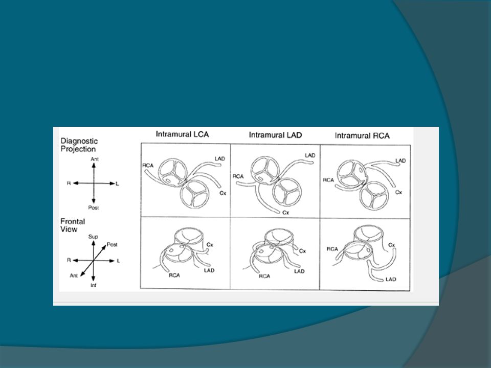
Coronary Anatomy The two aortic sinuses of Valsalva adjacent to the aorticopulmonary septum that “face” the pulmonary artery contain the ostia of the coronary arteries in more than 99% of cases

17
Coronary anatomy Usual-66.9 CX from RCA-16.1 Single RCA-3.9
Single LCA-1.7 Inverted-2.4 Intramural LCA-2.1 Other-1.6

19
SA node artery Origin and proximal course of artery may be variable; reaches the sinus node by the interatrial groove on the anterior surface of the heart, occasionally with an intramyocardial course in the anterosuperior rim of the fossa ovalis. can be damaged easily during balloon atrial septostomy, during surgical septectomy or when this portion of the septum is widely excised as in the Mustard or Senning atrial switch operation.

20
Coexisting Anomalies Nearly half of the hearts have no other anomaly except a PFO or a PDA. The VSD is the most frequent coexisting anomaly-40% to 45%. - perimembranous (33%) - inlet septum( 5%) - muscular (27%) - malalignment (30%) - conal septal hypoplasia type (5%)
 - inlet septum( 5%) - muscular (27%) - malalignment (30%) - conal septal hypoplasia type (5%)")
21
Malalignment VSD anterior malalignment of the infundibular septum is frequently associated with sub aortic stenosis, aortic arch hypoplasia, coarctation ,complete interruption of the aortic arch Posterior (leftward) malalignment is associated with varying degrees of LVOTO–subpulmonary stenosis, annular hypoplasia or even pulmonary valvar atresia
 malalignment is associated with varying degrees of LVOTO–subpulmonary stenosis, annular hypoplasia or even pulmonary valvar atresia.")
22
Subpulmonary Stenosis (25%)
Fixed -Circumferrential fibrous membrane /diaphragm - Fibromuscular ridge - Herniating tricuspid leaflet tissue - Anomalous MV septal attachments - Tissue tags from membranous septum Dynamic-associated with SAM

23
Subaortic Obstruction
Rightward and anterior displacement of the infundibular septum Associated aortic arch anomalies - hypoplasia - coarctation - interruption Asso. RV hypoplasia & tricuspid valve anomalies

24
TV anomalies Straddling/overriding of chordae
Overriding of the tricuspid annulus Abnormal chordal attatchments Dysplasia Accessory tissue Double orifice

25
MV anomalies Nearly 20% Functionally imp 4%
Cleft anterior mitral valve leaflet anomalous papillary muscles and chordae Straddling redundant tissue tags

26
Juxtaposition of atrial appendages
Both appendages or left + part of right are adjacent 2-6% Left > right -6x Female preponderance often additionally associated with major cardiac pathology, including dextrocardia, VSD, bilateral infundibulum, right ventricular hypoplasia and tricuspid stenosis or atresia.

27
HAEMODYNAMICS

28
Fetal and Post natal physiology

29
Fetal circulation

30
Fetal circulation in TGA

31
TGA with VSD in Fetus

32
TGA +VSD+PS IN FETUS

33
POSTNATAL PHYSIOLOGY OF TGA

34
Determinants of effective gas exchange
Effective ventilation Effective Pulmonary circulation Pulmonary blood flow Pulmonary vascular resistance Existence of a communication between pulmonary and systemic circuits Persistent fetal channel – PFO or DA Abnormal channels – ASD, VSD Effective delivery of oxygenated blood to the tissues

35
Definition of shunts Anatomical shunts
Left to Right: Blood flowing from left sided chambers to the right sided chambers Right to Left: Blood flowing from right sided chambers to the left sided chambers

36
Definition of shunts Physiological shunts
Left to right: The volume of oxygenated pulmonary venous return recirculated to pulmonary circulation (Qp – Qep) Right to left shunt: The volume of systemic venous return that contributes to cardiac output (reentering the systemic circulation) without having passed through the pulmonary circulation (Qs – Qep)
 Right to left shunt: The volume of systemic venous return that contributes to cardiac output (reentering the systemic circulation) without having passed through the pulmonary circulation (Qs – Qep)")
37
Definition of shunts Effective pulmonary blood flow (Qep):
The volume of systemic venous return that is effectively oxygenated in the lungs Effective systemic blood flow (Qes): The volume of oxygenated pulmonary venous return that enters the systemic circulation and perfuses the systemic capillary bed
: The volume of oxygenated pulmonary venous return that enters the systemic circulation and perfuses the systemic capillary bed.")
38
RIGHT HEART LEFT HEART Systemic venous Pulmonary venous return return
Anat R-L Anat L-R RIGHT HEART LEFT HEART Physio R-L Physio L-R LUNGS BODY

39
Right to Left Shunt Systole VSD Left to Right Shunt Diastole

40
TGA: Atrial and Ventricular level shunts
From LA to RA / LV to RV Anatomically left to right Physiologically, this volume of oxygenated blood enters systemic circulation. Hence, they contribute to Qes

41
TGA: Atrial and Ventricular level shunts
From RA to LA/ RV to LV Anatomically, right to left shunt Physiologically, this volume of systemic venous blood enters pulmonary circulation. Hence they contribute to Qep

42
TGA: Shunt at PDA level Aorta to PA flow:
Anatomically it is left to right Here the deoxygenated systemic venous blood enters pulmonary circulation. Hence, this volume contributes to Qep PA to Aorta flow: Anatomically it is right to left Here the oxygenated blood enters systemic circulation. Hence, this volume contributes to Qes Thus, the flow across the ductus is functionally opposite to that of flow across ASD or VSD in TGA

43
PDA Initially, bidirectional flow across the ductus
Later, once the PVR falls, the flow essentially becomes aorta to PA The pulmonary circulation becomes overloaded fast, especially if the PFO is restrictive

44
Unique feature Net inter-circulatory mixing volume is constant: net R-L, L-R, Qep and Qes are equal to each other Any major difference in the volumes would result in depletion of blood volume of one circulation at the expense of overloading the other circulation

45
Precise factors controlling intercirculatory exchange
SPECULATIVE, MULTIPLE LOCAL PRESSURE GRADIENTS Compliance of the cardiac chambers Phase of respiratory cycle Vascular resistances Heart rate Volume of blood flow

46
Flow across the communications “Rules of the Heart”
With only ASD, the flow has to be bidirectional If the flow is only or predominantly left to right across the ASD, it suggests presence of additional shunt (VSD or PDA) Unrestrictive VSD - flow is bidirectional Except in the initial few days, PDA flow is always left to right (Ao to PA). Presence of right to left flow across ductus may suggest the presence of coarctation of aorta
 Unrestrictive VSD - flow is bidirectional. Except in the initial few days, PDA flow is always left to right (Ao to PA). Presence of right to left flow across ductus may suggest the presence of coarctation of aorta.")
47
Factors influencing systemic saturation
Extent of inter-circulatory mixing and Total pulmonary blood flow High PBF results in increased oxygenated blood available in the left sided chambers for mixing: higher systemic SO2 if there is good mixing Reduced PBF will result in low systemic SO2 in spite of adequate anatomic shunts

48
Factors influencing systemic saturation
If there is delay in the fall of PVR (PPHN), hypoxemia will persist despite adequate ASD Need ECMO or urgent ASO Hypoxemia provokes a fall in SVR and increase the recirculating systemic volume Fall in SVR may deplete the pulmonary circulation further
, hypoxemia will persist despite adequate ASD. Need ECMO or urgent ASO. Hypoxemia provokes a fall in SVR and increase the recirculating systemic volume. Fall in SVR may deplete the pulmonary circulation further.")
49
Role of bronchopulmonary collaterals
Systemic arterial hypoxemia may stimulate development of bronchpulmonary collaterals Usually in TGA with solely a restrictive inter-atrial communication Prolonged survival of such infants may be due to this extra-cardiac site of shunting/mixing

50
History M:F – 4:1;unless juxtaposition of atrial appendages
Usually in multigravida-2X increase in > 3 pregnancies Familial recurrence-monogenic inheritance

51
CLINICAL MANIFESTATIONS

52
TGA PHYSIOLOGIC CLINICAL CLASSIFICATION
1. TGA (IVS OR SMALL VSD) with increased PBF and small ICS 2. TGA (VSD large) with increased PBF and large ICS 3. TGA(VSD and LVOTO), with restricted PBF 4. TGA(VSD and PVOD),with restricted PBF
 with increased PBF and small ICS 2. TGA (VSD large) with increased PBF and large ICS 3. TGA(VSD and LVOTO), with restricted PBF 4. TGA(VSD and PVOD),with restricted PBF")
53
Cyanosis As early as day 1 in pts with IVS(1st hr-56%;1st day-90%)
More intense if associated PS/atresia Mild if associated non restrictive VSD PS often responsible for hypercyanotic spells-intense cyanosis, tachypnea, extreme irritability and hypothermia Squatting is rare Reverse differrential cyanosis

54
CHF In patients with a large PDA
Large VSD – CHF develops within 1-3 wks

55
Mortality 1st week-30% 1st month-50% 1st year-90%
Depends on the degree of shunting Moderate PS improves survival Predilection for brain abscess but rare < 2 years

56
Arterial Pulse Bounding pulse
- due to large volume of highly unsaturated blood - Not due to PDA-since only systolic shunt from aorta to PA Diminished femoral pulses - CoA - Subaortic stenosis-anterior and rightward displacement of septum

57
Palpation Normal in neonates RV impulse
LV impulse – non restrictive VSD with low PVR Palpable S2 A2

58
Auscultation Loud A2 LV S3-mildly cyanosed patients,increased PBF,LV failure RV S3-deeply cyanosed patients, increased systemic flow, RV failure

59
Auscultation Ejection click-pulmonary;does not decrease with inspiration Aortic-subaortic stenosisdilated aortic root MSM-aortic:hypervolemic and hyperkinetic circulation Pulmonary: valvular- after few weeks of birth, progressively increases Subvalvular dynamic obstruction-3rd LICS and radiates to the right

60
Auscultation VSD: holosystolicshortensabolished PDA:
Systolic if large PDA since high PVR curtails diastolic flow Continuous if restrictive PDA MDM may be heard across AV valves

61
ECG Normal in first few days of life
RAE-increased pressure(CHF) or volume (hypervolemic systemic circulation) LAE-large ASD,increased PBF
 or volume (hypervolemic systemic circulation) LAE-large ASD,increased PBF.")
62
ECG RVH - NR VSD + high PVR/PS BVH - NR VSD + low PVR
Right precordial T waves not inverted but rather distinctly taller than the left sided T waves

64
CXR Absent thymic shadow after 12 hours of life
Narrow vascular pedicle - AP orientation of great vessels Right aoric arch % Egg on side appearance Juxtaposition-localised bulge along the mid left cardiac border which represents contiguous mass of the 2 appendages together PBF & Heart size inversely proportional

65
D-TGA with VSD

66
ECHO

67
Diagnosis Detection of shunt Detection of outflow obstructions Associated anomalies Coronary Anatomy

68
Parasternal long axis view showing the 2 great vessels parallel to each other. In a normal heart, the 2 great vessels should not appear together in any plane as they cross each other in their proximal course. The aorta here is anterior, not posterior as it normally should be.

69
Parasternal short axis view showing the aortic valve anterior to the pulmonary valve. .

70
Parasternal long axis with a tilt of the probe to show the entire length of the abnormally situated anterior aorta. The posteriorly situated pulmonary artery can be seen to bifurcate, a clue that the posterior vessel is not an aorta.

71
Subcostal view showing the pulmonary artery coming from the left ventricle and bifurcating as it travels distally.

74
Cardiac catheterization in TGA

75
Fallacies in application of Fick’s Principle in calculating shunts and flows in TGA
Oxygen consumption is not normal, so assumed values are unreliable Arteriovenous oxygen differences may be very small, so magnitudes of errors in calculated values would be very large. Effect / contribution of Bronchopulmonary collaterals to PBF – can result in overestimation.

77
TGA WITH NO ASSOCIATED DEFECTS IN NEWBORN

78
TGA WITH LARGE VSD IN NEWBORN

83
Management

84
Medical Prostaglandin E1 infusion should be started to improve arterial oxygen saturation by reopening the ductus. This should be continued throughout the cardiac catheterization and until the time of surgery. Oxygen should be administered for severe hypoxia. Oxygen may help lower pulmonary vascular resistance and increase PBF, resulting in increased systemic arterial oxygen saturation.

85
Role of PGE1 in TGA Considerable benefit in first few days till PVR is elevated, especially if PFO is small Enables bidirectional shunting, improves mixing If valve of FO is competent, it would result in increased LA pressure and pulmonary edema

86
Atrial Septostomy Before surgery, cardiac catheterization and a balloon atrial septostomy (i.e., the Rashkind procedure) are often carried out to have some flexibility in planning surgery. a balloon-tipped catheter is advanced into the left atrium (LA) through the PFO. The balloon is inflated with diluted radiopaque dye and abruptly with-drawn to the right atrium (RA) under fluoroscopic or echo monitoring.
 are often carried out to have some flexibility in planning surgery. a balloon-tipped catheter is advanced into the left atrium (LA) through the PFO. The balloon is inflated with diluted radiopaque dye and abruptly with-drawn to the right atrium (RA) under fluoroscopic or echo monitoring.")
87
Atrial Septostomy For older infants and those for whom the initial balloon atrial septostomy was only temporarily successful, blade atrial septostomy may be performed. Following this, the balloon procedure can be repeated for a better result.

88
Definitive Repair At three levels:
the atrial level : Senning or Mustard Sx ventricular level : Rastelli operation great artery level : arterial switch operation or Jatene operation

89
Atrial level Surgery Mustard operation: This oldest surgical technique redirects the pulmonary and systemic venous return at the atrial level by using either a pericardial or a prosthetic baffle.

90
Senning operation: This is a modification of the Mustard operation
Senning operation: This is a modification of the Mustard operation. It uses the atrial septal flap and the RA free wall to redirect the pulmonary and systemic venous return

91
Complications a.Obstruction to the pulmonary venous return (<5% of all cases) b.Obstruction to the systemic venous return (<5% of all cases) c.Residual intra-atrial baffle shunt (=20% of all cases) d.Tricuspid valve regurgitation (rare)
 d.Tricuspid valve regurgitation (rare)")
92
e.Absence of sinus rhythm (>50% of all cases) and frequent supraventricular arrhythmias
f.Depressed RV (i.e., systemic ventricular) function during exercise g.Sudden death attributable to arrhythmias (3% of survivors) h.Pulmonary vascular obstructive disease
 function during exercise. g.Sudden death attributable to arrhythmias (3% of survivors) h.Pulmonary vascular obstructive disease.")
93
Arterial switch operation (or Jatene operation)
")
96
Pre requisite An LV that can support the systemic circulation after surgery The LV pressure should be near systemic levels at the time of surgery, or the switch should be performed shortly after birth (i.e., before 2 weeks of age). In patients whose LV pressure is low, it can be raised by PA banding, either with or without a shunt, for 7 to 10 days (in cases of a rapid, two-stage switch operation) or for 5 to 9 months before undertaking the switch operation. LV pressure >85% and LV posterior wall thickness >4.5 mm appear to be satisfactory.
. In patients whose LV pressure is low, it can be raised by PA banding, either with or without a shunt, for 7 to 10 days (in cases of a rapid, two-stage switch operation) or for 5 to 9 months before undertaking the switch operation. LV pressure >85% and LV posterior wall thickness >4.5 mm appear to be satisfactory.")
97
Pre-op Coronary artery pattern amenable to transfer to the neoaorta without distortion or kinking. Risk is high when the left main or LAD coronary artery passes anteriorly between the aorta and the PA.

98
Pre-op The left ventricular inflow and outflow tracts must be free of significant structural abnormality. The right ventricular outflow tract should be free of significant stenosis. Mild pulmonary valve stenosis or dynamic or surgically remediable subpulmonary stenosis does not preclude a successful arterial switch operation. Patients with subaortic stenosis in association with a large VSD may receive the Damus-Kaye-Stansel operation

99
Anatomic variants that may impact operative mortality include
An intramural course of a coronary artery A retropulmonary course of the left coronary artery Multiple VSDs Coexisting abnormalities of the aortic Straddling AV valves Longer duration of global myocardial ischemic (cross-clamp) prolonged circulatory arrest times
 prolonged circulatory arrest times.")
100
Complications PA stenosis at the site of reconstruction - 5% to 10%
complete heart block - 5% to 10%. Aortic regurgitation (AR) late complication > 20% of patients especially PA banding An important cause of AR may be unequal size of the pulmonary cusps that leads to eccentric coaptation Coronary artery obstruction myocardial ischemia, infarction, and even death.
 late complication > 20% of patients especially PA banding. An important cause of AR may be unequal size of the pulmonary cusps that leads to eccentric coaptation. Coronary artery obstruction. myocardial ischemia, infarction, and even death.")
101
Rastelli operation In patients with VSD and severe PS
The LV is directed to the aorta by creating an intraventricular tunnel between the VSD and the aortic valve. A conduit is placed between the RV and the PA

102
Rastelli operation

103
Complications conduit obstruction (especially in those containing porcine heterograft valves) complete heart block (rarely occurs). This conduit needs to be replaced as the child grows.

104
Pulmonary Artery Banding
Transposition associated with large VSD without LVOTO To prevent Heart failure Pulmonary vascular disease Present Indications Presence of complex/multiple VSDs Coexisting medical conditions that cause a delay in surgery To train LV before switch in TGA/IVS

106
THANK U

107
MCQ

108
1. In a neonate with TGA false statement a
1.In a neonate with TGA false statement a.Ductal closure can precipitate severe desaturation b.The pulmonary and systemic circulations are arranged in series c.It may be necessary to create an ASD d.Immediate complete surgical repair is usually indicated

109
2. Most common coronary anomaly in TGA a. Single LAD b. Single RCA c
2.Most common coronary anomaly in TGA a.Single LAD b.Single RCA c.LCX from RCA d.Intramural LAD

110
3. Reverse differential cyanosis a. TGA with PDA+ high PVR b
3.Reverse differential cyanosis a.TGA with PDA+ high PVR b.PDA with reversal c.L TGA with reversal d.TGA+VSD+PS+PDA

111
4. Wrong statement in TGA a. TGA babies are predominantly males b
4.Wrong statement in TGA a.TGA babies are predominantly males b.Maternal diabetes may be associated c.Birth weight are normal d.Extracardiac anomalies are frequent

112
5. Surgical procedure of choice for a neonate with complete TGA a
5.Surgical procedure of choice for a neonate with complete TGA a.Atrial switch surgery b.Arterial switch surgery c.Rastelli operation d.Fontan operation

113
6. In TGA true statement a. A2 loud b
6.In TGA true statement a.A2 loud b.Arterial switch is the best option in the presence of pulmonary obstruction c.Cardiac catheterisation is the investigation of choice d.Egg on side in X ray always present

114
7. RVH in TGA at birth is defined by a. Monomorphic R wave in V1 b
7.RVH in TGA at birth is defined by a.Monomorphic R wave in V1 b.T upright after 1week in V1 c.RS ratio <1 in V6 d.Incomplete RBBB

116
b.1.2L/Min/m2 c.1.5L/Min/m2 d.0.5L/Min/m2
8. Effective pulmonary blood flow(Qep) a.0.9L/Min/m2 b.1.2L/Min/m2 c.1.5L/Min/m2 d.0.5L/Min/m2
 a.0.9L/Min/m2. b.1.2L/Min/m2. c.1.5L/Min/m2. d.0.5L/Min/m2.")
117
9. Anatomical right to left shunt a. 9L/Min/m2 b. 6L/Min/m2 c. 1
9.Anatomical right to left shunt a.0.9L/Min/m2 b.0.6L/Min/m2 c.1.5L/Min/m2 d.2L/Min/m2

118
10. Physiological right to left shunt equals to a. 11. 1L/Min/m2 b
10.Physiological right to left shunt equals to a.11.1L/Min/m2 b.15L/Min/m2 c.20L/Min/m2 d.7L/Min/m2

Similar presentations


 tricuspid valve 2. Hypoplastic right ventricle 3. Ventricular septal defect 4. Atrial.>")


