Download presentation
Presentation is loading. Please wait.

1
The Kidney

2
The kidneys are part of the urinary system and their job is to:
filter and cleanse the bloodstream of molecules like urea (waste product of used amino acids) and other molecular wastes osmoregulation (maintain the water to salt ratio within the body) Excretion is the removal from the body of waste products of metabolic pathways
 and other molecular wastes. osmoregulation (maintain the water to salt ratio within the body) Excretion is the removal from the body of waste products of metabolic pathways.")
4
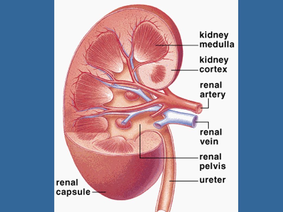
Renal artery – takes blood to the kidneys
Renal vein – drains blood from the kidneys Urine – fluid produced by the kidneys; it accumulates in the area called the renal pelvis Renal pelvis – collects the urine and drains it into a tube called the ureter, which then takes the urine to the bladder Renal cortex – the outer part of the kidney Renal medulla - the inside part of the kidney *Note: the cortex and medulla are packed with tubules and blood vessels for filtration and each kidney contains ~80km of tubules

5
Each nephron consists of:
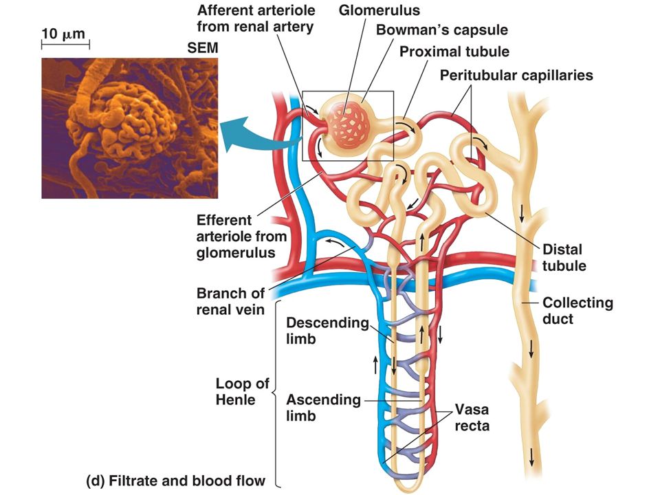
Nephrons The filtering units within the kidneys (each kidney is made up of ~1.25 million of them) Each nephron consists of: A capillary bed called the glomerulus, which filters substances from the blood A capsule surrounding the glomerulus called Bowman’s capsule A small tube extending from Bowman’s capsule with parts named in order: proximal convoluted tubule, loop of Henle, distal convoluted tubule A secondary capillary bed called the peritubular capillary bed which surrounds the tubules mentioned above
 Each nephron consists of: A capillary bed called the glomerulus, which filters substances from the blood. A capsule surrounding the glomerulus called Bowman’s capsule. A small tube extending from Bowman’s capsule with parts named in order: proximal convoluted tubule, loop of Henle, distal convoluted tubule. A secondary capillary bed called the peritubular capillary bed which surrounds the tubules mentioned above.")
8
Ultrafiltration Blood moving from the heart to the kidneys splits off into capillaries inside the glomerulus; these capillaries are fenestrated and open when blood is flowing at high pressure

9
Ultrafiltration is the process by which various substances are filtered out of the bloodstream through the glomerulus due to the unusually high blood pressure The fluids not filtered out by the glomerulus including blood cells and proteins exit Bowman’s capsule in the bloodstream The filtered fluid (filtrate) then passes through the basement membrane inside the glomerulus, then through the tubules so that the process of reabsorption can begin
 then passes through the basement membrane inside the glomerulus, then through the tubules so that the process of reabsorption can begin.")
10
Reabsorption of Molecules
The filtrate that leaves Bowman’s capsule contains substances that the body needs and cannot afford to lose in urine (H2O, salt ions, glucose) Most reabsorption within the nephron occurs in the proximal convoluted tubule; the reabsorbed nutrients re-enter the bloodstream by way of the surrounding peritubular capillary bed The inner lining of the proximal convoluted tubule contains microvilli in order to increase surface area for reabsoption; also cells of the tubule contain mitochondria to aid in active transport
 Most reabsorption within the nephron occurs in the proximal convoluted tubule; the reabsorbed nutrients re-enter the bloodstream by way of the surrounding peritubular capillary bed. The inner lining of the proximal convoluted tubule contains microvilli in order to increase surface area for reabsoption; also cells of the tubule contain mitochondria to aid in active transport.")
11
Methods of Reabsorption
Glucose – active transport moves all the glucose from the filtrate back into the bloodstream (in normal functioning kidney) Salt ions – the majority of salt ions (e.g., Na+, Cl-, K+) must be reabsorbed; ions move via active transport from tubules into capillaries Water – the movement of the salt ions causes the movement of water molecules to follow the same route via Osmosis (remember, water needs to move into high concentration solutions to help bring it back to equilibrium)
 Salt ions – the majority of salt ions (e.g., Na+, Cl-, K+) must be reabsorbed; ions move via active transport from tubules into capillaries. Water – the movement of the salt ions causes the movement of water molecules to follow the same route via Osmosis (remember, water needs to move into high concentration solutions to help bring it back to equilibrium)")
12
These mechanisms occur in the kidneys and counteract things like:
Osmoregulation It is the body’s response mechanisms which attempt to maintain homeostatic levels of water These mechanisms occur in the kidneys and counteract things like: Volume of H2O ingested through liquids and food Perspiration rate (exercise and temp dependent) Ventilation rate (exercise dependent) Amount of salt ingested via foods
 Ventilation rate (exercise dependent) Amount of salt ingested via foods.")
13
How the following parts help to maintain the water balance in blood:
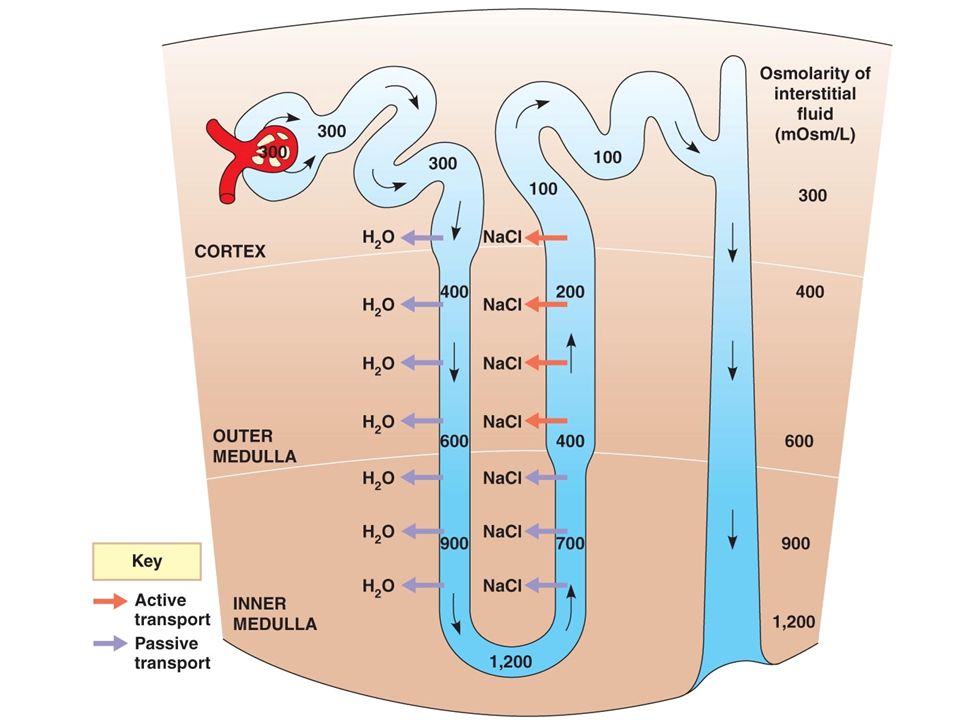
Loop of Henle – in descending loop water moves back into blood via osmosis and in ascending loop salt ions pumped out via active transport *Note: this loss of salt ions into the medulla aids in keeping the concentration gradient necessary for osmosis

15
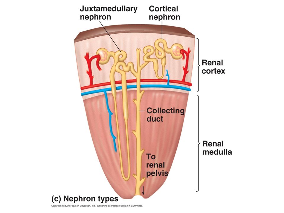
Medulla – the loop of Henle extends into the medulla region of the kidney; medulla is filled with ions (hypertonic compared to tubules) and this high level of concentration is what allows H2O molecules to continually diffuse from tubules back into blood Collecting duct – the wall of the collecting duct is permeable therefore H2O can pass from filtrate into blood; presence or absence of ADH (anti-diuretic hormone) determines the permeability of the walls
 determines the permeability of the walls.")
16
ADH – released from the pituitary gland when the concentration of solutes in the blood is too high; presence of ADH causes walls of collecting duct to be permeable to H2O (water moves into medulla tissue and then into peritubular capillaries to help make blood less concentrated); if no ADH is present then walls are impermeable and water stays in tubules and helps dilute urine
; if no ADH is present then walls are impermeable and water stays in tubules and helps dilute urine")
17
How do the kidneys change the blood? Molecule
Blood Plasma (from renal artery) Glomerular Filtrate (fluid entering tubules after ultrafiltration) Urine Proteins > 700 NONE Glucose > 90 >90 Urea 30 > 1800 *Note: all # are in mg/100 mL; need to understand the relationships, not the #
 Glomerular Filtrate (fluid entering tubules after ultrafiltration) Urine. Proteins. > 700. NONE. Glucose. > 90. >90. Urea. 30. > *Note: all # are in mg/100 mL; need to understand the relationships, not the #")
18
Diabetic’s Urine In healthy people, all the glucose that they eat is reabsorbed back into the bloodstream from the filtrate via active transport Active transport mechanisms have a maximum rate at which they can move substances and when the maximum is exceeded (like an undiagnosed diabetic who eats a carb-filled meal) then active transport cannot keep up and there will be glucose in the urine
 then active transport cannot keep up and there will be glucose in the urine.")
Similar presentations


 as urea and uric acid. B.Kidneys remove waste and water from blood.>")


![[Outer cortex, inner medulla, and renal pelvis]](/16/4931043/big_thumb.jpg "[Outer cortex, inner medulla, and renal pelvis]>")


 System>")


