Download presentation
Presentation is loading. Please wait.

1
Epidural and Subdural Hematoma
Kelly Kirby Jenna Baraki

2
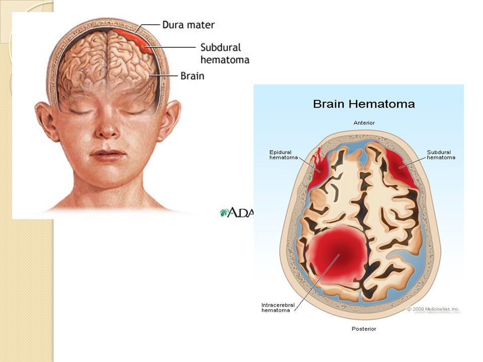
Epidural Hematoma Type of traumatic brain injury in which a build up of blood occurs between the dura mater and the skull.

3
Causes Due to trauma Potentially deadly because the build up of blood may increase pressure in the intracranial space and compress delicate brain tissue. Spontaneous hemorrhage is also known to cause this.

4
Specific treatments The blood may be removed surgically to get rid of the mass and reduce the pressure it puts on the brain.

5
Progression of the Disease
Epidermal Hematoma is considered to be the most serious complication of head injury.

6
Subdural Hematoma Collection of blood on the surface of the brain.
Form of traumatic brain injury. Blood gathers within the outermost meningeal layer between the dura mater which adheres to the skull.

7
Causes Most often caused by head injury when rapidly changing velocities within the skull may stretch and tear small veins. Subdural hemorrhage is a classic finding in shaken baby syndrome, in which similar shearing forces classically cause intra- and pre-retinal hemorrhages. Subdural hematoma is also commonly seen in the elderly and in alcoholics, who have evidence of cerebral atrophy.

8
Specific Treatments It is important that a patient receive medical assessment, including a complete neurological examination, after any head trauma. A CT scan or MRI scan will usually detect significant subdural hematomas. Other small subdural hematomas can be managed by inserting a temporary small catheter through a hole drilled through the skull and sucking out the hematoma; this procedure can be done at the bedside. Large or symptomatic hematomas require a craniotomy, the surgical opening of the skull.

9
Surgery A surgeon then opens the dura, removes the blood clot with suction or irrigation, and identifies and controls sites of bleeding. Postoperative complications include increased intracranial pressure, brain edema, new or recurrent bleeding, infection, and seizure. The injured vessels must be repaired.

Similar presentations


>")


