Download presentation
Presentation is loading. Please wait.

1
Urine analysis

2
Macroscopic urinalysis
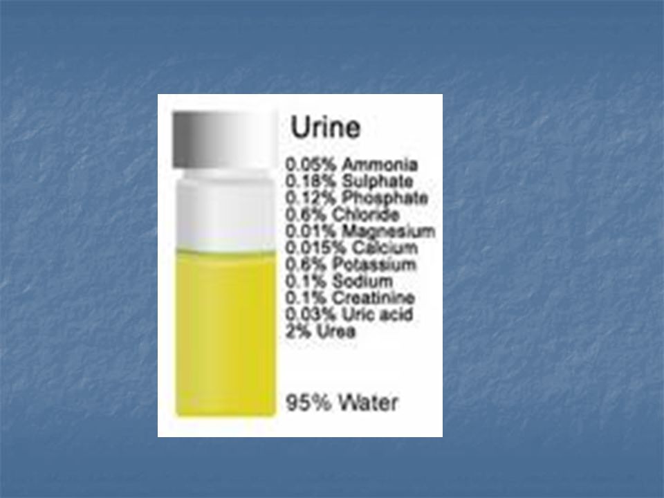
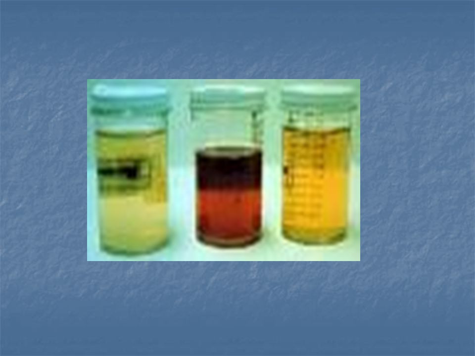
Is the direct visual observation of the urine, noting its quantity, color, clarity or cloudiness, etc Normal urine is typically light yellow and clear . Obvious abnormalities in the color, clarity, and cloudiness may suggest possibility of an infection, dehydration, blood in urine, liver disease, break down of muscle or red blood cells in the body.

5
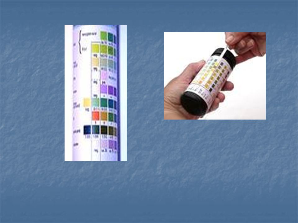
Dipstick chemical analysis
Urine dipstick is a narrow plastic strip which has several squares of different colors attached to it. Each small square represents a component of the test used to interpret urinalysis. The entire strip is dipped in the urine sample and color changes in each square are noted.

6
The squares on the dipstick represent the following components in the urine
Specific gravity Protein in the urine (mainly albumin) Glucose Ketone bodies Blood, leukocyte esterase (indicative of white blood cells in urine) Nitrite (suggestive of bacteria in urine) Bilirubin ( possible liver disease or red blood cell break down) Urobilinogen ( possible liver disease)
 Glucose. Ketone bodies. Blood, leukocyte esterase (indicative of white blood cells in urine) Nitrite (suggestive of bacteria in urine) Bilirubin ( possible liver disease or red blood cell break down) Urobilinogen ( possible liver disease)")
8
MICROSCOPIC URINALYSIS

9
MICROSCOPIC URINALYSIS
1. Red Blood Cells: Hematuria is the presence of abnormal numbers of red cells in urine due to: a. Glomerular damage b. Tumors c. Urinary tract stones d. Upper and lower urinary tract infections

10
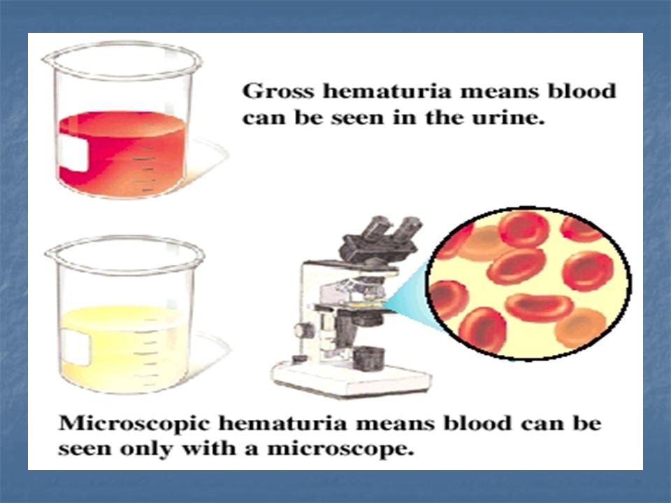
Hematuria Two Types of Hematuria
Gross hematuria means that the blood can be seen by the naked eye. The urine may look pinkish, brownish, or bright red.

11
Microscopic hematuria means that the urine is clear, but blood cells can be seen when urine is looked at under a microscope or tested in a lab.

13
RBC's may appear normally shaped, swollen by dilute urine (in fact, only cell ghosts and free hemoglobin may remain) or crenated by concentrated urine.
 or crenated by concentrated urine.")
14
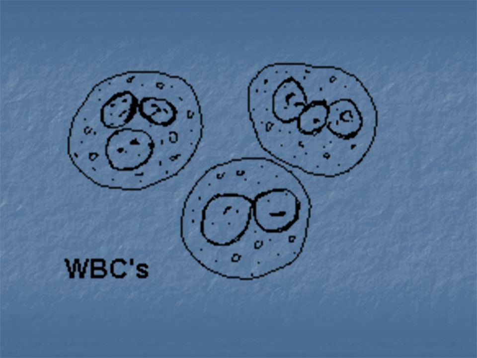
White Blood Cells Pyuria refers to the presence of abnormal numbers of leukocytes that may appear with infection in either the upper or lower urinary tract or with acute glomerulonephritis.

16
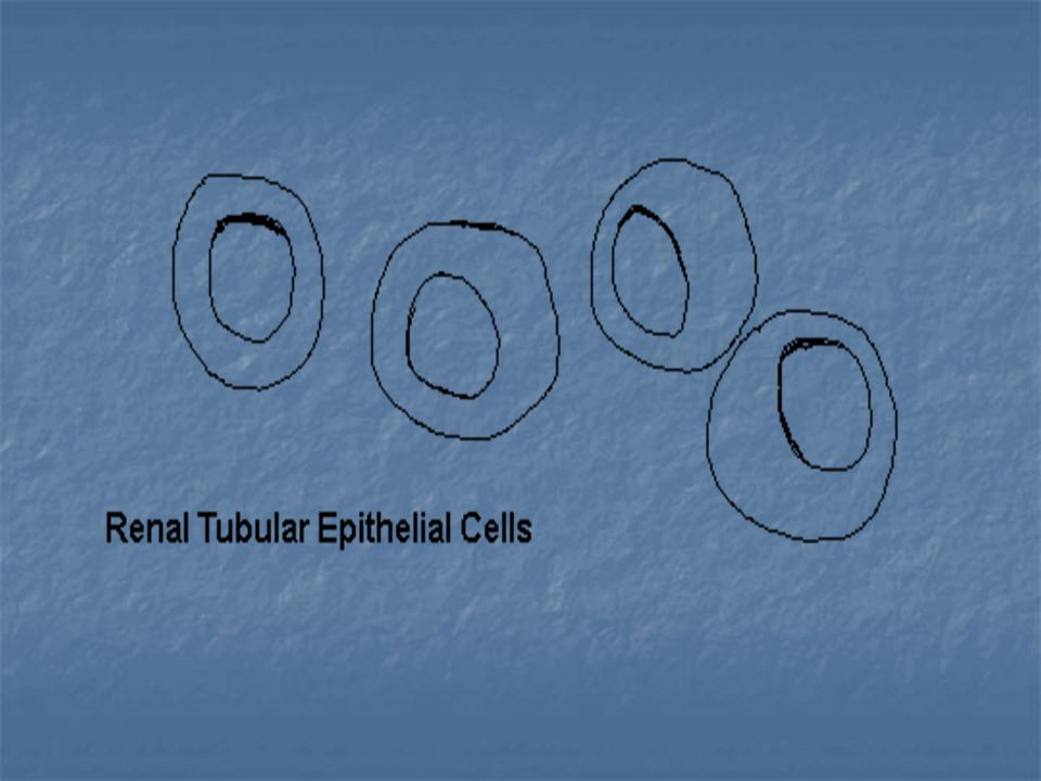
Epithelial Cells Renal tubular epithelial cells, contain a large round or oval nucleus and normally slough into the urine in small numbers. However, with nephrotic syndrome and in conditions leading to tubular degeneration, the number sloughed is increased

18
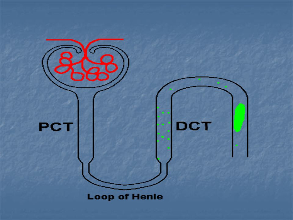
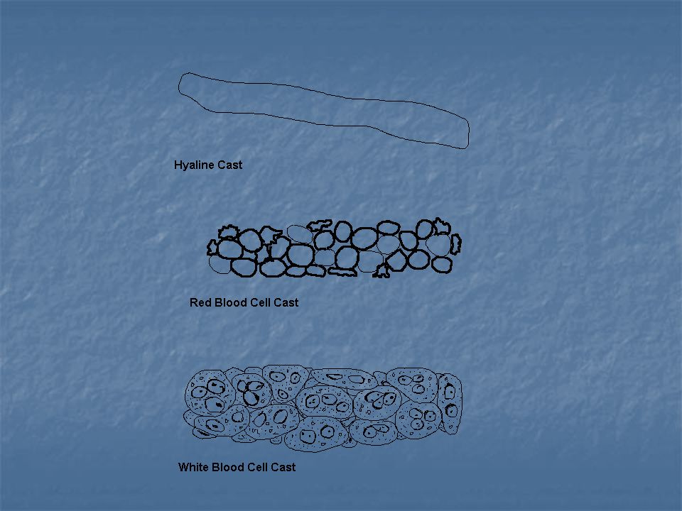
Casts Urinary casts are formed only in the distal convoluted tubule (DCT) or the collecting duct (distal nephron). The proximal convoluted tubule (PCT) and loop of Henle are not locations for cast formation. Hyaline casts are composed primarily of a mucoprotein (Tamm-Horsfall protein) secreted by tubule cells. The Tamm-Horsfall protein secretion (green dots) is illustrated in the diagram below, forming a hyaline cast in the collecting duct:
 and loop of Henle are not locations for cast formation. Hyaline casts are composed primarily of a mucoprotein (Tamm-Horsfall protein) secreted by tubule cells. The Tamm-Horsfall protein secretion (green dots) is illustrated in the diagram below, forming a hyaline cast in the collecting duct:")
20
The factors which favor protein cast formation are:
*low flow rate, *high salt concentration, *and low pH, all of which favor protein denaturation and precipitation, particularly that of the Tamm-Horsfall protein. Protein casts with long, thin tails formed at the junction of Henle's loop and the distal convoluted tubule are called cylindroids. Hyaline casts can be seen even in healthy patients

21
Red blood cells may stick together and form red blood cell casts
Red blood cells may stick together and form red blood cell casts. Such casts are indicative of glomerulonephritis, with leakage of RBC's from glomeruli, or severe tubular damage White blood cell casts are most typical for acute pyelonephritis, but they may also be present with glomerulonephritis. Their presence indicates inflammation of the kidney, because such casts will not form except in the kidney.

23
Bence Jones proteins Bence Jones proteins are small proteins found in the urine. Testing for these proteins is done to diagnose and monitor multiple myeloma and other similar diseases. Bence Jones proteins are considered the first tumor marker. A tumor marker is a substance, made by the body, that is linked to a certain cancer, or malignancy. Bence Jones proteins are made by plasma cells, a type of white blood cell. The presence of these proteins in a person's urine is associated with a malignancy of plasma cells.

24
Bence Jones protein cast (myeloma cast) from the urinary sediment of a patient with lambda-Bence Jones type multiple myeloma. Sternheimer stein, X200

25
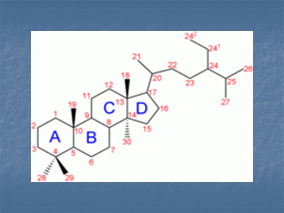
steroid, 17-keto- (17-KS), n steroidal compounds with a ketone (carbonyl) group at carbon 17. Derived from cortisol and adrenal and testicular androgen. Urinary neutral 17-ketosteroids represent the catabolic end products of the endocrine glands. Produced by the adrenal cortex and testes. Increased values occur in adrenogenital syndromes, adrenocortical carcinoma, Normal adult values for a 24-hour urine sample are 10 to 20 mg for men and 5 to 15 mg for women
 group at carbon 17. Derived from cortisol and adrenal and testicular androgen. Urinary neutral 17-ketosteroids represent the catabolic end products of the endocrine glands. Produced by the adrenal cortex and testes. Increased values occur in adrenogenital syndromes, adrenocortical carcinoma, Normal adult values for a 24-hour urine sample are 10 to 20 mg for men and 5 to 15 mg for women.")
26
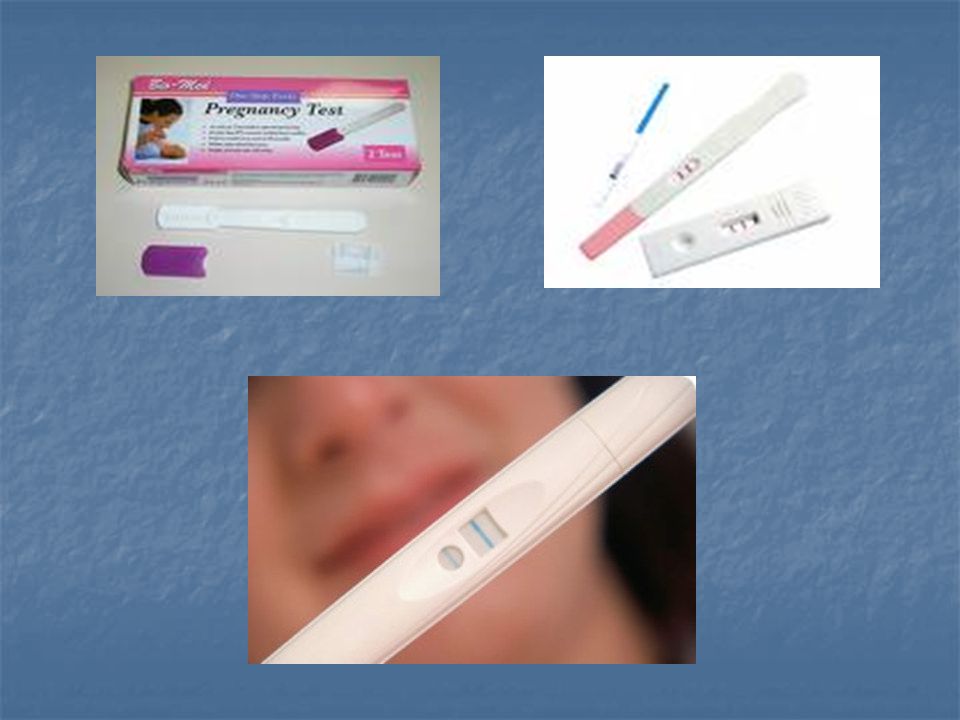
Pregnancy tests Home pregnancy tests consist of placing a drop of urine on a prepared chemical strip or placing the strip in the urine stream. The strip is designed to detect a pregnancy hormone called human chorionic gonadotropin (hCG).
.")
28
hCG is released into the body by the placenta when a woman is pregnant.
This hormone is also responsible for causing some of the initial symptoms of pregnancy such as breast tenderness and nausea. hCG levels usually become detectable in the urine about 10 days after conception

Similar presentations


 Ca+, K+, Na+ -Regulate acid/base.>")