Download presentation
Presentation is loading. Please wait.

1
Intro to Radiologic Technology (RADT A)
")
2
RTEC A INSTRUCTOR MINA COLUNGA, B.S.,RT., C.R.T.
Instructor, or WEB page:

3
WHY CHOOSE RADIOGRAPHY?

4
Is this a safe profession?
Why do you want to do this? Why are you taking this class?

5
Preconceived Ideas regarding the X-ray field
Elicit responses from students about what they think the x-ray field is all about.

7
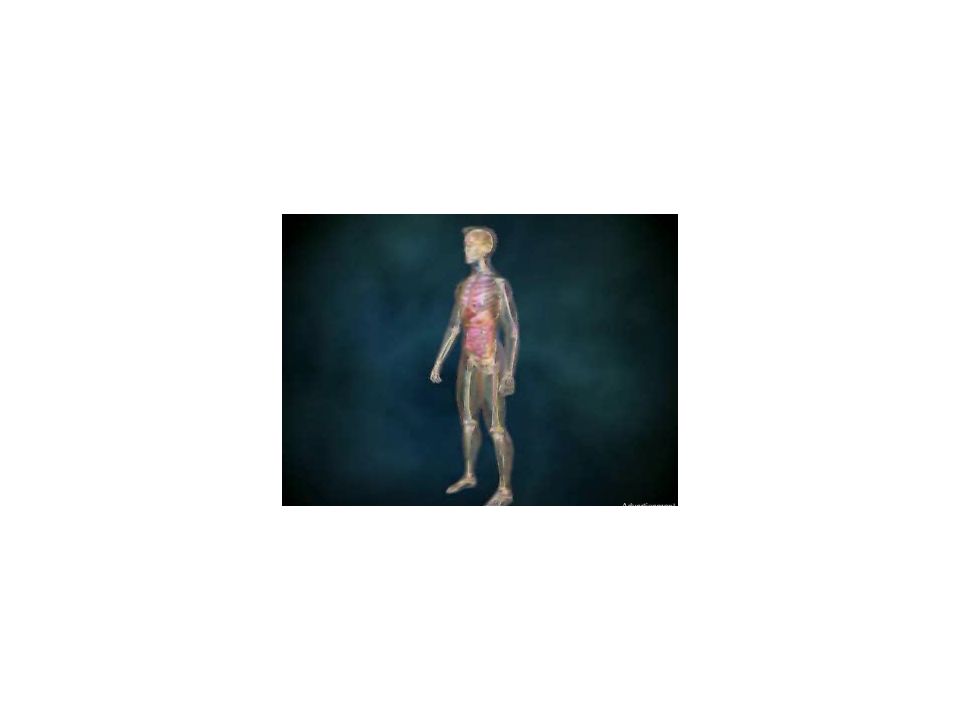
What is x-rays? X-rays are electromagnetic radiation with extremely short wavelengths. They can pass through many materials. Purpose of x-rays --To detect pneumonia, congestive heart failure, broken bones, tumors, and other abnormalities.

8
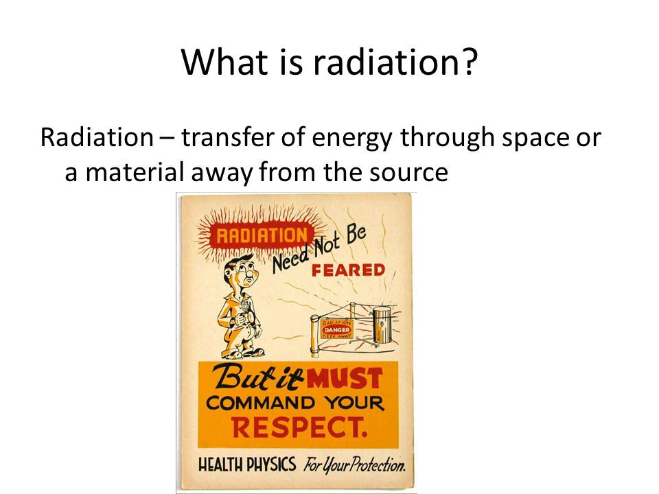
What is radiation? Radiation – transfer of energy through space or a material away from the source
9
Radiographic Terminology
Radiology- Medical specialty in which x-rays, radium, and radioactive substances are applied in the diagnosis and treatment of the patient Diagnostic Imaging- Medical specialty in which x-rays, radium, radioactive substances, sound waves, and radio frequencies are applied in the diagnosis and treatment of the patient Radiologist- Physician who applies any form of radiation in the diagnosis and treatment of disease.

10
Radiographic Terminology
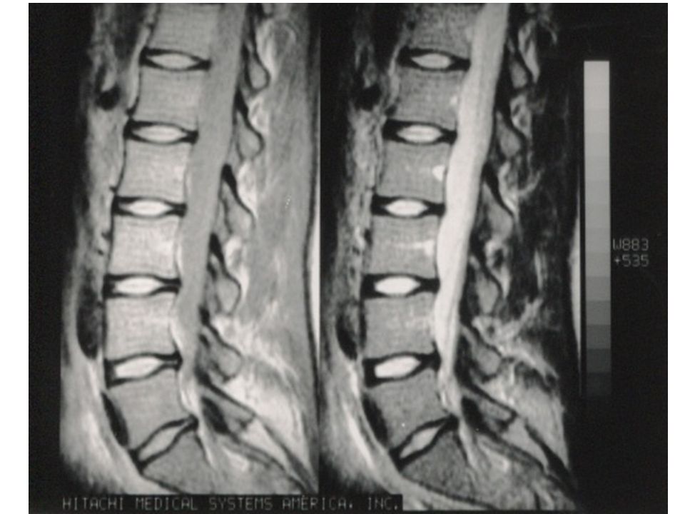
Radiographer- Skilled person qualified by education to provide patient services using imaging modalities as directed by a physician qualified to order and/or perform radiographic procedures. (X-ray Technologist) Radiograph- a photographic record produced by x-rays through an object.
 Radiograph- a photographic record produced by x-rays through an object.")
12
Types of Radiation Non-ionized Ionized ex: radio ex: x-rays, gamma

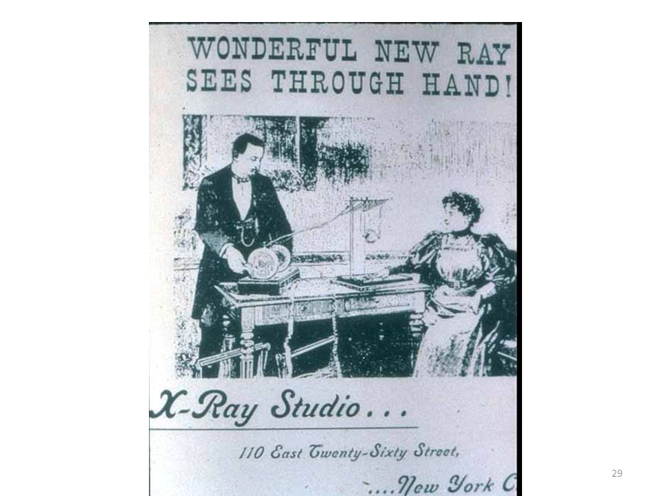
13
Electromagnetic Spectrum

14
History of Radiology

15
Historical Perspectives
November 8, 1895: Wilhelm Conrad Röntgen discovered x-rays German Physicist University of Wurtzburg

16
Wilhelm Röntgen in 1895 - discovered x-rays
Working with Crooke’s vacuum tube He found invisible rays were produced. These new rays could go through skin and flesh Give a picture of a person's bones. The discovery of X rays by the German physicist Wilhelm Roentgen in 1895 was an international sensation. Working with vacuum tubes, he bombarded a metal plate with high speed electrons. He found that invisible rays were produced. These mysterious new rays could go through skin and flesh and give a picture of a person's bones.

18
X-rays – the Basic Radiological Tool
Röntgen’s experimental apparatus -Crookes tube Roentgen’s experimental apparatus (Crookes tube) that led to the discovery of the new radiation on 8 Nov – he demonstrated that the radiation was not due to charged particles, but due to an as yet unknown source, hence “x” radiation or “x-rays” Known as “the radiograph of Bertha Roentgen’s hand” taken 22 Dec. 1895 Taken 22 Dec. 1895
 that led to the discovery of the new radiation on 8 Nov – he demonstrated that the radiation was not due to charged particles, but due to an as yet unknown source, hence x radiation or x-rays Known as the radiograph of Bertha Roentgen’s hand taken 22 Dec Taken 22 Dec")
19
First Radiograph Anna Bertha Röntgen 30 minute exposure .
those initial tubes were using very low energies for long periods of time

20
Collaborative Events Crookes tube Air evacuated glass tube
Cathode side Anode side Electrical supply Screen or board painted with barium platinocyanide Low light work area

22
“Willie Röntgen” Honored in 1901 with the first Nobel prize in physics for his efforts.

23
In the beginning…..

24
Early years in Radiologic Technology
Nurses or nurses aides taught how to “take an x-ray” NO special education Only “ON THE JOB” training Experience the best teacher The first Technologist is credited to be EDWARD C. JERMAN.

25
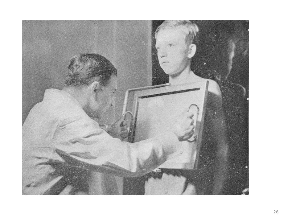
An early therapy session

27
In 30 years Developed from a technical trade to one of a professionalism Once thought that anyone could be trained to quickly = “push the buttons’ To now where it is considered a profession that requires analytical thinking and problem solving

30
X rays began to be used in industry and medicine
Years later, they noticed it can be harmful They could be harmful to: living tissue even cause cancer if the exposures were too great or too prolonged X rays soon found many valuable uses in medicine and in industry. It was, unfortunately, quite a few years later that a harmful side of X rays was also discovered. They could be harmful to living tissue, or even cause cancer if the exposures were too great or too prolonged

31
Early signs of possible damage from Radiation exposure
Skin dryness Erythema Ulcers formed

32
Acute: Ulceration

34
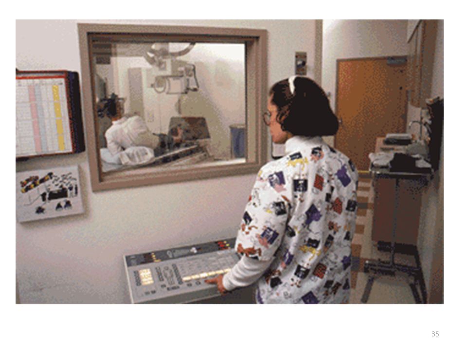
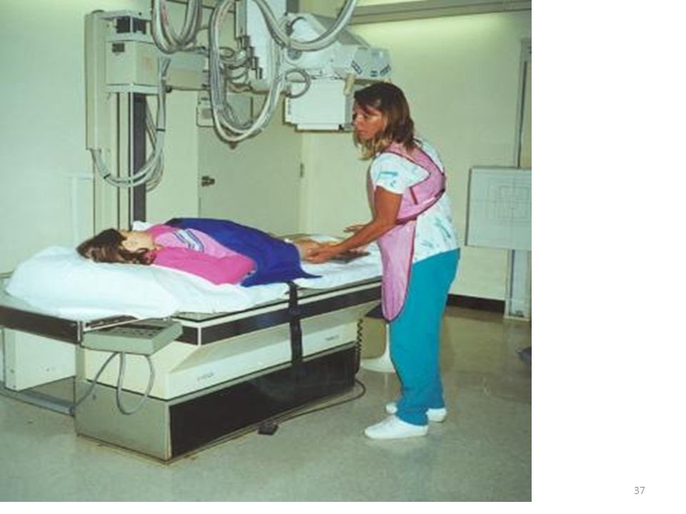
Radiologic Technologists
Practices RADIATION SAFETY TO SELF AND OTHERS

39
HISTORY REVIEW Who is this?

40
HISTORY REVIEW Wilhelm Conrad Röntgen

41
HISTORY REVIEW What did he discover?

42
HISTORY REVIEW He discovered x-rays

43
What were the series of events that led to the discovery?
HISTORY REVIEW What were the series of events that led to the discovery?

44
HISTORY REVIEW Crookes tube
With electrical supply 2) Screen coated with barium platinocyanide 3) Low light area
 Screen coated with barium platinocyanide. 3) Low light area.")
45
Accreditation, Certification, Registration, Licensing???
What is all that?

46
Accrediting Agencies for Schools (JRC’s)
Joint Review Committee on Education in Diagnostic Medical Sonography (JRCDMS) Joint Review Committee on Education in Nuclear Medicine Technology (JRCNMT) Joint Review Committee on Education in Radiologic Technology
 Joint Review Committee on Education in Nuclear Medicine Technology (JRCNMT) Joint Review Committee on Education in Radiologic Technology.")
47
Individual Certification
Take an exam Pay a fee You then get registered Nearly all hospitals require appropriate certifciation as a condition of employment.

48
National: Registry Agencies
American Registty of Diagnostic Medical Sonographers (ARDMS) American Registry of Radiologic Technologists Nuclear Medicine Certification Board
 American Registry of Radiologic Technologists. Nuclear Medicine Certification Board.")
49
State Licensing Agencies
Vary from state to state List of individual state requirement can be obtained at Must provide proof of certification Fill out paperwork Pay a fee Sometimes take an exam

50
Certification vs. License
ARRT National certification R.T. Must take an exam Pass with 75% Can take this after completing program CRT State Licensing Must pass ARRT or other equivalent national exam to get this Pay fee to get radiography license (R) Take fluoroscopy exam and pay a fee for (F) license
 Take fluoroscopy exam and pay a fee for (F) license.")
51
RADIOLOGIC TECHNOLOGY
It covers all of our individual disciplines.

52
RADIOLOGIC TECHNOLOGY
Radiography Mammography Computed Tomography Magnetic Resonance Imaging Quality Management Sonography Radiation Therapy Bone Densitometry Vascular Sonography Breast Sonography Cardiac Interventional Radiography Vascular Interventional radiography Radiologist Assistant Nuclear Medicine

53
5 Primary Certifications
Radiography (R) Nuclear Medicine Technology (NM) Radiation Therapy (T) Sonography (US) (RDMS) Magnetic Resonance Imaging (MR) candidates must have successfully completed a formal educational program in the respective discipline that is accredited by a mechanism acceptable to ARRT.
 Nuclear Medicine Technology (NM) Radiation Therapy (T) Sonography (US) (RDMS) Magnetic Resonance Imaging (MR) candidates must have successfully completed a formal educational program in the respective discipline that is accredited by a mechanism acceptable to ARRT.")
54
Post Primary Certifications
Mammography (M) Computed Tomography(CT) Magnetic Resonance Imaging (MR) or (MRI) Note: Both a primary and post-primary track Quality Management (QM) Cardiac-Interventional Radiography (CI) Vascular-Interventional Radiography (VI) Sonography (US) or (RDMS) Note: Both a primary and post-primary track Vascular Sonography (VS) Breast Sonography (BS) Bone Densitometry (BD) Registered Radiologist Assistant (RA)
 Computed Tomography(CT) Magnetic Resonance Imaging (MR) or (MRI) Note: Both a primary and post-primary track. Quality Management (QM) Cardiac-Interventional Radiography (CI) Vascular-Interventional Radiography (VI) Sonography (US) or (RDMS) Note: Both a primary and post-primary track. Vascular Sonography (VS) Breast Sonography (BS) Bone Densitometry (BD) Registered Radiologist Assistant (RA)")
55
MRI and Sonography are PRIMARY and POST PRIMARY
Can get formal education On the job training if you have a primary certification in radiography, nuclear medicine or radiation therapy meet clinical requirements.

56
Individual Disciplines of Radiology

57
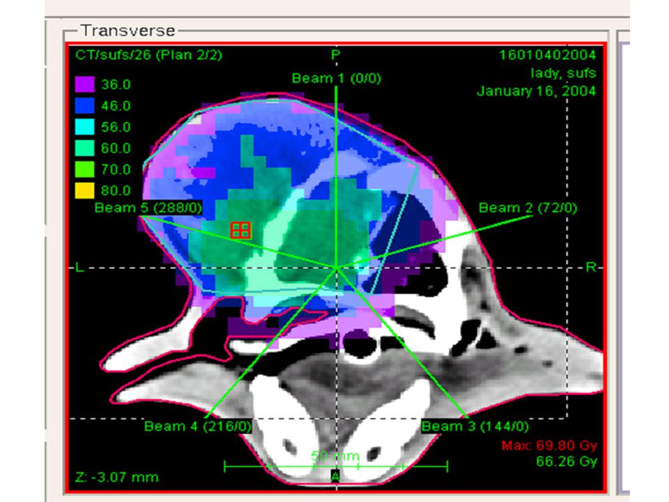
Radiography : Primary Certification
Mina Colunga R.T. (R) Mina Colunga Registered Technologist in the specialty of Radiography
 Mina Colunga Registered Technologist in the specialty of Radiography.")
58
RADIOGRAPHY Diagnostic Radiology Technologist Radiographer
Technician (Limited Licensure) Specializing in the use of x-rays to create images of the body including the skeletal system,chest and abdomen
 Specializing in the use of x-rays to create images of the body including the skeletal system,chest and abdomen.")
59
Two Types of x-ray examinations
Radiography Fluoroscopy

60
Fluoroscope 1898 by Thomas Edison

62
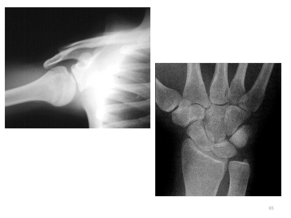
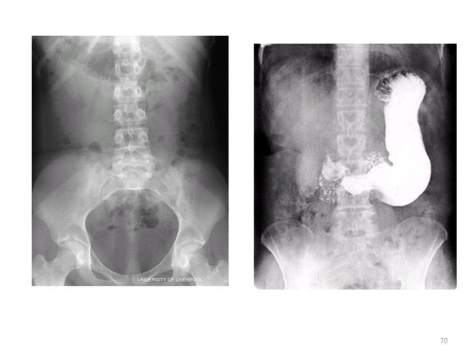
Types of Diagnostic Exams
Chest Extremities Skull/ Facial Spine Gastrointestinal Interventional

63
All types of EXAMS & PEOPLE
Infants Elderly All classes All ethnicity All backgrounds Head to toes Trauma Special procedures Critical patients Walk ins Surgery

64
Uses Ionizing Radiation to create images of the human body

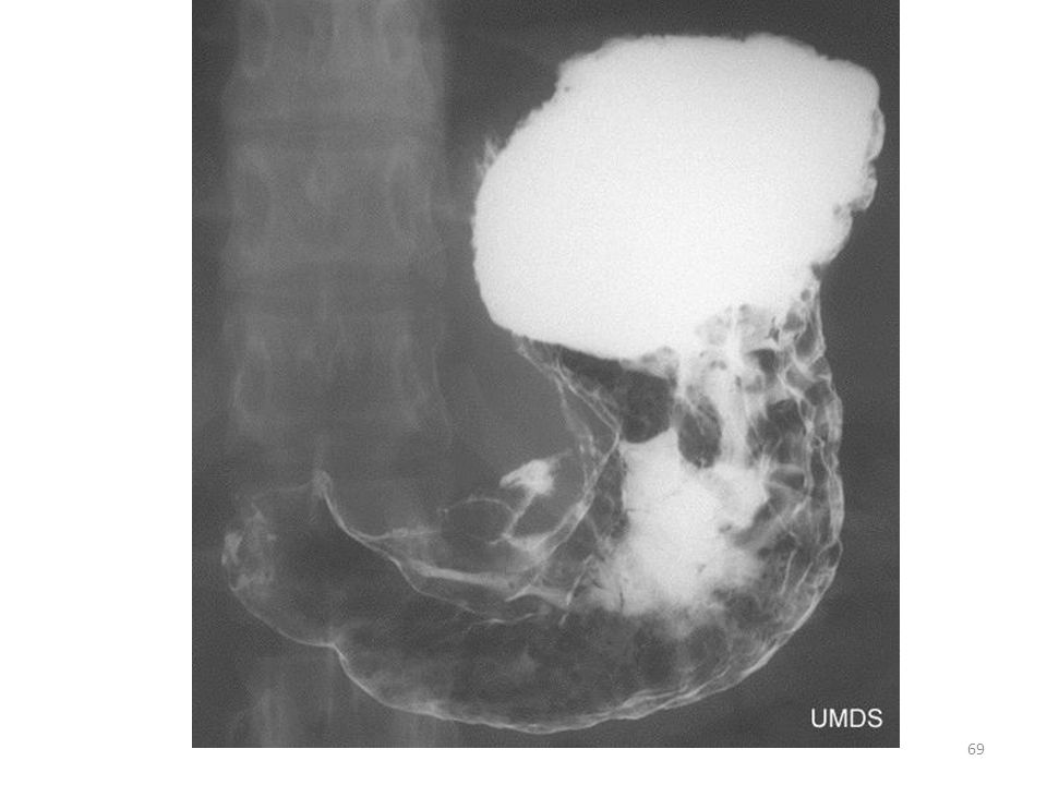
67
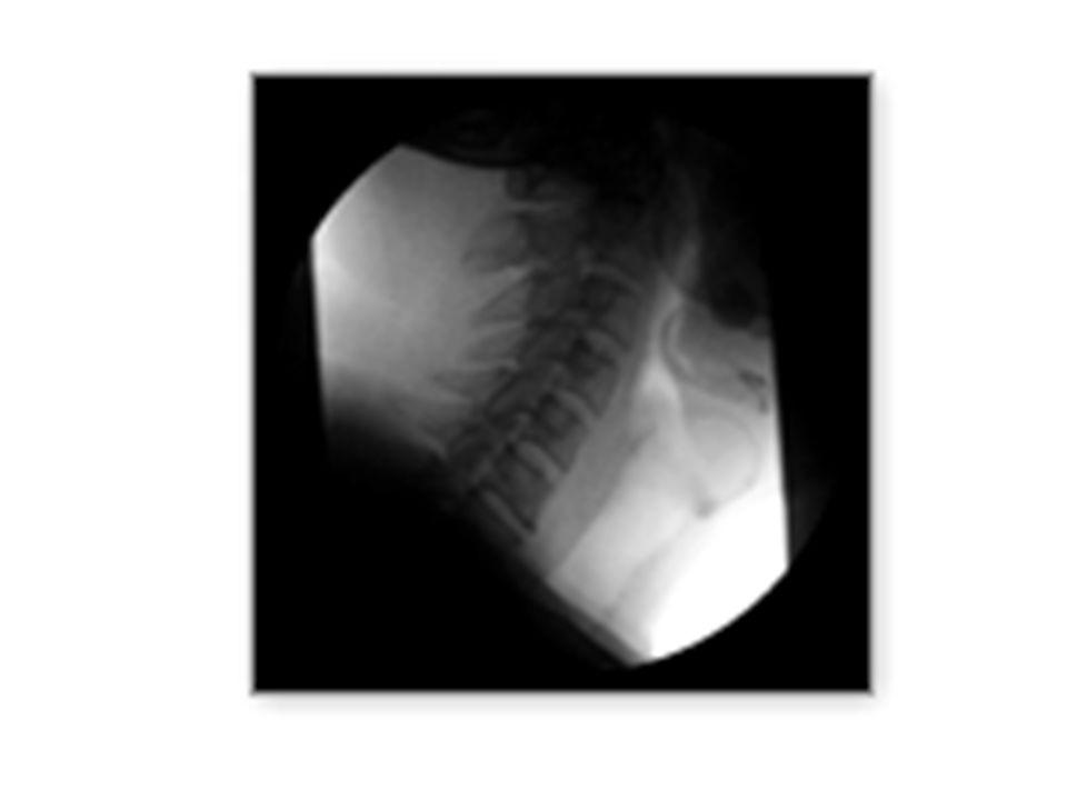
Flouroscopy- xrays in motion

68
Fluoroscopy

73
Beyond Diagnostic Radiography
Ultrasound (sonography) Angiography Computerized tomography (CT) Magnetic Resonance Imaging (MRI) Positron Emission Tomography (PET) Nuclear Medicine Mammography Radiation Therapy
 Angiography. Computerized tomography (CT) Magnetic Resonance Imaging (MRI) Positron Emission Tomography (PET) Nuclear Medicine. Mammography. Radiation Therapy.")
74
Beyond Diagnostic Radiography
Ultrasound (sonography) Angiography Computerized tomography (CT) Magnetic Resonance Imaging (MRI) Positron Emission Tomography (PET) Nuclear Medicine Mammography Radiation Therapy
 Angiography. Computerized tomography (CT) Magnetic Resonance Imaging (MRI) Positron Emission Tomography (PET) Nuclear Medicine. Mammography. Radiation Therapy.")
76
SALARY RANGES RT’s New R.T. (R) = $ 23 -$40 per hour
ON-CALL + O.T. $48,000 – $83,000 YR Advance disciplines R.T. (CT), (T), (NM), (S), (M), etc $ 30 - $50 PER HOUR
, (T), (NM), (S), (M), etc. $ 30 - $50 PER HOUR.")
77
Bone Densitometry (BD) – Post primary certification
1) Must have primary certification in radiography, nuclear medicine or radiation therapy 2) Meet clinical requirements
 Must have primary certification in radiography, nuclear medicine or radiation therapy. 2) Meet clinical requirements.")
78
Bone Densitometry- measures mineral content and density of bones

79
Low Doses of Radiation

80
--uses high frequency sound waves
Career in Radiography Ultrasound (sonography) --uses high frequency sound waves
 --uses high frequency sound waves.")
81
Ultrasound beam is transmitted and reflected –
as special crystal at the end of the transducer can determine the type of tissue Determines depth

82
Uses SOUND WAVES (NOT X-RAYS)
“real time” images

83
ULTRASOUND uses a technique similar to Navy SONAR to produce diagnostic images.

87
U/S & the “real thing”

88
Vascular Sonography cd

90
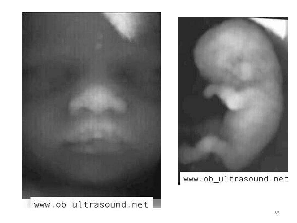
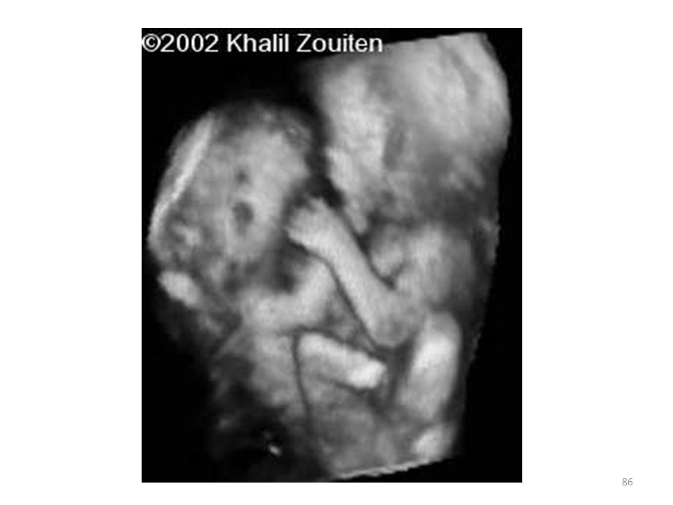
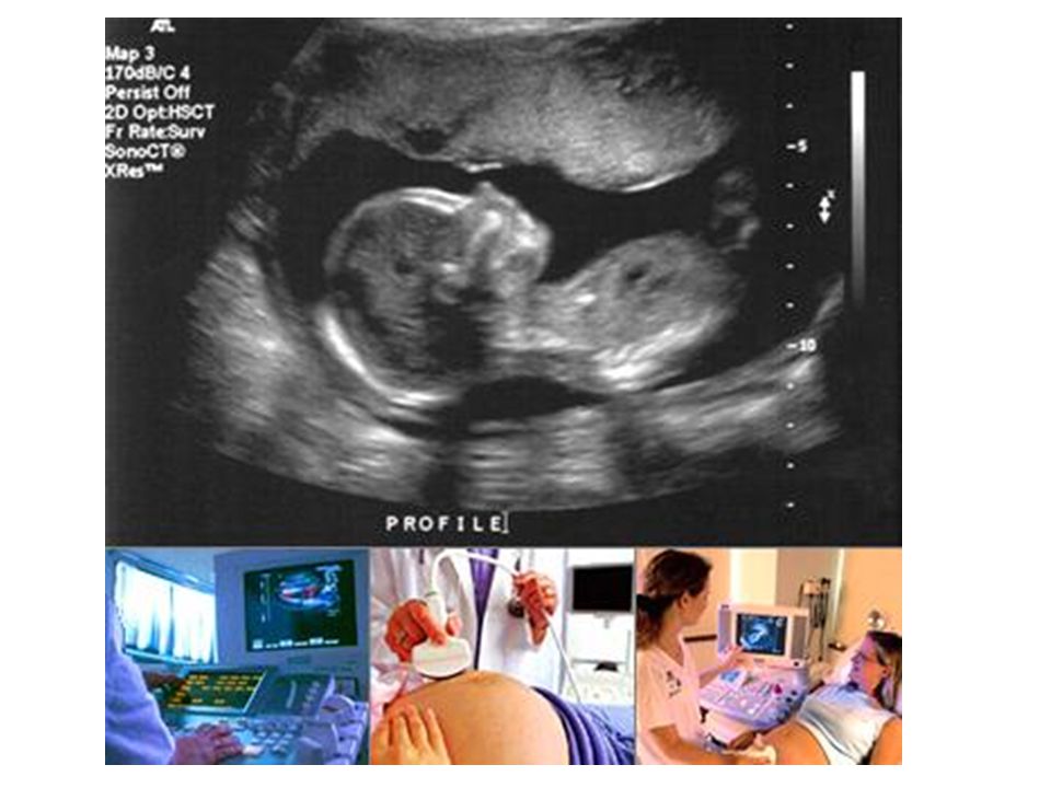
Obstetric Ultrasound is the use of ultrasound scans in pregnancy
Obstetric Ultrasound is the use of ultrasound scans in pregnancy. Since its introduction in the late 1950’s ultrasonography has become a very useful diagnostic tool in Obstetrics. Currently used equipments are known as real-time scanners, with which a continuous picture of the moving fetus can be depicted on a monitor screen. Very high frequency sound waves of between 3.5 to 7.0 megahertz (i.e. 3.5 to 7 million cycles per second) are generally used for this purpose. They are emitted from a transducer which is placed in contact with the maternal abdomen, and is moved to "look at" (likened to a light shined from a torch) any particular content of the uterus. Repetitive arrays of ultrasound beams scan the fetus in thin slices and are reflected back onto the same transducer. The information obtained from different reflections are recomposed back into a picture on the monitor screen (a sonogram, or ultrasonogram). Movements such as fetal heart beat and malformations in the fetus can be assessed and measurements can be made accurately on the images displayed on the screen. Such measurements form the cornerstone in the assessment of gestational age, size and growth in the fetus
 are generally used for this purpose. They are emitted from a transducer which is placed in contact with the maternal abdomen, and is moved to look at (likened to a light shined from a torch) any particular content of the uterus. Repetitive arrays of ultrasound beams scan the fetus in thin slices and are reflected back onto the same transducer. The information obtained from different reflections are recomposed back into a picture on the monitor screen (a sonogram, or ultrasonogram). Movements such as fetal heart beat and malformations in the fetus can be assessed and measurements can be made accurately on the images displayed on the screen. Such measurements form the cornerstone in the assessment of gestational age, size and growth in the fetus.")
92
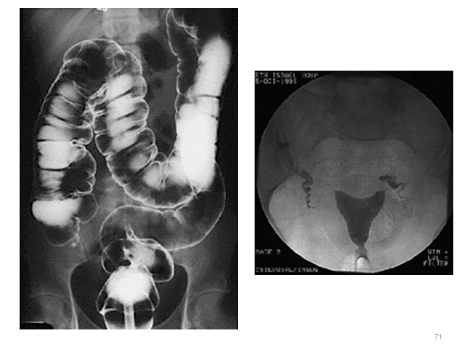
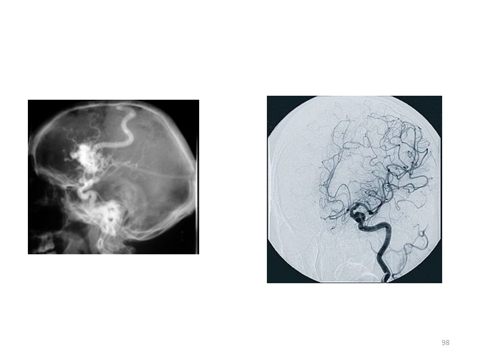
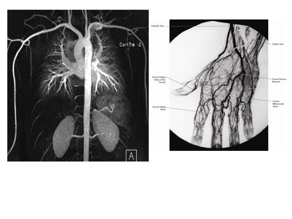
Angiography

93
ANGIOGRAPHY is a specialized radiographic examination where the images of the blood vessels of the body are demonstrated by injection of contrast media

95
SUB SPECIALITY IN ANGIOGRAPHY
Cardiovascular Interventional Technology Vascular Interventional Technology Must have certification in diagnostic radiography in order to be trained and certified in these special procedures.

96
Cardiac Interventional Radiography (CI)- Primary certification
Mike Smith, RT (R) (CI) Must have primary certification in radiography 2) Meet clinical requirements
 (CI) Must have primary certification in radiography. 2) Meet clinical requirements.")
97
Vascular Interventional Radiography (VI)- Post primary certification
JOE CAR, RT (R) (VI) Must have primary certification in radiography Meet clinical requirements
 (VI) Must have primary certification in radiography. Meet clinical requirements.")
99
99

100
Angiogram A medical imaging technique using x-ray and contrast agent to visualize the inside of blood vessels and organs of the body. Angiography or arteriography is a medical imaging technique used to visualize the inside, or lumen, of blood vessels and organs of the body, with particular interest in the arteries, veins and the heart chambers. This is traditionally done by injecting a radio-opaque contrast agent into the blood vessel and imaging using X-ray based techniques such as fluoroscopy.

103
Also known as CT, Cat Scans
Computed Tomography Also known as CT, Cat Scans

104
Computed Tomography Uses ionized radiation to obtain cross sectional images Designated by CT Jennifer Smith, R.T. (R) (CT) Must have primary certification in radiography, nuclear medicine or radiation therapy Meet clinical requirements

105
Computed Tomography Able to do 3D reconstruction

107
MRI Magnetic Resonance Imaging

108
MRI SIGNAL PRODUCTION Uses Magnet field radio waves

109
MRI Uses magnetic and radio waves to create images
Can be whole body or cross sectional Designated by MRI Jeremy Assef, R.T., CRT, (MRI) Magnetic resonance imaging (MRI), is primarily a medical imaging technique most commonly used in radiology to visualize the structure and function of the body. It provides detailed images of the body in any plane. MRI provides much greater contrast between the different soft tissues of the body than computed tomography (CT) does, making it especially useful in neurological (brain), musculoskeletal, cardiovascular, and oncological (cancer) imaging. Unlike CT, it uses no ionizing radiation, but uses a powerful magnetic field to align the nuclear magnetization of (usually) hydrogen atoms in water in the body. Radiofrequency fields are used to systematically alter the alignment of this magnetization, causing the hydrogen nuclei to produce a rotating magnetic field detectable by the scanner. This signal can be manipulated by additional magnetic fields to build up enough information to construct an image of the body.
 Magnetic resonance imaging (MRI), is primarily a medical imaging technique most commonly used in radiology to visualize the structure and function of the body. It provides detailed images of the body in any plane. MRI provides much greater contrast between the different soft tissues of the body than computed tomography (CT) does, making it especially useful in neurological (brain), musculoskeletal, cardiovascular, and oncological (cancer) imaging. Unlike CT, it uses no ionizing radiation, but uses a powerful magnetic field to align the nuclear magnetization of (usually) hydrogen atoms in water in the body. Radiofrequency fields are used to systematically alter the alignment of this magnetization, causing the hydrogen nuclei to produce a rotating magnetic field detectable by the scanner. This signal can be manipulated by additional magnetic fields to build up enough information to construct an image of the body.")
113
Which one is MRI? CT?

114
Look for the signs….

115
What are the differences between MRI and CT?
Uses magnets and radiowaves Cannot be used on patients who have metal in their body Slow Uses ionizing radiation Can be used on any patient Fast

116
Which is better?

117
What are the similarities between CT and MRI?

118
Nuclear Medicine

119
Primary or Post primary certification
Nuclear Medicine Uses radioactive isotopes to produce images Radiation comes from within the patient Primary or Post primary certification

121
PET scan

122
Mammography

123
Mammography Breast imaging using ionized radiation

124
Radiation Therapy Medical dosimetrists are involved in treatment planning and dose calculations 1-4 year program

127
Radiation therapy

128
Primary certification
Radiation Therapy Involved the treatment of diseases Use high level of ionized radiation (megavolt) to kill cancerous cells Primary certification
 to kill cancerous cells. Primary certification.")
130
Additional Opportunities
Education Administration Management (QM) Commercial Radiologist Assistant = RA Sales Application specialist
 Commercial. Radiologist Assistant = RA. Sales. Application specialist.")
131
Radiologist Assistant (RA)
Still not widely accepted Must have a primary certification in radiography Must meet clinical requirements

132
TRAVELING TECHNOLOGIST = SEE THE WORLD AND GET $$$

133
Other working opportunities…
Registry (local) Registry (out of state) X rays taken around the world !!
 Registry (out of state) X rays taken around the world !!")
134
Variety of Work Settings
physicians offices, clinical outpatient facilities, free standing imaging centers, mobile imaging centers portable services to rehabs Mammo’s to under privileged areas Urgent care

135
RA Radiology Assistant (Like PA) LLU PART OF RADIOLOGIST GROUP
Still not widely accepted

136
Diagnostic Imaging Modalities
Questions ? Diagnostic Imaging Modalities

Similar presentations


 PowerPoint.>")


 CT scanning or (CAT scanning) is using X-rays to create a 3D image of the inside of an object. CT stands for computed tomography.>")
 Medical Professional –In a challenging field Safely uses x-rays to image anatomy –Positions –Using sophisticated.>")


 RADIOLOGIC TECHNOLOGIST –RADIOGRAPHER –X-RAY TECHNOLOGIST “ TECHNICIAN” –Limited License RADIOLOGIST –Medical.>")


