Download presentation
Presentation is loading. Please wait.

1
The Urinary System

2
Functions of the Kidneys
Filter nearly 1200 ml of blood per minute Return needed substances back to body Regulate the volume and chemical makeup of the blood (maintaining pH, water and salt concentrations) Production of renin and erythropoietin Metabolizing vitamin D to its active form
 Production of renin and erythropoietin. Metabolizing vitamin D to its active form.")
3
Gross Anatomy Kidneys: two bean-shaped organs that filter blood
Ureters: two tubes that drain urine away from the kidneys Urinary Bladder: stores urine until micturition occurs; composed of transitional epithelium to allow for stretching Urethra: drains urine from bladder and transports it outside; in females, only carries urine; carries urine and semen in males

4
Gross Anatomy Two Kidneys Two Ureters Urinary Bladder Urethra

5
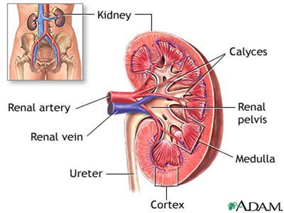
External Kidney Anatomy
Located in a retroperitoneal position Renal Hilus: region where ureters, renal blood vessels, lymphatics, and nerves all join together and exit/enter kidney Connective Tissue Layers Renal Capsule (prevents spread of infection) Adipose Capsule (fatty mass for shock absorbtion) Renal Fascia (outer layer that anchors kidney to surrounding areas)
 Adipose Capsule (fatty mass for shock absorbtion) Renal Fascia (outer layer that anchors kidney to surrounding areas)")
6
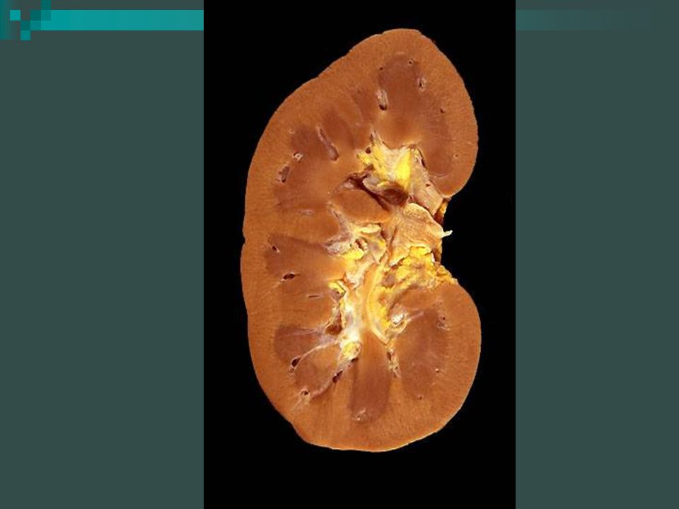
Internal Kidney Anatomy
Renal Cortex Renal Medulla With renal pyramids Renal columns Renal Pelvis With major calyces and minor calyces

9
Arterial Blood Supply of the Kidney
Renal Arteries Segmental arteries Lobar arteries Interlobar arteries Arcuate arteries Interlobular arteries Afferent arterioles Glomerular capillaries Efferent arterioles Peritubular capillaries

10
Venous Blood Supply of the Kidney
Renal Veins Peritubular venules Interlobular veins Arcuate veins Interlobar veins Renal vein Inferior vena cava

11
Nephron Anatomy and Structure
Nephrons: functional unit of the kidney Renal Corpuscle: Glomerulus (capillary bed made up of fenestrated capillaries for filtration, allows filtrate to pass into the Bowman’s capsule) Bowman’s Capsule (collection tubule surrounding glomerulus) Three types of cells inside renal corpuscle: Juxtaglomerular cells (mechanoreceptors in afferent arteriole sensing changes in BP, secrete renin) Macula Densa cells (osmoreceptors responding to solute concentrations and flow rate, vasoconstriction or vasodilation) Mesangial cells (posses phagocytic and contractile abilities, increase surface area for absorption)
 Bowman’s Capsule (collection tubule surrounding glomerulus) Three types of cells inside renal corpuscle: Juxtaglomerular cells (mechanoreceptors in afferent arteriole sensing changes in BP, secrete renin) Macula Densa cells (osmoreceptors responding to solute concentrations and flow rate, vasoconstriction or vasodilation) Mesangial cells (posses phagocytic and contractile abilities, increase surface area for absorption)")
12
Juxtaglomerular cells in the kidney respond to changes in blood pressure and plasma sodium concen. Decrease in either one will cause these cells to make renin. Renin breaks down a plasma protein called angiotensinogen which in turn releases a substance called angiotensin-I. Another enzyme, angiotensin –converting enzyme; ACE, produced in the lungs converts angiotensin-I to angiotensin –II which is carried off in the blood. When angiotensin-II reaches the adrenal cortex it stimulates the release of aldosterone. Angiotensin-II is also a powerful vasoconstrictor used in regulating BP.

13
Renal Corpuscle

14
Nephron Anatomy and Structure
Renal Tubule: Proximal convoluted tubule (contain brush border cuboidal epithelium for absorption and secretion in the cortex) Loop of Henle (ascending and descending branches in the medulla) Distal convoluted tubule (non-brush border cuboidal epithelium cells, more secretion than absorption) Proximal/Distal refer to location relative to the loop
 Loop of Henle (ascending and descending branches in the medulla) Distal convoluted tubule (non-brush border cuboidal epithelium cells, more secretion than absorption) Proximal/Distal refer to location relative to the loop.")
15
Renal Tubule

16
Types of Nephrons Cortical: make up 85% of the nephron content and found solely in the cortex Juxtamedullary: their loops of Henle dip down into the medulla; associated with vasa recta (regions where the efferent arteriole does not break up into peritubular capillaries) produce concentrated urine
 produce concentrated urine.")
17
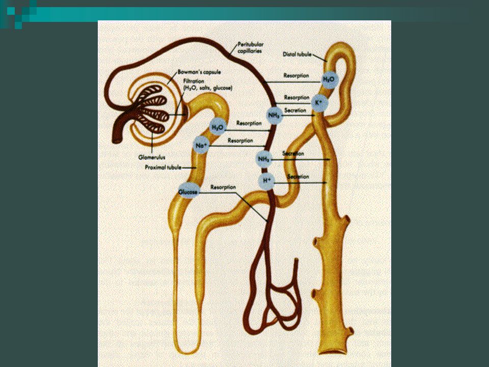
Non-selective filtration
Renal Physiology Occurs in Three Steps: Non-selective filtration Tubular reabsorption Tubular secretion

18
Overview of Renal Physiology
FILTRATION REABSORPTION SECRETION

19
Non-Selective Filtration
(1) Unfiltered blood enters glomerulus via afferent arteriole (2) Inside the glomerulus, hydrostatic pressure is high, pushing the filtrate (everything except proteins and blood cells) into the Bowman’s capsule (3) Efferent arterioles transport filtered blood to the capillary beds Glomerulus unique because arterioles bring blood to the capillary bed and take it away Glomerular Filtration Rate: the amount of filtrate formed in both kidneys per minute (125 mL/ min); majority is reabsorbed in the renal tubule (124 mL)
 Unfiltered blood enters glomerulus via afferent arteriole. (2) Inside the glomerulus, hydrostatic pressure is high, pushing the filtrate (everything except proteins and blood cells) into the Bowman’s capsule. (3) Efferent arterioles transport filtered blood to the capillary beds. Glomerulus unique because arterioles bring blood to the capillary bed and take it away. Glomerular Filtration Rate: the amount of filtrate formed in both kidneys per minute (125 mL/ min); majority is reabsorbed in the renal tubule (124 mL)")
20
Non-selective Filtration

21
Tubular Reabsorption Water, ions, and other substances reabsorbed into the blood via the peritubular capillaries Na+ moved out of tubule into the blood via facilitated diffusion Glucose, amino acids, lactic acid, vitamins, and most cations absorbed by secondary active transport (energy used from Na+/K+ pump) Some ions (e.g. K+ and Cl-) move through the intercellular spaces to leave the tubules in the interstitial spaces and then simply diffuse out Most substances (urea, some drugs, fat-soluble vitamins) diffuse directly from the lumen of the tubules and into the peritubular capillary network 98-99% of filtrate is reabsorbed
 Some ions (e.g. K+ and Cl-) move through the intercellular spaces to leave the tubules in the interstitial spaces and then simply diffuse out. Most substances (urea, some drugs, fat-soluble vitamins) diffuse directly from the lumen of the tubules and into the peritubular capillary network % of filtrate is reabsorbed.")
23
Tubular Reabsorption

24
Tubular Reabsorption

25
Tubular Secretion Involves the movement of substances out of blood (peritubular capillaries) and into the filtrate Substances can move by active or passive means Substances commonly secreted: K+, H+, ammonia, by-products of drugs and penicillin, and creatinine and hormones Final fluid draining from DCT into collecting ducts called urine Urine drains into renal pelvis and then merges into the ureters and is sent by peristalsis to the bladder

26
Tubular Secretion

27
Regulation of Urine Volume
(1) The descending limb of loop of Henle is impermeable to solutes and permeable to water (thus osmolarity increases) (2) The ascending limb is permeable to solutes, but not to water (thus osmolarity decreases) (3) The collecting ducts in the deep medullary regions are permeable to urea Results in the concentration of urine
 The descending limb of loop of Henle is impermeable to solutes and permeable to water (thus osmolarity increases) (2) The ascending limb is permeable to solutes, but not to water (thus osmolarity decreases) (3) The collecting ducts in the deep medullary regions are permeable to urea. Results in the concentration of urine.")
28
Effects of Hormones on Urine Formation
Antidiuretic hormone (ADH) Aldosterone Atrial natriuretic hormone (ANH) Renin and Angiotensin II
 Aldosterone. Atrial natriuretic hormone (ANH) Renin and Angiotensin II.")
29
Urine Formation Urine composition Urine characteristics 90-95% water
Solutes constitute the other 5% Metabolic wastes (urea, uric acid, and creatinine) Ions (Na+, K+, PO43-, SO42-, Ca2+, Mg2+) Toxins and pigments (urochrome) Hormones Urine characteristics Yellow in color Aromatic or ammonia pH slightly acidic (can vary from 4.5 to 8.0) Specific gravity to 1.035
 Ions (Na+, K+, PO43-, SO42-, Ca2+, Mg2+) Toxins and pigments (urochrome) Hormones. Urine characteristics. Yellow in color. Aromatic or ammonia. pH slightly acidic (can vary from 4.5 to 8.0) Specific gravity to")
30
Abnormal Urine Constituents

31
Excretion and Micturition
Bladder serves as a urine storage organ Openings for both ureters and urethra located on bladder interior (region bordered by these three openings called the trigone) Very distensible and can change shape to accommodate for urine Urethra conveys urine outside the body Two sphincters (internal-involuntary and external-voluntary) regulate urine flow outside the body
 Very distensible and can change shape to accommodate for urine. Urethra conveys urine outside the body. Two sphincters (internal-involuntary and external-voluntary) regulate urine flow outside the body.")
32
The Bladder

Similar presentations


 System>")


>")