Download presentation
Presentation is loading. Please wait.

2
Brain Basics

3
BRAIN ON DRUGS?

4
What is your nervous system?

5
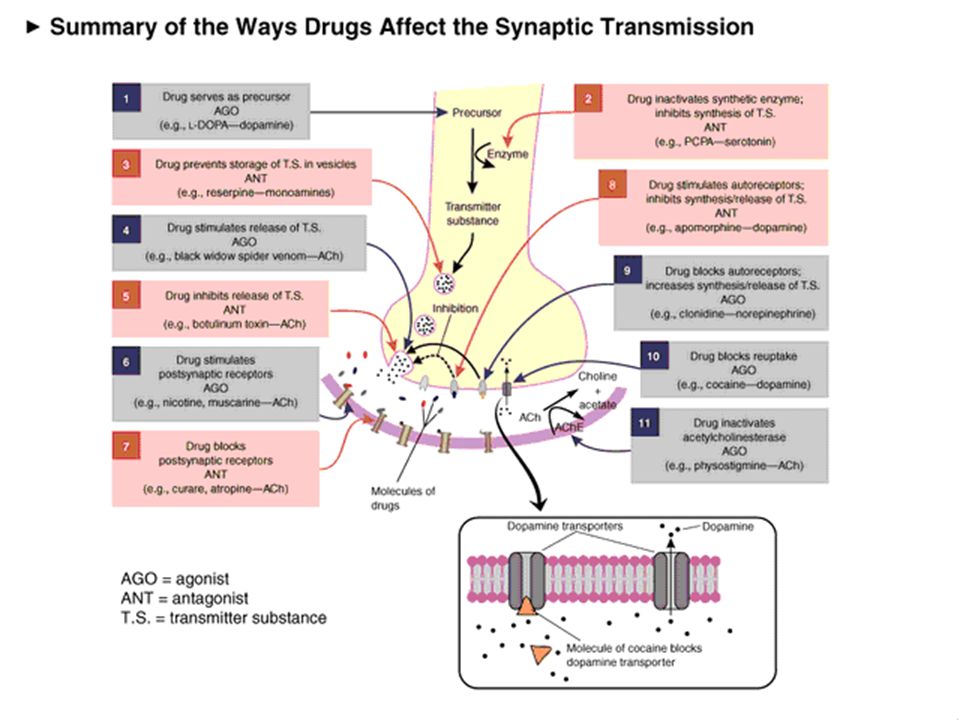
Neurons “communicate” with each other using neurotransmitters
Slide 7: Summary of neuronal transmission Use the example with 2 neurons making contact to summarize neuronal transmission. Point to the cell on the top and indicate that electrical impulses flow in the direction toward the terminal. Remind the students what happens when impulses reach the terminal; neurotransmitters are released, they bind to their receptors, and new impulses are generated in the cell on the bottom. Explain that this is how information travels from neuron to neuron.

6
Neurotransmitters convey “messages” across the synapse
Slide 7: The synapse and synaptic neurotransmission Describe the synapse and the process of chemical neurotransmission. As an electrical impulse arrives at the terminal, it triggers vesicles containing a neurotransmitter, such as dopamine (in blue), to move toward the terminal membrane . The vesicles fuse with the terminal membrane to release their contents (in this case, dopamine). Once inside the synaptic cleft (the space between the 2 neurons) the dopamine can bind to specific proteins called dopamine receptors (in pink) on the membrane of a neighboring neuron. This is illustrated in more detail on the next slide.
, to move toward the terminal membrane . The vesicles fuse with the terminal membrane to release their contents (in this case, dopamine). Once inside the synaptic cleft (the space between the 2 neurons) the dopamine can bind to specific proteins called dopamine receptors (in pink) on the membrane of a neighboring neuron. This is illustrated in more detail on the next slide.")
8
Dopamine/Opioids: Brain’s incentive reward systems
Slide 11: The reward pathway Tell your audience that this is a view of the brain cut down the middle. An important part of the reward pathway is shown and the major structures are highlighted: the ventral tegmental area (VTA), the nucleus accumbens and the prefrontal cortex. The VTA is connected to both the nucleus accumbens and the prefrontal cortex via this pathway and it sends information to these structures via its neurons. The neurons of the VTA contain the neurotransmitter dopamine which is released in the nucleus accumbens and in the prefrontal cortex (point to each of these structures). Reiterate that this pathway is activated by a rewarding stimulus. [Note: the pathway shown here is not the only pathway activated by rewards, other structures are involved too, but only this part of the pathway is shown for simplicity.]
, the nucleus accumbens and the prefrontal cortex. The VTA is connected to both the nucleus accumbens and the prefrontal cortex via this pathway and it sends information to these structures via its neurons. The neurons of the VTA contain the neurotransmitter dopamine which is released in the nucleus accumbens and in the prefrontal cortex (point to each of these structures). Reiterate that this pathway is activated by a rewarding stimulus. [Note: the pathway shown here is not the only pathway activated by rewards, other structures are involved too, but only this part of the pathway is shown for simplicity.]")
9
Activation of reward center produces a “wanting” and “liking” response

10
Natural events activate these reward systems

11
DA Concentration (% Baseline)
Natural Events Elevate Dopamine Levels FOOD SEX 200 200 NAc shell 150 150 DA Concentration (% Baseline) 100 100 15 5 10 Copulation Frequency % of Basal DA Output Empty 50 Box Feeding Natural rewards increase dopamine neurotransmission. For example, eating something that you enjoy or engaging in sexual behavior can cause dopamine levels to increase. In these graphs, dopamine is being measured inside the brains of animals, its increase shown in response to food or sex cues. This basic mechanism has been carefully shaped and calibrated by evolution to reward normal activities critical for survival. 60 120 180 Female Present Time (min) 1 2 3 4 5 6 7 8 Sample Number Mounts Intromissions Ejaculations Di Chiara et al., Neuroscience, 1999. Fiorino and Phillips, J. Neuroscience, 1997.
 Copulation Frequency. % of Basal DA Output. Empty. 50. Box. Feeding. Natural rewards increase dopamine neurotransmission. For example, eating something that you enjoy or engaging in sexual behavior can cause dopamine levels to increase. In these graphs, dopamine is being measured inside the brains of animals, its increase shown in response to food or sex cues. This basic mechanism has been carefully shaped and calibrated by evolution to reward normal activities critical for survival Female Present. Time (min) Sample. Number. Mounts. Intromissions. Ejaculations. Di Chiara et al., Neuroscience, Fiorino and Phillips, J. Neuroscience,")
12
Some drugs activate your reward systems since they act on the same receptors

13
BUT only when your brain is on drugs.
Drugs make your brain really happy….. BUT only when your brain is on drugs. Normal Brain Brain on Drugs

14
Effects of Drugs on Dopamine Release
100 200 300 400 500 600 700 800 900 1000 1100 1 2 3 4 5 hr Time After Amphetamine % of Basal Release DA DOPAC HVA Accumbens AMPHETAMINE 100 200 300 400 1 2 3 4 5 hr Time After Cocaine % of Basal Release DA DOPAC HVA Accumbens COCAINE 100 150 200 250 1 2 3 hr Time After Nicotine % of Basal Release Accumbens Caudate NICOTINE 100 150 200 250 1 2 3 4 5hr Time After Morphine % of Basal Release Accumbens 0.5 1.0 2.5 10 Dose (mg/kg) MORPHINE Drugs of abuse increase dopamine neurotransmission. All the drugs depicted in this slide have different mechanisms of action; however, all increase activity in the reward pathway by increasing dopamine neurotransmission. Because drugs activate these brain regionsusually more effectively than natural rewardsthey have an inherent risk of being abused. Di Chiara and Imperato, PNAS, 1988
 MORPHINE. Drugs of abuse increase dopamine neurotransmission. All the drugs depicted in this slide have different mechanisms of action; however, all increase activity in the reward pathway by increasing dopamine neurotransmission. Because drugs activate these brain regionsusually more effectively than natural rewardsthey have an inherent risk of being abused. Di Chiara and Imperato, PNAS,")
15
Repeated use of drugs trigger compensatory processes and saturate the brain’s reward systems
individual can become conditioned/habituated/adapted to the intense level of drug-induced pleasure (develops tolerance or sensitization) the normal level of natural rewards are no longer experienced as very pleasurable, and after chronic use, the brain’s reward systems becomes so changed that nothing is pleasurable – not even the drugs!
 the normal level of natural rewards are no longer experienced as very pleasurable, and. after chronic use, the brain’s reward systems becomes so changed that nothing is pleasurable – not even the drugs!")
16
Homeostatic Changes to Postsynaptic Receptor Density as a Function
of the Amount of Neurotransmitter Released Upregulation Downregulation

17
Chronic drug taking ….reorganizes the liking and wanting systems
Brain on drugs after tolerance Brain on drugs for an extended period … drugs may no longer be pleasurable but you still want them…

18
Drugs can change your brain so that natural events are no longer pleasurable

19
The brain now has a disease… it’s a different brain under constant stress
When the “switch” gets flips depends on …. Addicted your brain chemistry…. your drug history…. your drug history…. and other factors Normal

20
Even 80 days following detox, a methamphetamine user’s dopamine transporter system (right) hasn’t recovered to normal levels (left)
 hasn’t recovered to normal levels (left)")
21
Cocaine has long lasting effects
Normal Cocaine Abuser (10 da) Slide 8: Long-term effects of drug abuse. This PET scan shows us that once addicted to a drug like cocaine, the brain is affected for a long, long time. In other words, once addicted, the brain is literally changed. Let’s see how... In this slide, the level of brain function is indicated in yellow. The top row shows a normal-functioning brain without drugs. You can see a lot of brain activity. In other words, there is a lot of yellow color. The middle row shows a cocaine addict’s brain after 10 days without any cocaine use at all. What is happening here? [Pause for response.] Less yellow means less normal activity occurring in the brain—even after the cocaine abuser has abstained from the drug for 10 days. The third row shows the same addict’s brain after 100 days without any cocaine. We can see a little more yellow, so there is some improvement— more brain activity—at this point. But the addict’s brain is still not back to a normal level of functioning. . . more than 3 months later. Scientists are concerned that there may be areas in the brain that never fully recover from drug abuse and addiction. Photo courtesy of Nora Volkow, Ph.D. Volkow ND, Hitzemann R, Wang C-I, Fowler IS, Wolf AP, Dewey SL. Long-term frontal brain metabolic changes in cocaine abusers. Synapse 11: , 1992; Volkow ND, Fowler JS, Wang G-J, Hitzemann R, Logan J, Schlyer D, Dewey 5, Wolf AP. Decreased dopamine D2 receptor availability is associated with reduced frontal metabolism in cocaine abusers. Synapse 14: , 1993. Cocaine Abuser (100 da)
 Slide 8: Long-term effects of drug abuse. This PET scan shows us that once addicted to a drug like cocaine, the brain is affected for a long, long time. In other words, once addicted, the brain is literally changed. Let’s see how... In this slide, the level of brain function is indicated in yellow. The top row shows a normal-functioning brain without drugs. You can see a lot of brain activity. In other words, there is a lot of yellow color. The middle row shows a cocaine addict’s brain after 10 days without any cocaine use at all. What is happening here [Pause for response.] Less yellow means less normal activity occurring in the brain—even after the cocaine abuser has abstained from the drug for 10 days. The third row shows the same addict’s brain after 100 days without any cocaine. We can see a little more yellow, so there is some improvement— more brain activity—at this point. But the addict’s brain is still not back to a normal level of functioning. . . more than 3 months later. Scientists are concerned that there may be areas in the brain that never fully recover from drug abuse and addiction. Photo courtesy of Nora Volkow, Ph.D. Volkow ND, Hitzemann R, Wang C-I, Fowler IS, Wolf AP, Dewey. SL. Long-term frontal brain metabolic changes in cocaine abusers. Synapse 11: , 1992; Volkow ND, Fowler JS, Wang G-J, Hitzemann R, Logan J, Schlyer D, Dewey 5, Wolf AP. Decreased dopamine D2. receptor availability is associated with reduced frontal metabolism in cocaine abusers. Synapse 14: , Cocaine Abuser (100 da)")
22
At high enough doses, Ecstasy destroys nerve fibers
Slide 17: Long-term Effects in Monkeys A very important experiment was performed in monkeys to determine if Ecstasy can actually damage neurons. Monkeys were given Ecstasy twice a day for 4 days (control monkeys were given saline). One group of monkeys’ brains were removed 2 weeks later for analysis and another group of monkeys lived for an additional 7 years before their brains were removed. Scientists examined the brains for the presence of serotonin. This slide shows the presence of serotonin in neurons of the neocortex from 3 typical monkeys. On the left, the monkey who did not receive any Ecstasy had a lot of serotonin (in pink) in the neocortex. Two weeks after a monkey received Ecstasy, most of the serotonin was gone (point to the middle panel), suggesting that the serotonin neuron terminals were destroyed (there was no destruction of the serotonin cell bodies arising back in the brainstem). Point to the right hand panel and show students that this damage appeared to be long-term because 7 years later there was some recovery, but it was not complete (in fact the pattern of regrowth of serotonin terminals was abnormal– point out one of the areas where the pink lines are running sideways). Scientists found similar changes in limbic areas of the brain such as the hippocampus and amygdala. The monkey experiments are an important reminder that humans may suffer the same fate, although this still remains to be demonstrated. Tell the students how difficult it is to do this same kind of experiment in humans because it requires removing pieces of the brain to look for the loss of the serotonin neurons. Image courtesy of Dr. GA Ricaurte, Johns Hopkins University School of Medicine
. One group of monkeys’ brains were removed 2 weeks later for analysis and another group of monkeys lived for an additional 7 years before their brains were removed. Scientists examined the brains for the presence of serotonin. This slide shows the presence of serotonin in neurons of the neocortex from 3 typical monkeys. On the left, the monkey who did not receive any Ecstasy had a lot of serotonin (in pink) in the neocortex. Two weeks after a monkey received Ecstasy, most of the serotonin was gone (point to the middle panel), suggesting that the serotonin neuron terminals were destroyed (there was no destruction of the serotonin cell bodies arising back in the brainstem). Point to the right hand panel and show students that this damage appeared to be long-term because 7 years later there was some recovery, but it was not complete (in fact the pattern of regrowth of serotonin terminals was abnormal– point out one of the areas where the pink lines are running sideways). Scientists found similar changes in limbic areas of the brain such as the hippocampus and amygdala. The monkey experiments are an important reminder that humans may suffer the same fate, although this still remains to be demonstrated. Tell the students how difficult it is to do this same kind of experiment in humans because it requires removing pieces of the brain to look for the loss of the serotonin neurons. Image courtesy of Dr. GA Ricaurte, Johns Hopkins University School of Medicine.")
23
DA Receptors and the Response to
Methylphenidate (MP) High DA receptor high Dopamine receptor level low Low DA receptor The level of dopamine receptors in the brain can influence whether a person likes or dislikes the effects of a drug. Dopamine is one of many chemicals normally found in the brain and is involved in the perception of pleasure. Most abused drugs directly or indirectly increase dopamine in brain regions related to reward and motivation. Dopamine receptors are the sites on nerve cells where dopamine attaches to regulate their function. The graph on the right side of this slide shows that individuals with a lot of dopamine receptors (top picture on the left; dopamine receptors are in yellow/red) tend to experience methylphenidate (a stimulant) as unpleasant, whereas persons with lower numbers of dopamine receptors (bottom picture on the left) find it pleasurable. This biological difference can influence the chances of re-exposure to a drug, and thus the risk of abuse or addiction. As a group, subjects with low receptor levels found MP pleasant while those with high levels found MP unpleasant Adapted from Volkow et al., Am. J. Psychiatry, 1999.
 High DA. receptor. high. Dopamine receptor level. low. Low DA. receptor. The level of dopamine receptors in the brain can influence whether a person likes or dislikes the effects of a drug. Dopamine is one of many chemicals normally found in the brain and is involved in the perception of pleasure. Most abused drugs directly or indirectly increase dopamine in brain regions related to reward and motivation. Dopamine receptors are the sites on nerve cells where dopamine attaches to regulate their function. The graph on the right side of this slide shows that individuals with a lot of dopamine receptors (top picture on the left; dopamine receptors are in yellow/red) tend to experience methylphenidate (a stimulant) as unpleasant, whereas persons with lower numbers of dopamine receptors (bottom picture on the left) find it pleasurable. This biological difference can influence the chances of re-exposure to a drug, and thus the risk of abuse or addiction. As a group, subjects with low receptor levels found MP pleasant while those with high levels found MP unpleasant. Adapted from Volkow et al., Am. J. Psychiatry,")
24
Drugs not only affect the brain, but also affect the body

26
Gross Brain Anatomy Forebrain Midbrain Hindbrain
(Brainstem = Midbrain + Hindbrain - Cerebellum)
")
27
Hindbrain Medulla Pons Cerebellum

28
Medulla caudal end of brainstem; rostral end of spinal cord
connects rest of brain to spinal cord life support functions (heart rate, respiration)
")
29
Pons ventral side of cerebellum levels of consciousness, sleep,
arousal, control of autonomic functions, sleep, relay info to cerebellum

30
Cerebellum coordination of voluntary movement learning motor behaviors
involved in cognition timing of motor output

31
Midbrain rostral end of brainstem: reticular formation, superior/inferior colliculi arousal, wakefulness information modulation source cells for some important neurotransmitters (biogenic amines)
")
32
Forebrain Cerebral Cortex Thalamus Hypothalamus Basal Ganglia
Limbic System

33
Thalamus relays information from diverse areas to cerebral cortex
integrates sensory information regulates sleep-wakefulness

34
Hypothalamus homeostatic control (e.g. body temperature, cardiovascular system, food and water intake) regulates autonomic and endocrine systems Infundibulum connects hypothalamus to pituitary glands

35
Basal Ganglia voluntary movement, posture

36
Limbic System Medial Forebrain Bundle Hippocampus Amygdala
collection of various nerves running upstream through midbrain involved in reinforcement Hippocampus Amygdala Nucleus Accumbens

37
Hippocampus medial side of temporal lobe
consolidation of short term memory into more permanent memory recollection of spatial relationships

38
Amygdala inferior medial temporal lobe
emotional feelings, fear, behavior, perception

39
Nucleus Accumbens very important in reinforcement and addiction
regulation of movement cognitive aspects of motor control

40
Mu receptor distribution
5HT1a receptor distribution

41
Cerebral Cortex Frontal lobe Parietal lobe Temporal lobe
Occipital lobe

42
Occipital Lobe vision

43
Parietal Lobe body sensation (touch, pain, etc.) speech reception
spatial relationships

44
Temporal Lobe hearing memory emotion vision

45
Frontal Lobe planned motor behavior speech production higher cognition
social reasoning

Similar presentations