Download presentation
Presentation is loading. Please wait.

1
Tissue Types: Epithelial Connective Muscular Nervous
Tissues – a group of similar cells working together to perform a function Tissue Types: Epithelial Connective Muscular Nervous

2
Histology The study of tissues at the microscopic level

3
Histology The human body is composed of approximately 200 distinctly different types of cells. These cells are organized into four basic categories of tissues that, in turn, are assembled to form organs. Tissues can be distinguished from each other by variations in cell size, shape, organization, and function.

4
How Do Cells Form Tissues?
Remember adhesion proteins embedded in the cell membrane!

5
Cell Junctions- When cells are touching each other they can be joined in 1 of 3 ways
Tight Junctions – sealed together by attachment proteins so that nothing can pass between the cells (if something passes thru, it must go thru the bilayer not between them) Typically join cells that form sheetlike layers; digestive tract Desmosomes – tight junctions with extra protein reinforcement so that the cells won’t rip apart when stretched (in skin). The protein reinforcements bind the attachment proteins to the cytoskeleton of both attaching cells Gap Junctions – where the cells attach, there are small tubes connecting the two cells together so that ions can pass freely between the cells (cardiac muscle in heart and smooth muscle in digestive tract); cytoplasm becomes linked together.
 Typically join cells that form sheetlike layers; digestive tract. Desmosomes – tight junctions with extra protein reinforcement so that the cells won’t rip apart when stretched (in skin). The protein reinforcements bind the attachment proteins to the cytoskeleton of both attaching cells. Gap Junctions – where the cells attach, there are small tubes connecting the two cells together so that ions can pass freely between the cells (cardiac muscle in heart and smooth muscle in digestive tract); cytoplasm becomes linked together.")
6
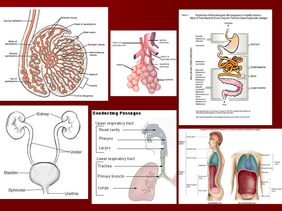
Epithelial Tissue Location – Lines a space - forms inner lining of body cavities, covers body surface and organs, lines hollow organs and glands Specific Examples: Skin Lines whole digestive and respiratory passages (mucous membranes) Lines whole urinary system Membranes lining spinal cord and brain (Meniges) Lines the inside of the heart cavities Capillary walls Alveoli in lungs Lines the uterus in females, epididymis in males Makes up the serous membranes (enclosed cavities like pericardium, pleura, and peritonium) Prostate gland & other glands
 Lines whole urinary system. Membranes lining spinal cord and brain (Meniges) Lines the inside of the heart cavities. Capillary walls. Alveoli in lungs. Lines the uterus in females, epididymis in males. Makes up the serous membranes (enclosed cavities like pericardium, pleura, and peritonium) Prostate gland & other glands.")
8
Functions: Protection - from leakage or from things getting in that aren’t supposed to Absorption - nutrients Secretion – mucous, pepsin, HCl, milk Filtration – oxygen and carbon dioxide

9
Characteristics: (structure = function)
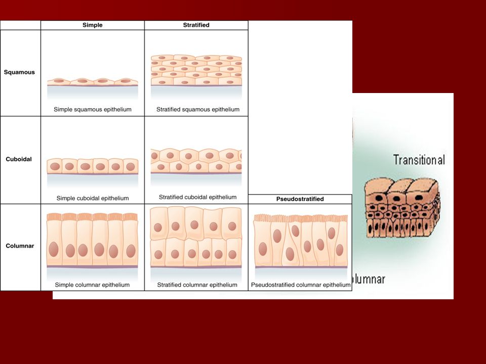
Fit closely together (tight junctions or desmosomes) – sealed so that nothing can pass between the cells Two adjectives describe all epithelial tissue Cells are square, rectangular or “squished” Cuboidal, columnar, squamous Tissue is found in one or multiple layers Simple vs. stratified
 – sealed so that nothing can pass between the cells. Two adjectives describe all epithelial tissue. Cells are square, rectangular or squished Cuboidal, columnar, squamous. Tissue is found in one or multiple layers. Simple vs. stratified.")
11
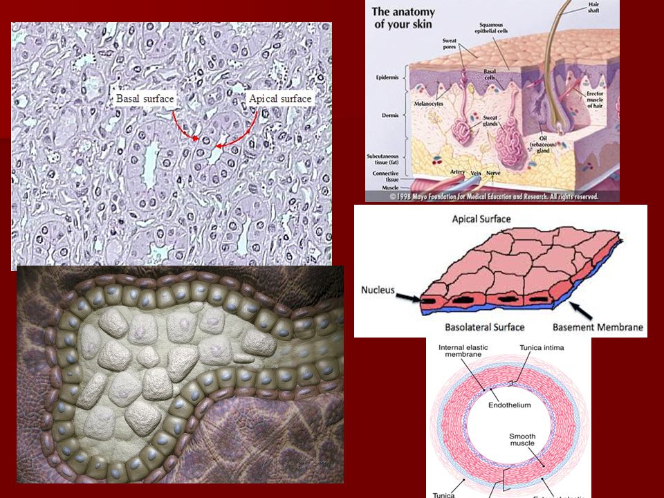
Characteristics: Basement membrane – thin (deepest layer of epithelial tissue), nonliving layer of collagen protein (created by cells in the connective tissue) that anchors epithelium to underlying connective tissue. Apical surface - free surface that is exposed to the outside of body or organ, or the inside of a cavity, gland or tube (lumen)
, nonliving layer of collagen protein (created by cells in the connective tissue) that anchors epithelium to underlying connective tissue. Apical surface - free surface that is exposed to the outside of body or organ, or the inside of a cavity, gland or tube (lumen)")
14
Characteristics: Avascular - no direct connection to blood vessels; must get nutrients from diffusion from underlying capillaries in connective tissue

15
Side Note: Cancer Cancer cells secrete a substance that dissolves basement membranes, enabling the cells to invade tissue layers. Cancer cells also produce less adhesion proteins, or none, which allows them to spread into surrounding tissue. (metastasis)
")
16
Modified/Specialized Cells
Microvilli – tiny , cylindrical processes extending from the free surface of epithelial cells Increases surface area of cell membrane which is exposed to substances being absorbed. small intestines

17
Modified/Specialized Cells
Goblet Cells and cilia - in the trachea, goblet epithelial cells secrete mucus on the free surface of tissue which provides lubrication to trap bacteria and dust (ciliated epithelium then sweeps it away from lungs).
.")
18
Two Types of Epithelial Tissues
Glandular Epithelium Covering/lining Epithelium covers the outside surfaces of the body and lines internal organs. secretes hormones or other products (mucous, milk). Consists of one or many cells
. Consists of one or many cells.")
19
Two Types: Endocrine vs. Exocrine Glands
Exocrine - epithelial tissue that secretes their substances (sweat, saliva, milk, stomach acid, and digestive enzymes) into tubes, or ducts, which carry the secretions to the epithelial surface of cavity or skin Endocrine - epithelial tissue secretes products (hormones) into tissue fluid or blood
 into tubes, or ducts, which carry the secretions to the epithelial surface of cavity or skin. Endocrine - epithelial tissue secretes products (hormones) into tissue fluid or blood.")
20
Two Types: Endocrine vs. Exocrine Glands
Endocrine Glands Exocrine Glands

21
Types of Epithelium Simple Squamous – flat, 1-layer, easily damaged walls of blood vessel lung alveoli Locations: Capillary walls, alveoli of the lungs Function: diffusion and filtration

22
Types of Epithelium Simple Cuboidal – square, 1-layer
kidney's tubules cut such that they appear as rings of cells around empty spaces Locations: lining kidney tubules, salivary ducts, pancreatic ducts Functions: secretion, excretion, and absorption

23
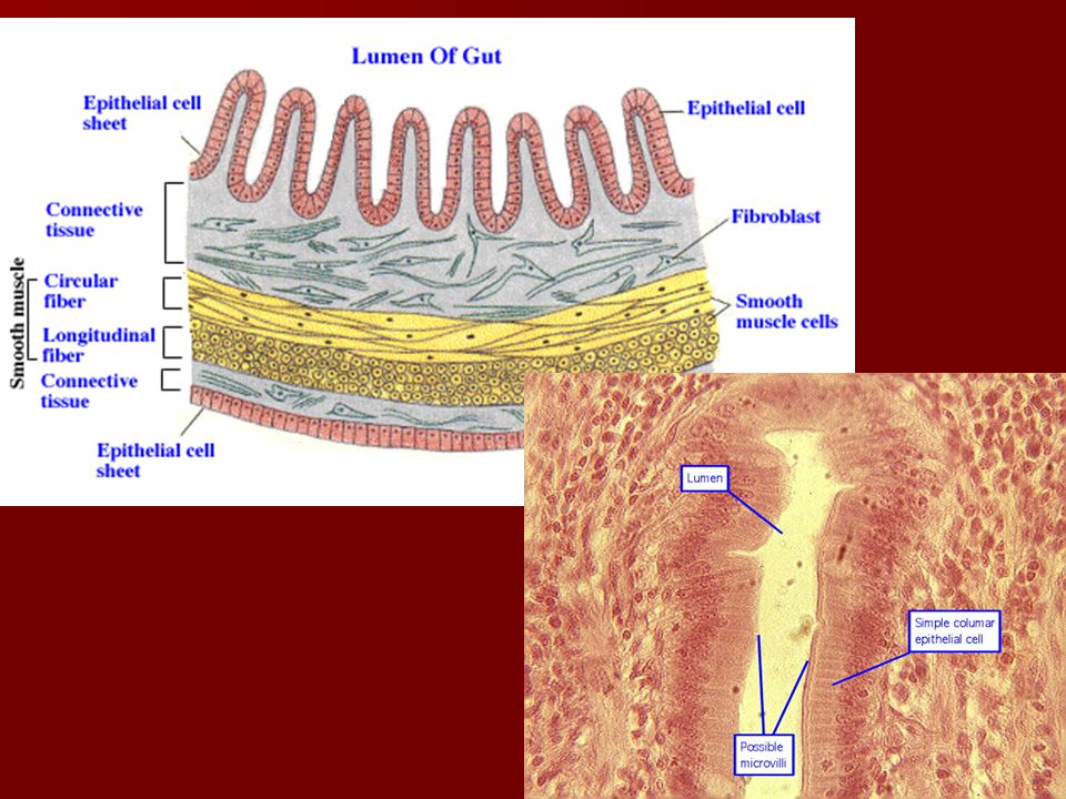
Types of Epithelium Simple Columnar – tall rectangular, 1-layer, thick to protect underlying tissue; used for absorption Ciliated – cells that line the throat and digestive tract. Funct: trap and move "pollutants" to the mouth where they are swallowed. Cells lining the uterine tube to uterus. Funct: sweeps egg along Nonciliated – lines uterus and portions of digestive tract (stomach and small intestines), Funct: secretes digestive fluids, absorbs nutrients, protection. ) Microvilli – minute surface extensions found in small intestine, to increase surface area for absorbing substances
, Funct: secretes digestive fluids, absorbs nutrients, protection. ) Microvilli – minute surface extensions found in small intestine, to increase surface area for absorbing substances.")
24
Types of Epithelium Statified Squamous – many layers, resistant to damage. Location: where there is a lot of abrasion – mouth, outermost layer of skin, esophagus, anal canal. Function: protection skin

25
Types of Epithelium Transitional – Stratified, many layers –range from flat to tall cells that can extend or compress in response to body movement, act as barrier. Location: inner lining of bladder and ureters. Funct: allows to distend and contract without compromising it. bladder

26
Outer Covering View -skin -organs

27
Cross section view -glands -tubes

28
Take Note! Multiple Tissues are On One Slide
Left to right Epithelial covering, dense fibrous CT, epithelial glands, dense fibrous CT, cartilage

29
Connective Tissue Location - found everywhere
most abundant tissue by weight (proteins!) Major supporting tissue of the body Holds everything together
 Major supporting tissue of the body. Holds everything together.")
30
Types of Connective Tissue (CT)
Ordinary CT – loose vs. dense depending on the relative number of cells, fibers and ground substance Loose CT Areolar Reticular Elastic Dense CT Regular & Irregular Specialized CT Cartilage Bone Blood Adipose

31
Connective Tissue - Functions
Protection (can cushion) Serve as frameworks Support Bind structures Fill spaces Store fat Produce blood cells Remember – structure relates to function!
 Serve as frameworks. Support. Bind structures. Fill spaces. Store fat. Produce blood. cells. Remember – structure relates to function!")
32
CT Characteristics: Cells are not directly connected to each other (can be fixed cells or wandering cells) Surrounded by an extracellular matrix (ECM) (protein fibers, other molecules and interstitial fluid) that is made and secreted by the cells Matrix may be jelly-like, liquid, hard, fibrous The kinds and amounts of fiber and ground substance determine the character of the matrix, which in turn defines the kind of connective tissue. (structure = function)
 (protein fibers, other molecules and interstitial fluid) that is made and secreted by the cells. Matrix may be jelly-like, liquid, hard, fibrous. The kinds and amounts of fiber and ground substance determine the character of the matrix, which in turn defines the kind of connective tissue. (structure = function)")
33
CT Characteristics Most connective tissue has a good blood supply
Tendons and ligaments have minimal blood supply and cartilage is avascular. Given this information, why does it makes sense that sports injuries involving tendons and ligaments take a long time to heal? Given this information, why are knee replacements prevalent in old age?

34
Connective Tissue - Cell Types
CT contain a variety of cells Fibroblasts – most common type in both loose and dense connective tissues, star-shaped, produce fibers (ex: a protein, collagen) and secrete it from the cell to create the ECM of CT Mast Cells – release histamine to promote inflammation and allergy/immune response. Macrophages – originate as WBCs, play role in immunity by carrying out phagocytosis – engulf foreign and dead cells.
 and secrete it from the cell to create the ECM of CT. Mast Cells – release histamine to promote inflammation and allergy/immune response. Macrophages – originate as WBCs, play role in immunity by carrying out phagocytosis – engulf foreign and dead cells.")
35
Extracellular Matrix Fibers: Fibroblasts produce 3 proteins that make up ECM
Collagen fibers – thick threads of protein collagen, major structural protein of body, tough and flexible, can resist pulling force. (p. 157 collagen disorders) Dense CT – many collagen fibers (tendons-connect muscle to bone & ligaments – connect bone to bone) Loose CT – sparse collagen fibers (holds internal organs in position) Elastic fibers - made of springlike protein elastin, strong and stretchable, can resume form (vocal cords and air passages) Reticular fibers - made of thin collagen fibers, highly branched to form delicate supporting networks (spleen)
 Dense CT – many collagen fibers (tendons-connect muscle to bone & ligaments – connect bone to bone) Loose CT – sparse collagen fibers (holds internal organs in position) Elastic fibers - made of springlike protein elastin, strong and stretchable, can resume form (vocal cords and air passages) Reticular fibers - made of thin collagen fibers, highly branched to form delicate supporting networks (spleen)")
36
Types of Connective Tissue:
Bone – most rigid connective tissue cells (osteoblasts -premature cells /osteocytes -mature cells) Lacuna – blank space that cells sit in Lacuna is surrounded by a matrix made of collagen fibers and hardened due to calcium between the cells. contains canals to connect to blood vessels Location: skeleton Function: framework/support, attachement point for muscles, protection, contains red marrow to form RBCs.
 Lacuna – blank space that cells sit in. Lacuna is surrounded by a matrix made of collagen fibers and hardened due to calcium between the cells. contains canals to connect to blood vessels. Location: skeleton. Function: framework/support, attachement point for muscles, protection, contains red marrow to form RBCs.")
37
-Osteoblasts (osteocytes) are bone cells that sit in an open
space called a lacuna. -This allows for a blood supply with nutrients to reach the cells. -Good blood supply allows bone to heal when injured as opposed to avascular tissue like cartilage.

38
Microscope Pics Cross section plane – see circle pattern
Frontal plane – no circle pattern

39
Types of Connective Tissue:
Cartilage: 3 types - hyaline, elastic and fibrous cells in lacunae (Chondrocytes) surrounded by semi-hard/gel like matrix (collagen or elastic fibers) cartilage is tough but flexible, avascular (reason for knee/hip replacements), and without nerves.
 surrounded by semi-hard/gel like matrix (collagen or elastic fibers) cartilage is tough but flexible, avascular (reason for knee/hip replacements), and without nerves.")
40
Locations: joints, between vertebrae (discs), connecting ribs to sternum, ears, nose
, connecting ribs to sternum, ears, nose")
41
Cartilage Microscope Pics
Elastic – more flexible because of elastic fibers, Location: ear, epiglottis, larynx Function: flexible support Fibrous – very tough, many collagen fibers Location: pubic symphysis, intervertebral discs in spinal column, knees Function: supports, withstands compression, “shock absorber”

42
Cartilage Microscope Pics
Hyaline – extremely fine collagen fibers, looks glassy Location: ends of bones and in joints, soft part of nose, rings in trachea, fetal skeleton, connecting ribs to sternum Function: precursor to bone, support

43
Types of Connective Tissue:
Dense Fibrous CT (regular and irregular) – few fibroblasts and many collagen fibers made by the fibroblasts, closely packed vs. randomly organized fibers Location/Function: Regular - tendons and ligaments; Fibers run one direction and get pulled on in one direction; great tensile strength; binds body parts (ex: achilles tendon) Irregular – dermis (inner skin); Sustain tension exerted from different directions. Looks like rope = very strong
 – few fibroblasts and many collagen fibers made by the fibroblasts, closely packed vs. randomly organized fibers. Location/Function: Regular - tendons and ligaments; Fibers run one direction and get pulled on. in one direction; great tensile strength; binds body parts (ex: achilles tendon) Irregular – dermis (inner skin); Sustain tension exerted from. different directions. Looks like rope = very strong.")
44
Types of Connective Tissue:
Areolar (loose fibrous) – few fibroblasts, loose network of collagen and elastic fibers (strong and elastic), lots of liquid space (variety of cells with immune functions) Locations: beneath the skin and around blood vessels, muscles and nerves Functions: binds one tissue to another (connects skin to underlying muscle), protection and nourishment to the organs & structures it binds, & stores "body fluid"
 – few fibroblasts, loose network of collagen and elastic fibers (strong and elastic), lots of liquid space (variety of cells with immune functions) Locations: beneath the skin and around blood vessels, muscles and nerves. Functions: binds one tissue to another (connects skin to underlying muscle), protection and nourishment to the organs & structures it binds, & stores body fluid")
45
Types of Connective Tissue:
Reticular (loose fibrous) – fibroblasts, thin collagen fibers (reticular fibers) woven into a 3D mesh, and liquid Locations: spleen, lymph nodes, liver Function: gives support to soft organs
 – fibroblasts, thin collagen fibers (reticular fibers) woven into a 3D mesh, and liquid. Locations: spleen, lymph nodes, liver. Function: gives support to soft organs.")
46
Types of Connective Tissue:
Elastic CT – consists of elastic and collagen fibers and fibroblast cells Location: large arteries, bronchial tubes Function: Elastic fibers can stretch 1 and 1/2 times their length and then recoil, provide elasticity to tissues. aorta

47
Types of Connective Tissue:
Adipose – fat storing tissue – cells with a big oil vacuole (looks like bubble wrap) Cells – adipocytes; store fat in droplets in their cytoplasm; accumulate fat, enlarge, pushing nuclei to one side of cell; called signet cells because resemble that type of class ring
 Cells – adipocytes; store fat in droplets in their cytoplasm; accumulate fat, enlarge, pushing nuclei to one side of cell; called signet cells because resemble that type of class ring.")
48
Adipose Location – beneath skin, between muscles, around kidneys, behind eyeballs, abdominal membranes, surface of heart, around joints Function – insulates, cushions, lines whole body underneath skin, energy storage (*Function in adipose tissue is less about the matrix and more about the cells)
")
49
-Born with certain number of fat cells
-Amount of adipose tissue over time reflects diet (store excess calories as fat) or endocrine disorder.
 or endocrine disorder.")
50
Types of Connective Tissue:
Blood – cells (red, white and platelets) in a liquid matrix (plasma), protein “fibers” are soluble that form during clotting. Function: Carries oxygen, carbon dioxide, nutrients, and wastes Carries immune cells and antibodies to infected or damaged area Carries minerals Has lots of proteins for osmotic balance
 in a liquid matrix (plasma), protein fibers are soluble that form during clotting. Function: Carries oxygen, carbon dioxide, nutrients, and wastes. Carries immune cells and antibodies to infected or damaged area. Carries minerals. Has lots of proteins for osmotic balance.")
51
Structure/Function Blood - cells and extracellular matrix are of equal importance Liquid matrix allows for material to be dissolved and transported (ex – hormones, glucose, amino acids) Cells carry material that can not dissolve in liquid (ex – oxygen) Red blood cells White blood cells and Platelets all reside in Plasma (liquid matrix)
 Cells carry material that can not dissolve in liquid (ex – oxygen) Red blood cells. White blood cells and. Platelets all reside. in Plasma (liquid. matrix)")
52
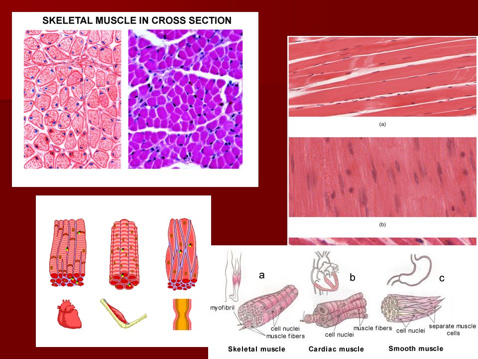
Muscle Tissue Shape: Muscle cells are also called muscle fibers because the cells are elongated (cylindrical or spindle shaped) Function: Length of cell allows for contraction; shorten and thicken Three types

53
Skeletal Muscle Location – attached to bones
Functions – voluntary movement (head, trunk, limbs, facial expressions, write, talk, sing, chew, breathe) and temperature regulation Characteristics: Long (up to 2 inches) and cylindrical Multinucleated Striated (alternating light and dark markings due to pattern of actin and myosin) Voluntary control
 and temperature regulation. Characteristics: Long (up to 2 inches) and cylindrical. Multinucleated. Striated (alternating light and dark markings due to pattern of actin and myosin) Voluntary control.")
54
Skeletal Muscle

55
Cardiac Muscle Location – Heart only
Function – Involuntary pumping of blood from heart into blood vessels Characteristics: Cells: long, cylindrical, branched Single nucleus Striated Involuntarily Controlled Has intercalated discs (gap junctions) – ions that set off contraction spread from cell to cell quickly
 – ions that set off contraction spread from cell to cell quickly.")
56
Cardiac Muscle Intercalated Disc →

57
Smooth Muscle Location: Walls of hollow organs (stomach, intestines, bladder, uterus, blood vessels), glands Function: Squeeze out the contents of an organ such as food through digestive tract or urine from bladder, constricts blood vessels, or squeeze hormones down a duct to the blood Characteristics Spindle-shaped Single central nucleus No striations Involuntarily controlled

58
Smooth Muscle vs. Dense CT
Nuclei cells are between the fibers Long stretched nuclei inside the cells

60
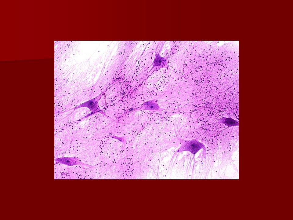
Nervous Tissue Location: Brain, spinal cord, peripheral nerves
Functions: Communicate quickly with other cells Send information from the brain and spinal cord to nervous tissue, muscle tissue, glands Carry input from sensory receptors to the brain and spinal cord

61
Characteristics of Nervous Tissue
Large cell body with axon which is a long extension of cytoplasm (may be up to 3 ft.) Many of the long axons are covered in myelin (a fat) to improve conductivity Cell body also has many short extensions connecting to other close nerve cells called dendrites
 Many of the long axons are covered in myelin (a fat) to improve conductivity. Cell body also has many short extensions connecting to other close nerve cells called dendrites.")
62
Neuron Cell Body Dendrites Axon

64
References http://www.cliffsnotes.com/study_guide/
Holes Human Anatomy and Physiology

Similar presentations


 a. Epithelial b. Connective c. Muscular.>")


