Download presentation
Presentation is loading. Please wait.

1
Required readings: Biomechanics and Motor Control of Human Movement (class text) by D.A. Winter, pp.
2
Next Class Reading assignment Exam on anthropometry
Biomechanics of Skeletal Muscle by T. Lorenz and M. Campello (adapted from M. I. Pitman and L. Peterson; pp EMG by W. Herzog, A. C. S. Guimaraes, and Y. T. Zhang; pp Surface Electromyography: Detecting and Recording The Use of Surface Electromyography in Biomechanics Exam on anthropometry Turn in EMG abstract Prepare short presentation on EMG research article Laboratory experiment on EMG Hour assigned

3
Advanced Biomechanics of Physical Activity (KIN 831)
Muscle – Structure, Function, and Electromechanical Characteristics Material included in this presentation is derived primarily from two sources: Jensen, C. R., Schultz, G. W., Bangerter, B. L. (1983). Applied kinesiology and biomechanics. New York: McGraw-Hill Nigg, B. M. & Herzog, W. (1994). Biomechanics of the musculo-skeletal system. New York: Wiley & Sons Nordin, M. & Frankel, V. H. (1989). Basic Biomechanics of the Musculoskeletal System. (2nd ed.). Philadelphia: Lea & Febiger Winter, D.A. (1990). Biomechanical and motor control of human movement. (2nd ed.). New York: Wiley & Sons
. Applied kinesiology and biomechanics. New York: McGraw-Hill. Nigg, B. M. & Herzog, W. (1994). Biomechanics of the musculo-skeletal system. New York: Wiley & Sons. Nordin, M. & Frankel, V. H. (1989). Basic Biomechanics of the Musculoskeletal System. (2nd ed.). Philadelphia: Lea. & Febiger. Winter, D.A. (1990). Biomechanical and motor control of human movement. (2nd ed.). New York: Wiley & Sons.")
4
Introduction Muscular system consists of three muscle types: cardiac, smooth, and skeletal Skeletal muscle most abundant tissue in the human body (40-45% of total body weight) Human body has more than 430 pairs of skeletal muscle; most vigorous movement produced by 80 pairs
 Human body has more than 430 pairs of skeletal muscle; most vigorous movement produced by 80 pairs.")
5
Introduction (continued)
Skeletal muscles provide strength and protection for the skeleton, enable bones to move, provide the maintenance of body posture against gravity Skeletal muscles perform both dynamic and static work

6
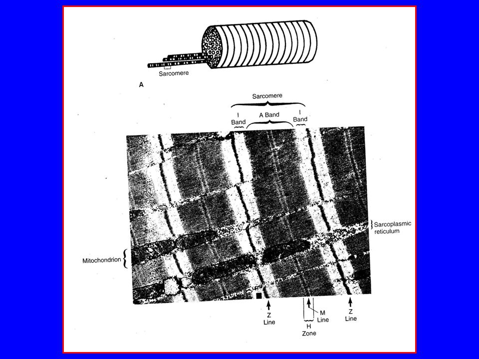
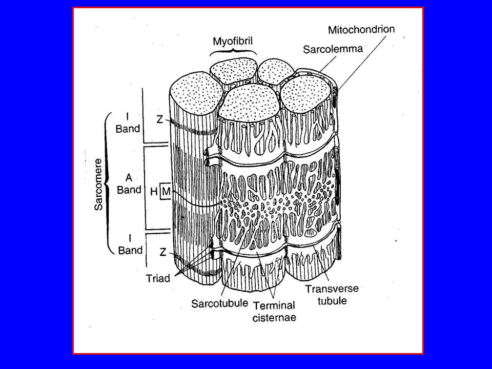
Muscle Structure Structural unit of skeletal muscle is the multinucleated muscle cell or fiber (thickness: m, length: 1-30 cm Muscle fibers consist of myofibrils (sarcomeres in series: basic contractile unit of muscle) Myofibrils consist of myofilaments (actin and myosin)
 Myofibrils consist of myofilaments (actin and myosin)")
7
Microscopic-Macroscopic Structure of Skeletal Muscle

8
Muscle Structure (continued)
Composition of sarcomere Z line to Z line ( m in length) Thin filaments (actin: 5 nm in diameter) Thick filaments (myosin: 15 nm in diameter) Myofilaments in parallel with sarcomere Sarcomeres in series within myofibrils
 Thin filaments (actin: 5 nm in diameter) Thick filaments (myosin: 15 nm in diameter) Myofilaments in parallel with sarcomere. Sarcomeres in series within myofibrils.")
10
Muscle Structure (continued)
Motor unit Functional unit of muscle contraction Composed of motor neuron and all muscle cells (fibers) innervated by motor neuron Follows “all-or-none” principle – impulse from motor neuron will cause contraction in all muscle fibers it innervates or none
 innervated by motor neuron. Follows all-or-none principle – impulse from motor neuron will cause contraction in all muscle fibers it innervates or none.")
12
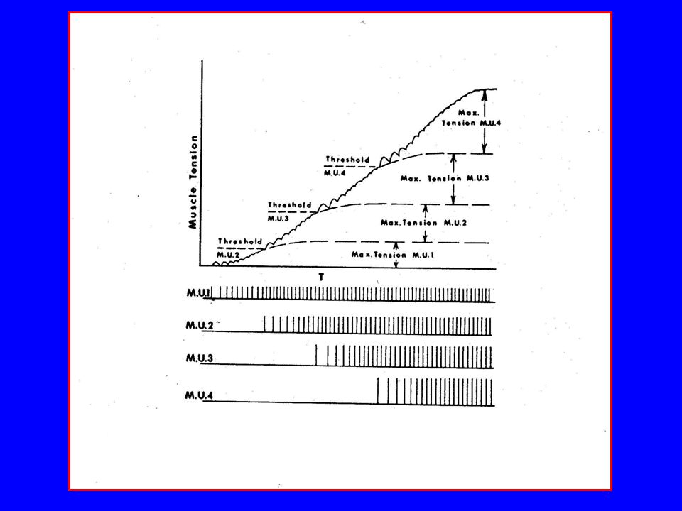
Smallest MU recruited at lowest stimulation frequency
As frequency of stimulation of smallest MU increases, force of its contraction increases As frequency of stimulation continues to increase, but not before maximum contraction of smallest MU, another MU will be recruited Etc.

13
Size Principle Smallest motor units recruited first
Smallest motor units recruited with lower stimulation frequencies Smallest motor units with relatively low levels of tension provide for finer control of movement Larger motor units recruited later with increased frequency of stimulation and increased need for greater tension

14
Size Principle Tension is reduced by the reverse process
Successive reduction of firing rates Dropping out of larger units first

15
Muscle Structure (continued)
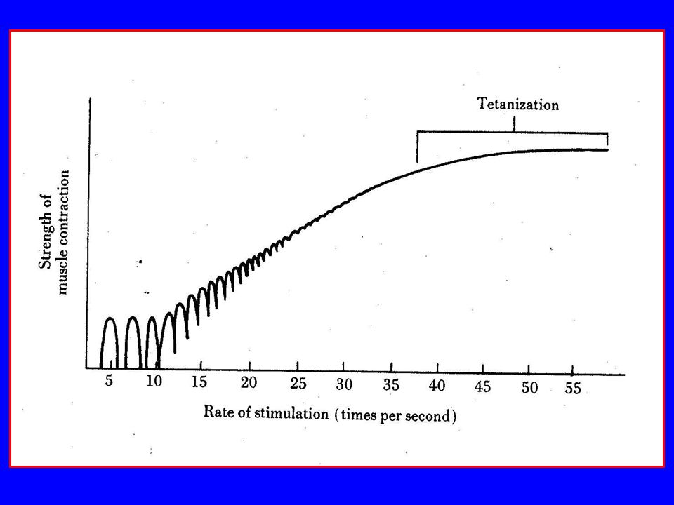
Motor unit Vary in ratio of muscle fibers/motor neuron Fine control – few fibers (e.g., muscles of eye and fingers, as few as 3-6/motor neuron), tetanize at higher frequencies Gross control – many fibers (e.g., gastrocnemius, 2000/motor neuron), tetanize at lower frequencies Fibers of motor unit dispersed throughout muscle
, tetanize at higher frequencies. Gross control – many fibers (e.g., gastrocnemius, 2000/motor neuron), tetanize at lower frequencies. Fibers of motor unit dispersed throughout muscle.")
17
Motor Unit Tonic units – smaller, slow twitch, rich in mitochondria, highly capillarized, high aerobic metabolism, low peak tension, long time to peak (60-120ms) Phasic units – larger, fast twitch, poorly capillarized, rely on anaerobic metabolism, high peak tension, short time to peak (10-50ms)
 Phasic units – larger, fast twitch, poorly capillarized, rely on anaerobic metabolism, high peak tension, short time to peak (10-50ms)")
18
Muscle Structure (continued)
Motor unit (continued) Weakest voluntary contraction is a twitch (single contraction of a motor unit) Twitch times for tension to reach maximum varies by muscle and person Twitch times for maximum tension are shorter in the upper extremity muscles (≈40-50ms) than in the lower extremity muscles (≈70-80ms)
 Weakest voluntary contraction is a twitch (single contraction of a motor unit) Twitch times for tension to reach maximum varies by muscle and person. Twitch times for maximum tension are shorter in the upper extremity muscles (≈40-50ms) than in the lower extremity muscles (≈70-80ms)")
19
Motor Unit Twitch

20
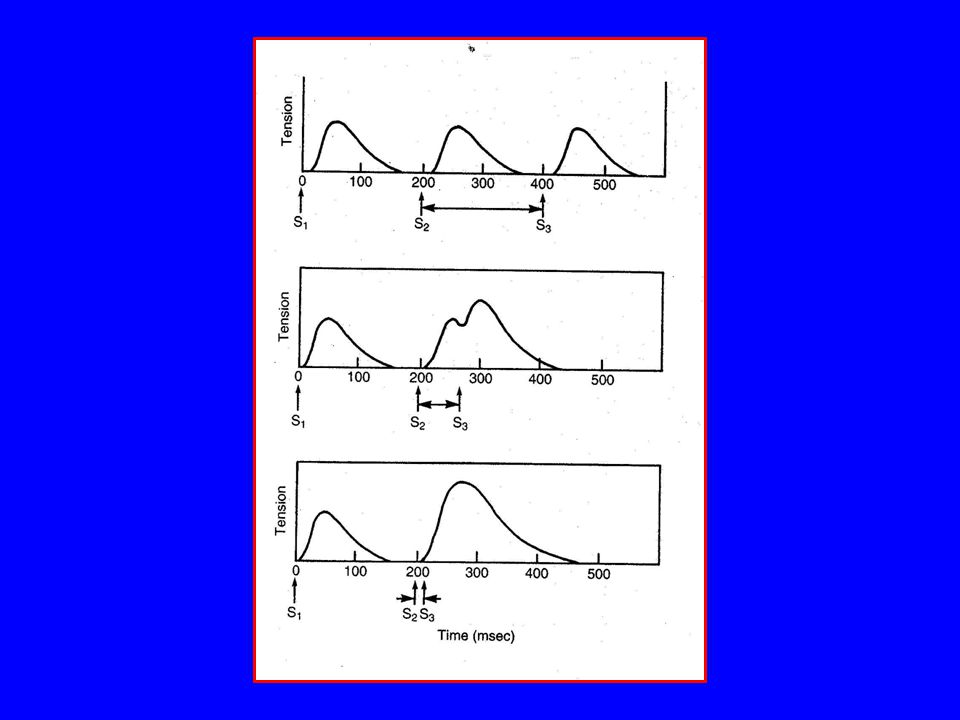
Shape of Graded Contraction

21
Shape of Graded Contraction
Shape and time period of voluntary tension curve in building up maximum tension Due to delay between each MU action potential and maximum twitch tension Related to the size principle of recruitment of motor units Turn-on times ≈ 200ms Shape and time period of voluntary relaxation curve in reducing tension Related to shape of individual muscle twitches Related to the size principle in reverse Due to stored elastic energy of muscle Turn-off times ≈ 300ms

22
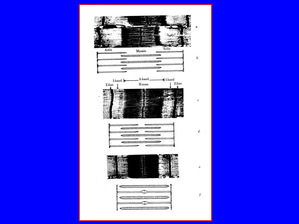
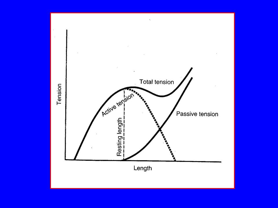
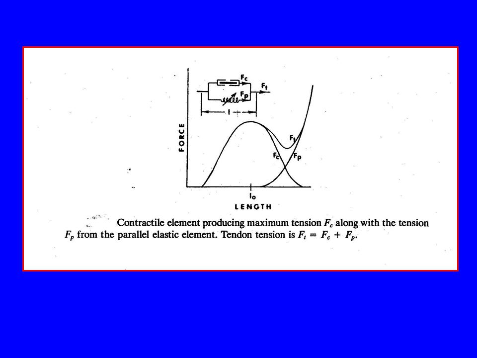
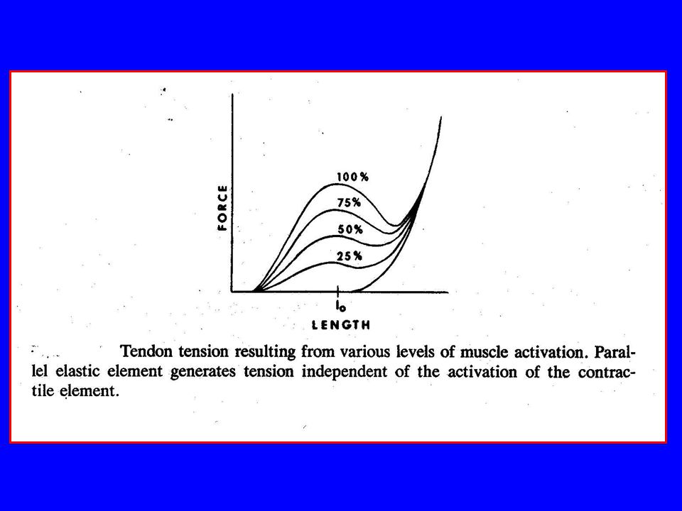
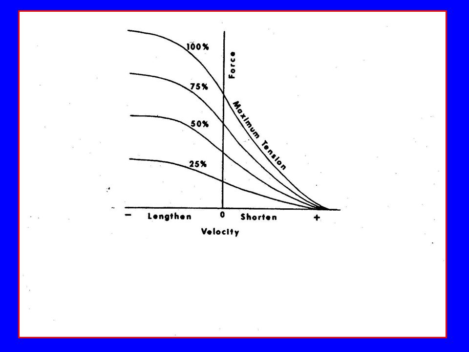
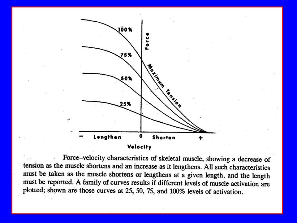
Force Production – Length-Tension Relationship
Force of contraction in a single fiber determined by overlap of actin and myosin (i.e., structural alterations in sarcomere) (see figure) Force of contraction for whole muscle must account for active (contractile) and passive (series and parallel elastic elements) components
 (see figure) Force of contraction for whole muscle must account for active (contractile) and passive (series and parallel elastic elements) components.")
25
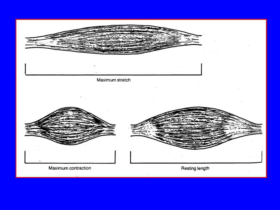
Parallel Connective Tissue
Parallel elastic component Tissues surrounding contractile elements Acts like elastic band Slack when muscle at resting length of less Non-linear force length curve Sarcolemma, endomysium, perimysium, and epimysium forms parallel elastic element of skeletal muscle

29
Series Elastic Tissue Tissues in series with contractile component
Tendon forms series elastic element of skeletal muscle Endomysium, perimysium, and epimysium continuous with connective tissue of tendon Lengthen slightly under isometric contraction (≈ 3-7% of muscle length) Potential mechanism for stored elastic energy (i.e., function in prestretch of muscle prior to explosive concentric contraction)
 Potential mechanism for stored elastic energy (i.e., function in prestretch of muscle prior to explosive concentric contraction)")
30
Isometric Contraction

31
Musculotendinous Unit
Tendon and connective tissues in muscle (sarcolemma, endomysium, perimysium, and epimysium) are viscoelastic Viscoelastic structures help determine mechanical characteristics of muscles during contraction and passive extension
 are viscoelastic. Viscoelastic structures help determine mechanical characteristics of muscles during contraction and passive extension.")
32
Musculotendinous Unit (continued)
Functions of elastic elements of muscle Keep “ready” state for muscle contraction Contribute to smooth contraction Reduce force buildup on muscle and may prevent or reduce muscle injury Viscoelastic property may help muscle absorb, store, and return energy

33
Muscle Model

34
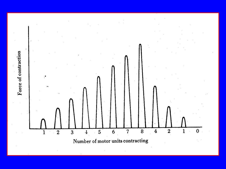
Force Production – Gradation of Contraction
Synchronization (number of motor units active at one time) – more force potential Size of motor units – motor units with larger number of fibers have greater force potential Type of motor units – type IIA and IIB force potential, type I force potential
 – more force potential. Size of motor units – motor units with larger number of fibers have greater force potential. Type of motor units – type IIA and IIB force potential, type I force potential.")
36
Force Production – Gradation of Contraction (continued)
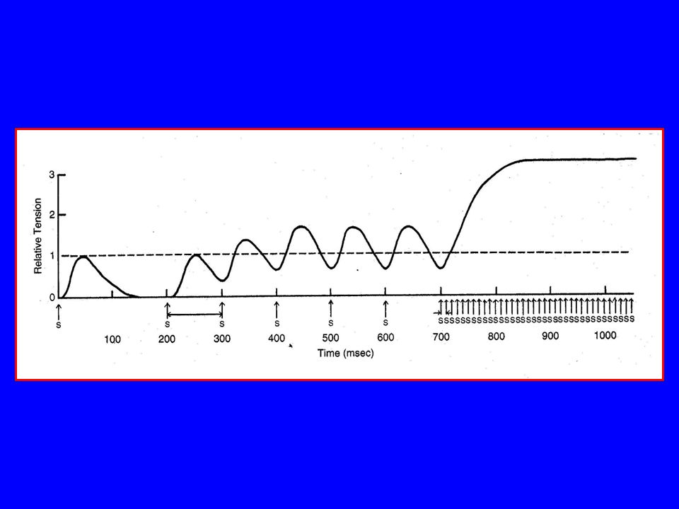
Summation – increase frequency of stimulation, to some limit, increases the force of contraction

40
Force Production – Gradation of Contraction (continued)
Size principle – tension increase Smallest motor units recruited first and largest last Increased frequency of stimulation force of contraction of motor unit Low tension movements can be achieved in finely graded steps Increases frequency of stimulation recruitment of additional and larger motor units Movements requiring large forces are accomplished by recruiting larger and more forceful motor units Size principle – tension decrease Last recruited motor units drop out first

41
Types of Muscle Contraction
Type of Contraction Definition Work Concentric Force of muscle contraction resistance Positive work; muscle moment and angular velocity of joint in same direction Eccentric Force of muscle contraction resistance Negative work; muscle moment and angular velocity of joint in opposite direction Isokinetic Force of muscle contraction = resistance; constant angular velocity; special case is isometric contraction Isometric Force of muscle contraction resistance; series elastic component stretch = shortening of contractile element (few to 7% of resting length of muscle) No mechanical work; physiological work
 No mechanical work; physiological work.")
42
Force Production – Length-Tension Relationship
Difficult to study length-tension relationship Difficult to isolate single agonist Moment arm of muscle changes as joint angle changes Modeling may facilitate this type of study

43
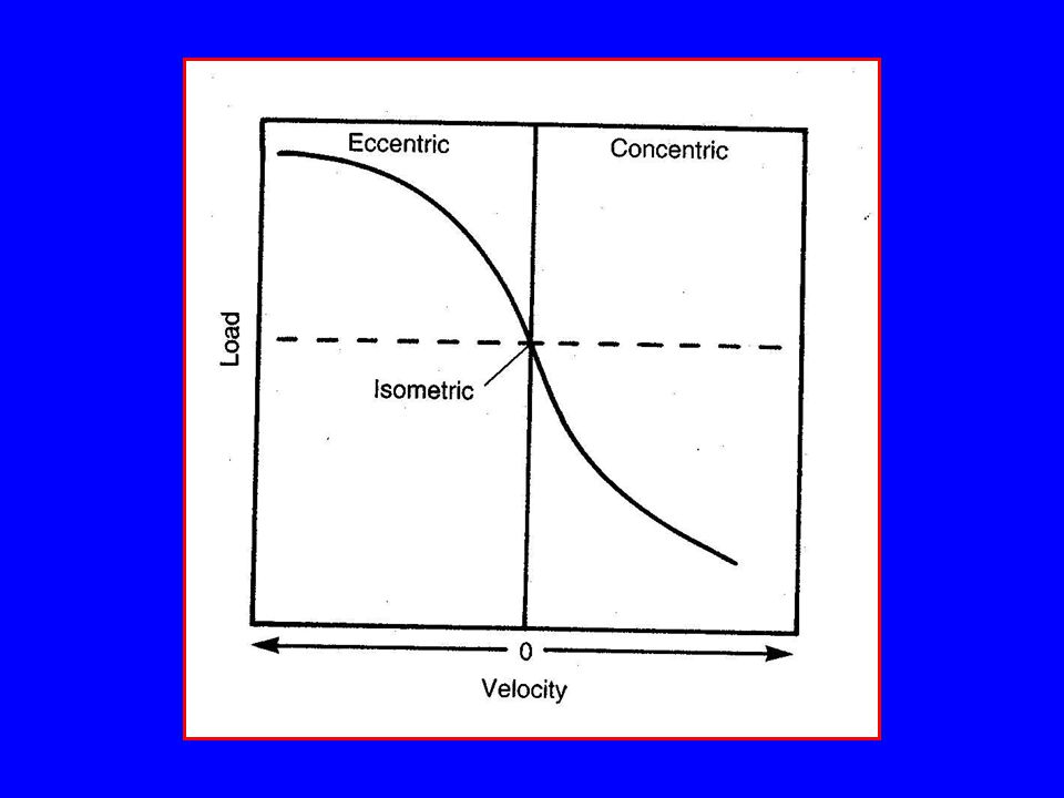
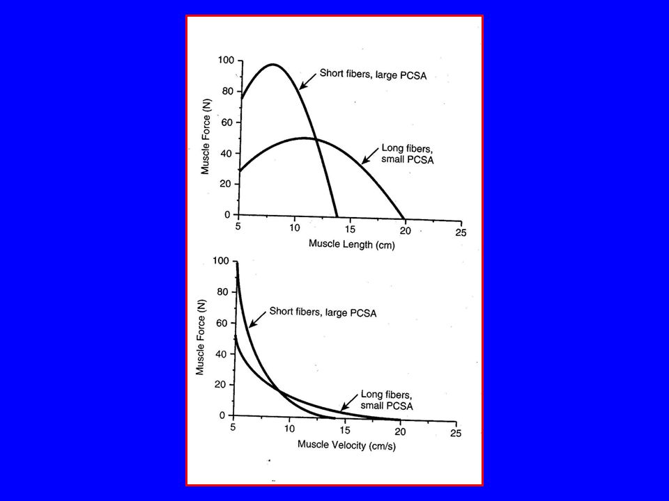
Force Production – Load-Velocity Relationship
Concentric contraction (muscle shortening) occurs when the force of contraction is greater than the resistance (positive work) Velocity of concentric contraction inversely related to difference between force of contraction and external load Zero velocity occurs (no change in muscle length) when force of contraction equals resistance (no mechanical work)
 occurs when the force of contraction is greater than the resistance (positive work) Velocity of concentric contraction inversely related to difference between force of contraction and external load. Zero velocity occurs (no change in muscle length) when force of contraction equals resistance (no mechanical work)")
45
Force Production – Load-Velocity Relationship
Eccentric contraction (muscle lengthening) occurs when the force of contraction is less than the resistance (negative work) Velocity of eccentric contraction is directly related to the difference between force of contraction and external load
 occurs when the force of contraction is less than the resistance (negative work) Velocity of eccentric contraction is directly related to the difference between force of contraction and external load.")
48
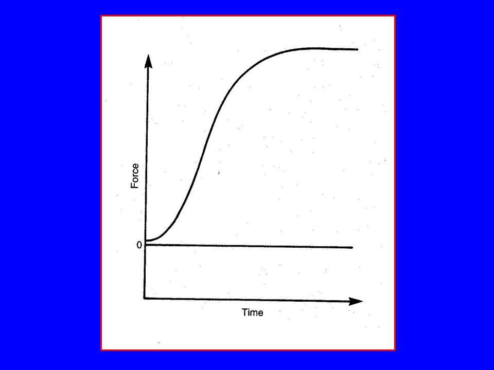
Force Production – Force-Time Relationship
In isometric contractions, greater force can be developed to maximum contractile force, with greater time Increased time permits greater force generation and transmission through the parallel elastic elements to the series elastic elements (tendon) Maximum contractile force may be generated in the contractile component of muscle in 10 msec; transmission to the tendon may take 300msec
 Maximum contractile force may be generated in the contractile component of muscle in 10 msec; transmission to the tendon may take 300msec.")
50
3-D Relationship of Force-Velocity-Length

51
3-D Relationship of Force-Velocity-Length

52
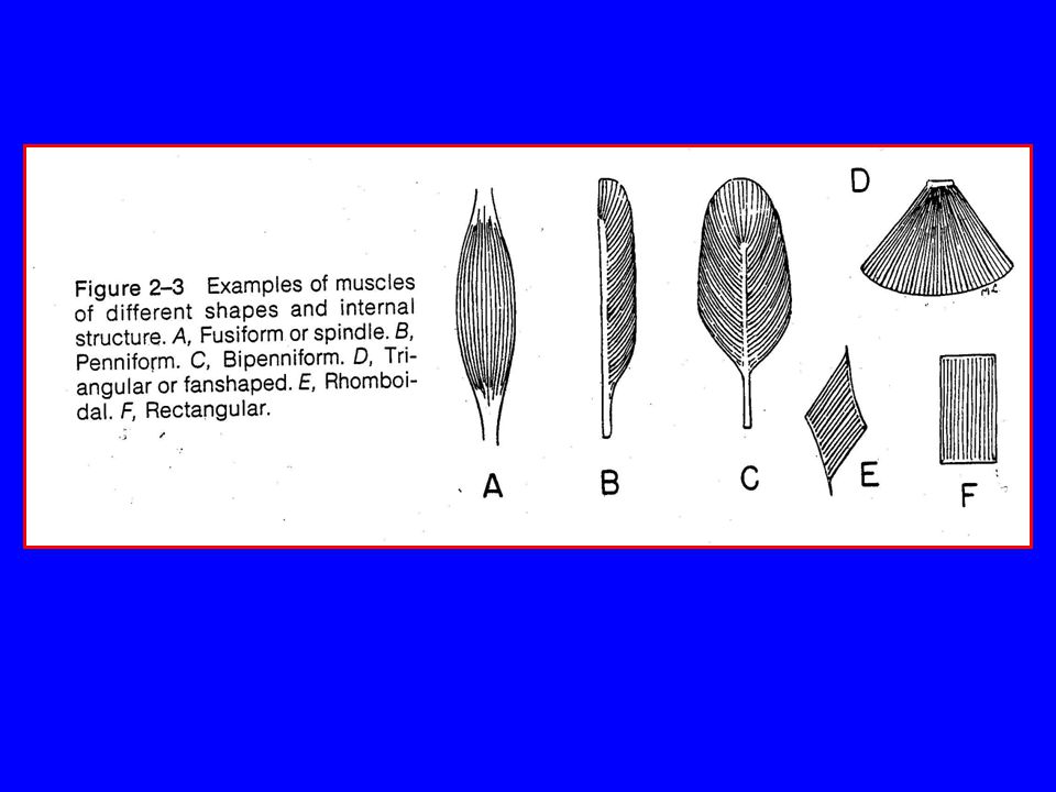
Effect of Muscle Architecture on Contraction
Fusiform muscle Fibers parallel to long axis of muscle Many sarcomeres make up long myofibrils Advantage for length of contraction Example: sartorius muscle Force of contraction along long axis of muscle of force of contraction of all muscle fibers Tends to have smaller physiological cross sectional area (see figure)
")
53
Fusiform Fiber Arrangement
Fa = force of contraction of muscle fiber parallel to longitudinal axis of muscle Fa = sum of all muscle fiber contractions parallel to long axis of muscle Fa

54
Effect of Muscle Architecture on Contraction (continued)
Pennate muscle Fibers arranged obliquely to long axis of muscle (pennation angle) Uni-, bi-, and multi-pennate Advantage for force of contraction Example: rectus femoris (bi-pennate) Tends to have larger physiological cross sectional area
 Uni-, bi-, and multi-pennate. Advantage for force of contraction. Example: rectus femoris (bi-pennate) Tends to have larger physiological cross sectional area.")
55
Pennate Fiber Arrangement
Fa = force of contraction of muscle fiber parallel to longitudinal axis of muscle Fm = force of contraction of muscle fiber = pennation angle Fa = (cos )(Fm) Fa = sum of all muscle fiber contractions parallel to long axis of muscle Fa Fm
(Fm) Fa = sum of all muscle fiber contractions parallel to long axis of muscle. Fa. Fm. ")
58
Effect of Muscle Architecture on Contraction (continued)
Force of muscle contraction proportional to physiological cross sectional area (PCSA); sum of the cross sectional area of myofibrils Velocity and excursion (working range or amplitude) of muscle is proportional to length of myofiblril
; sum of the cross sectional area of myofibrils. Velocity and excursion (working range or amplitude) of muscle is proportional to length of myofiblril.")
60
Muscle Fiber Types Type I Slow-Twitch Oxidative (SO) Type IIA
Fast-Twitch Oxidative-Glycolytic (FOG) Type IIB Fast-Twitch Glycolytic (FG) Speed of contraction Slow Fast Primary source of ATP production Oxidative phosphorylation Anaerobic glycolysis Glycolytic enzyme activity Low Intermediate High Capillaries Many Few Myoglobin content Glycogen content Fiber diameter Small Large Rate of fatigue
 Type IIB. Fast-Twitch Glycolytic (FG) Speed of contraction. Slow. Fast. Primary source of ATP production. Oxidative phosphorylation. Anaerobic glycolysis. Glycolytic enzyme activity. Low. Intermediate. High. Capillaries. Many. Few. Myoglobin content. Glycogen content. Fiber diameter. Small. Large. Rate of fatigue.")
61
Muscle Fiber Types (continued)
Smaller slow twitch motor units are characterized as tonic units, red in appearance, smaller muscle fibers, fibers rich in mitochondria, highly capillarized, high capacity for aerobic metabolism, and produce low peak tension in a long time to peak (60-120ms). Larger fast twitch motor units are characterized as phasic units, white in appearance, larger muscle fibers, less mitochondria, poorly capillarized, rely on anaerobic metabolism, and produce large peak tensions in shorter periods of time (10-50ms).
. Larger fast twitch motor units are characterized as phasic units, white in appearance, larger muscle fibers, less mitochondria, poorly capillarized, rely on anaerobic metabolism, and produce large peak tensions in shorter periods of time (10-50ms).")
62
Muscle Fiber Types (continued)
Nerve innervating muscle fiber determines its type; possible to change fiber type by changing innervations of fiber All fibers of motor unit are of same type Fiber type distribution in muscle genetically determined Average population distribution: 50-55% type I 30-35% type IIA 15% type IIB

63
Muscle Fiber Types (continued)
Fiber composition of muscle relates to function (e.g., soleus – posture muscle, high percentage type I) Muscles mixed in fiber type composition Natural selection of athletes at top levels of competition
 Muscles mixed in fiber type composition. Natural selection of athletes at top levels of competition.")
64
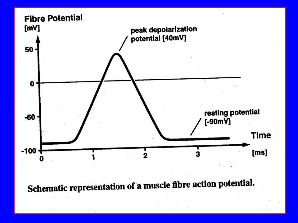
Electrical Signals of Muscle Fibers
At rest, action potential of muscle fiber -90 mV;caused by concentrations of ions outside and inside fiber (resting state) With sufficient stimulation, potential inside cell raised to mV (depolarization); associated with transverse tubular system and sarcoplasmic reticulum; causes contraction of fiber Return to resting state (repolarization) Electrical signals from the motor units (motor unit action potential, muap) can be recorded (EMG) via electrodes
 With sufficient stimulation, potential inside cell raised to mV (depolarization); associated with transverse tubular system and sarcoplasmic reticulum; causes contraction of fiber. Return to resting state (repolarization) Electrical signals from the motor units (motor unit action potential, muap) can be recorded (EMG) via electrodes.")
Similar presentations


 Muscle Composition endomysium – loose CT surrounding each fiber perimysium – dense CT that bundles multiple.>")