Download presentation
Presentation is loading. Please wait.

1
Muscle Tissue and Organization
Muscular System Muscle Tissue and Organization

2
Muscle Tissue Muscle tissue is one of the 4 primary tissue types
Three types of muscle tissue Skeletal – moves the body Cardiac – heart muscle Smooth – moves fluid and solids through the digestive tract

3
Characteristics of Muscle Types
SKELETAL Voluntary Striated Multinucleate CARDIAC Involuntary Single nuclei Intercalated discs SMOOTH Not striated Single nucleus

4
Features of Muscle Tissue
Contractility – ability to shorten and pull Excitability – responds to stimuli Elasticity – muscle can rebound to its original shape after contraction Extensibility – ability to contract over a range of resting lengths

5
Functions of Skeletal Muscle
Movement Posture Stabilize joints Support soft tissue Generation of heat Regulate entrances and exits (orifices)
")
6
Muscle Attachment TENDONS attach muscle to bone Dense regular CT
Each muscle has an ORIGIN and INSERTION, and a specific ACTION The origin remains stationary while the insertion moves

7
Connective Tissue of Muscle
Skeletal muscle has three layers of connective tissue 1. EPIMYSIUM – dense irregular CT that surrounds the entire muscle 2. PERIMYSIUM – divides muscle into compartments or bundles of muscle fibers called FASCICLES 3. ENDOMYSIUM – surrounds each muscle fiber (muscle cell)
")
10
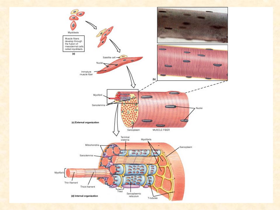
Muscle Cell Terminology
Muscle cells are very long; muscle fibers SARCOLEMMA – cell membrane SARCOPLASM – cytoplasm Sarcoplasm is filled with thousands of MYOFIBRILS that are responsible for contraction Myofibrils are composed of MYOFILAMENTS Myofilaments are composed of the proteins ACTIN and MYOSIN

12
Sarcomere Organization of thick(myosin) and thin filaments(actin) in the myofibrils Movements of these filaments causes muscle contraction Sliding filament theory 1954 Sir Andrew Huxley and Rolf Niedeigerke Myosin heads bind to the actin and pull or “slide” the actin past the myosin to shorten the sarcomere

13
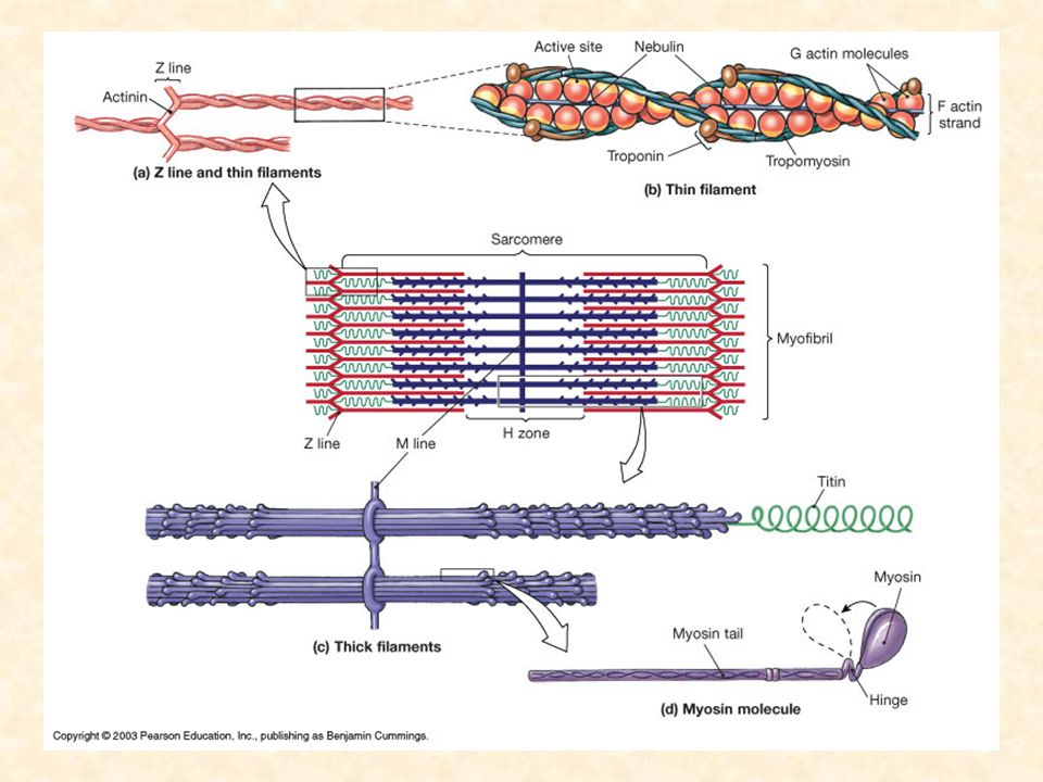
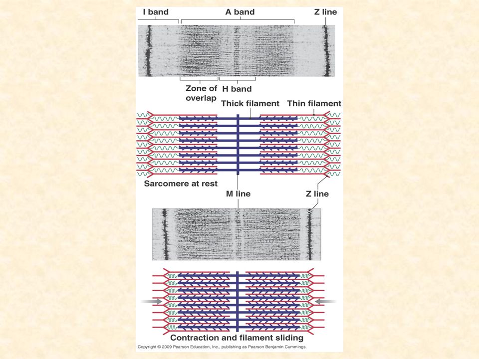
Sarcomere Z line = end of sarcomere I band = thin (actin) filaments
H zone = thick (myosin) filaments A band = zone of overlap, thick and thin
 filaments. A band = zone of overlap, thick and thin.")
14
Thin Filaments Twisted strands of globular G actin molecules
Each molecule of G actin has an active site that can bind to a myosin molecule Thin filaments also have two other proteins associated it Tropomyosin – covers active sites on actin Troponin - holds tropomyosin in place

15
Thick filaments Bundles of myosin molecules
About 500 myosin molecules per bundle Myosin molecules have heads that can cross bridge to actin active sites The binding of myosin heads to actin result in muscle contraction

17
Figure 9.4b Sarcomere Structure

18
Muscle contraction-Sliding filament theory
Contraction exerts a pull – tension Interaction between actin and myosin triggered by calcium ions and presence of ATP Sliding filament theory: H band and I band get smaller Zone of overlap gets larger Z lines move closer together Width of A band remains constant

19
Sliding Filament Theory
Myosin heads cross bridge to the actin active sites Myosin attachment “pulls” the actin toward the center of the sarcomere Contraction begins with release of Ca2+ from the terminal cisternae of the sarcoplasmic reticulum The release of ions is the result of electrical stimulation of the muscle fiber

20
Terminal cisternae and T tubules

21
T-tubules The t-tubules distribute the electrical signal for contraction deep into the muscle fiber As the signal travels the terminal cisternae release calcium ions Release of calcium cause the troponin molecule to change shape Change in troponin causes a change in the position of tropomyosin, myosin can bind to action and contraction occurs!

22
Nervous System Control of Contraction
Skeletal muscle fibers are controlled by a motor neuron Place where the nerve fiber and muscle meet is called the neuromuscular junction Synaptic terminal – end of axon Acetylcholine – neurotransmitter Motor unit – all the muscle fiber controlled by a single motor neuron

23
Neuromuscular Junction

24
Neuromuscular junction

25
Muscle contraction

26
Muscle relaxation

28
Sarcomere contraction

29
Sliding filaments

30
Sliding filaments Sarcomere shortens, fiber contracts

31
Other components of the sarcomere
Sarcoplasmic reticulum – stores Ca+² ions for muscle contraction Transverse tubules – carry impulse to stimulate and coordinate contraction

32
Sarcoplasmic reticulum

33
Types of Skeletal Muscle Fibers
Fast fibers Short duration, rapid fatigue Anaerobic metabolism Few mitochondria Brief periods of intense exercise Slow fibers Longer duration Aerobic metabolism Myoglobin present for oxygen binding Marathon running Intermediate fibers Greater resistance to fatigue Similar to fast fibers but with more mitochondria

35
Organization of Muscle Fibers
Parallel Convergent Unipennate Bipennate Multipennate Circular

38
Muscles and Leverage Muscles “work” by leverage, moving at a joint
In the body the bone is the lever, the joint is the fulcrum Three types of levers in the body First-class Second-class Third-class (most common)
")
42
Muscle Actions Prime movers Synergist Antagonists
Muscle mainly responsible for producing a certain action Biceps brachii is prime mover for elbow flexion Synergist Assists the prime mover Antagonists Action opposite of prime mover Triceps brachii is antagonist of biceps brachii

Similar presentations


 –sheet or band of fibrous C.T. under.>")


